Retrograde Intramedullary Nailing for Distal Femur Fracture with Osteoporosis
Jihyeung Kim, MD, Seung-Baik Kang, MD, Kyungpyo Nam, MD, Seung Hwan Rhee, MD, Jong Won Won, MD, Hyuk-Soo Han, MD
Department of Orthopedic Surgery, Seoul Metropolitan Government Seoul National University Boramae Medical Center, Seoul, Korea
Received December 15, 2011; Accepted April 9, 2012 Correspondence to: Seung-Baik Kang, MD
Department of Orthopedic Surgery, Seoul Metropolitan Government Seoul National University Boramae Medical Center, 20 Boramae-ro 5-gil, Dongjak-gu, Seoul 156-707, Korea
Tel: +82-2-870-2313, Fax: +82-2-870-3866 E-mail: [email protected]
Epidemiologic study in the United Kingdom found distal femur fracture accounts for 0.4% of adult fractures, with an average patient age of 61 years.1) A population based study2) in Olmsted County, Minnesota, USA suggested that non-hip femoral fractures increased between 1984 and 2007, largely because of an increase in moderate trauma fractures in older women over this period. In their study, distal femur fracture comprised 29% of cases. Kan- nus et al.3) also reported a rising problem of osteoporotic
Background: The incidence of distal femur fracture in the elderly has been increasing recently, and commonly occurs with osteo- porosis. Retrograde intramedullary nailing has been considered a good surgical option for distal femur fracture. The purpose of the present study was to present our surgical results with retrograde intramedullary nailing for distal femur fractures with osteoporo- sis.
Methods: Thirteen patients diagnosed with extra-articular distal femur fracture and osteoporosis and managed with retrograde intramedullary nailing were retrospectively reviewed. Cement augmentation was used in four patients, shape memory alloy was used in eight patients and both were used in one patient. All patients were followed up for more than 2 years. Radiologic align- ments were scored and Tegner and the Lysholm activity score was used for a functional assessment.
Results: The average time to clinical union was 13 weeks (range, 10 to 15 weeks). In 12 of our cases, the total alignment scores were excellent. At the last follow-up, the mean range of motion was 116° (range, 110° to 125°). The average functional score at postoperative 1 year was 2.6 (range, 1 to 5).
Conclusions: Retrograde intramedullary nailing is a good surgical option for distal femur fracture with osteoporosis. Cement augmentation and shape memory alloy can also be used for added mechanical stability. This surgical technique is very useful for distal femur fracture with osteoporosis as it promotes fracture healing and early rehabilitation.
Keywords: Distal femur fracture, Osteoporosis, Intramedullary nailing, Cement augmentation, Shape memory alloy
knee fractures in elderly women. Since the incidence of distal femur fracture has been increasing recently and commonly occurs with osteoporosis, we should pay more attention to the management of distal femur fracture with osteoporosis.
Compared to plate osteosynthesis, intramedullary fixation requires less extensive dissection and is better biomechanically. In a biomechanical study, the stiffness of the intramedullary nail was better than a dynamic con- dylar screw or a locking compression plate.4) Retrograde intramedullary nailing is a good surgical option in the management of distal femur fracture with osteoporosis.
Scheerlinck et al.5) reported good surgical results for twelve elderly and osteoporotic patients with a femoral supracon- dylar nail.
However, in the treatment of osteoporotic distal fe- mur fracture, it is very difficult to obtain sufficient implant
Copyright © 2012 by The Korean Orthopaedic Association
This is an Open Access article distributed under the terms of the Creative Commons Attribution Non-Commercial License (http://creativecommons.org/licenses/by-nc/3.0) which permits unrestricted non-commercial use, distribution, and reproduction in any medium, provided the original work is properly cited.
Clinics in Orthopedic Surgery • pISSN 2005-291X eISSN 2005-4408
anchorage. Wahnert et al.6) suggested that distal locking has a major impact on the implant anchorage in osteopo- rotic bone, and concluded that a supracondylar nail should be considered for mobile patients where early postopera- tive mobilization for rehabilitation is required. Cement augmentation has been used to stabilize intramedullary nailing. Dall’Oca et al.7) managed osteoporotic intertro- chanteric fracture with cement augmentation to improve mechanical stability of the implant. Roth et al.8) suggested cement augmented proximal screws to improve stability in the management of the proximal tibia fracture with os- teoporosis. Shape memory alloy (Bio-smart, Ulsan, Korea) has also been used with retrograde intramedullary nailing in the management of periprosthetic supracondylar frac- tures.9) Our study was undertaken to analyze clinical out- comes of retrograde intramedullary nailing with cement augmentation and/or shape memory alloy for distal femur fracture with osteoporosis.
METHODS
We retrospectively reviewed 20 patients who were admit- ted to our hospital between January 2005 and October 2009 for extra-articular distal femur fracture with osteo- porosis and tracked for more than 2 years. Bone mineral density was evaluated with biplanar radiographs and
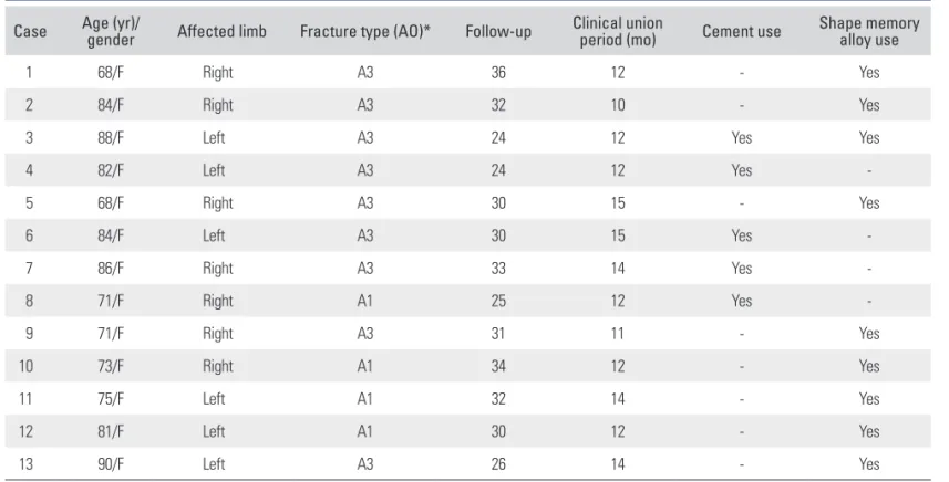
dual-energy X-ray absorptiometry (DEXA) scanning in the hip and lumbar spine. In all of our cases, the lowest T-score was less than -2.5 standard deviation. Of the 20, 16 patients were managed with retrograde intramedullary nailing (AIM titanium supracondylar nail, Depuy ACE, Leeds, UK). Cement augmentation or shape memory alloy (Bio-smart) was combined with retrograde intramedullary nailing in 13 patients to improve mechanical stability. We included these 13 patients in this study (Table 1). Their mean age was 79 years (range, 68 to 90 years) and all were females. Four cases were type A1 and 9 cases were type A3 according to the AO Foundation and Orthopaedic Trauma Association (AO-OTA) classification system.10) The Insti- tutional Review Board of our hospital reviewed and ap- proved this study.
All operations were performed by one orthopedic surgeon. The patient was placed in the supine position on a radiolucent operating table. A midline incision and me- dial parapatellar approach were used. A guide wire was in- serted under C-arm fluoroscopy and the medullary cavity was reamed. When we inserted distal interlocking screws, we checked the bone stock quality in the distal fragment.
We added cement augmentation or shape memory alloy, if rigid fixations between distal fragment and distal in- terlocking screws were not achieved due to configuration and location of the fracture or severe comminution. If the
Table 1. Patient Demographics and Clinical Data Case Age (yr)/
gender Affected limb Fracture type (AO)* Follow-up Clinical union
period (mo) Cement use Shape memory alloy use
1 68/F Right A3 36 12 - Yes
2 84/F Right A3 32 10 - Yes
3 88/F Left A3 24 12 Yes Yes
4 82/F Left A3 24 12 Yes -
5 68/F Right A3 30 15 - Yes
6 84/F Left A3 30 15 Yes -
7 86/F Right A3 33 14 Yes -
8 71/F Right A1 25 12 Yes -
9 71/F Right A3 31 11 - Yes
10 73/F Right A1 34 12 - Yes
11 75/F Left A1 32 14 - Yes
12 81/F Left A1 30 12 - Yes
13 90/F Left A3 26 14 - Yes
*Fracture classification in the AO Foundation and Orthopaedic Trauma Association.
fracture line was located above the flare of the femoral condyle and the configuration of the fracture was spiral or long oblique, we used shape memory alloy (Fig. 1, Table 1).
Even in the A3 type fracture, if the configuration of major fragments was spiral or long oblique, we applied shape memory alloy. When we applied shape memory alloy, we paid close attention to preserve periosteum and applied shape memory alloy outside of the periosteum. To improve mechanical stability of the bone implant construct and to prevent pulling out of the interlocking screw, we used ce- ment augmentation (Figs. 2 and 3, Table 1). In the A3 type fracture with anterior cortical comminution, we also used cement augmentation (Fig. 3). From the 1st postoperative day, joint exercise using a continuous passive motion ma- chine was started and weight bearing was allowed at the 6th postoperative week.
Postoperative assessments were done on an outpa- tient basis at the 4th, 8th, 12th, and 24th postoperative week and annually thereafter. Clinical fracture union was defined by bridging callus formation on the anteropos-
Fig. 1. (A) Preoperative radiographs of the left knee in an 81-year-old woman. (B) Radiographs taken 1 year after surgery.
We performed retrograde intramedullary nailing and used shape memory alloy to reinforce mechanical stability. Bony union was achieved without complication.
Fig. 2. (A) Preoperative radiographs of the right knee in a 71-year-old woman. (B) Radiographs taken 1 year after surgery.
We performed retrograde intramedullary nailing with cement augmentation. We achieved good mechanical stability of the bone implant construct and recommended early rehabilitation.
Fig. 3. Intraoperative photograph of cement augmentation. There were anterior cortical defects in the distal femur and comminution in the medial aspect of the distal fragment. We performed cement augmentation in the anterior and medial aspect of the distal femur for mechancial stability and prevention of loosening of the interlocking screw.
terior (AP) and lateral radiographs and pain free weight bearing. If we used cement augmentation for filling in the anterior cortex with severe comminution, we evaluated bridging callus formation in the medial and lateral cortices on the AP radiograph and posterior cortex on the lateral radiograph. The alignments of the distal femurs were measured on the postoperative 1 year radiographs (short- ening, varus/valgus, antecurvatum/retrocurvatum). And the measurements were scored and assessed as showed in Table 2.11) Tegner and Lysholm activity score was used for functional assessments.12)
RESULTS
The postoperative follow-up period was 30 months (range, 24 to 36 months). The average time to clinical union was 13 weeks (range, 10 to 15 weeks). In the assessment of the radiographic alignment, the average score for shortening was 3.8 (range, 2 to 4). That of varus/valgus alignments was 3.4 (range, 1 to 4) and that of antecurvatum/retrocur- vatum was 3.6 (range 2 to 4). In 12 of our cases, the total alignment scores were excellent. At the last follow-up, the mean range of motion was 116° (range, 110° to 125°). The average pre-injured functional score was 4.2 (range 2 to 5) and the average functional score at the 1 year postopera- tive time point was 2.6 (range, 1 to 5).
Postoperatively, there were no complications such as infection, neurovascular injury and implant failure. All patients were satisfied with the surgical results.
DISCUSSION
Distal femur fracture with osteoporosis is one of the un- solved problems in orthopedic and trauma surgery. The incidence of this fracture is bimodal, appearing mostly in young men and old women.13) Recently the incidence of osteoporotic knee fractures has been rising,3) and more than 50% of patients with distal femur facture occur in el- derly patients.2) In the management of distal femur fracture
with osteoporosis, early rehabilitation is very important for satisfactory surgical results since most are elderly pa- tients. For early rehabilitation, mechanical stability should be maintained. There are several surgical options such as an angled blade plate, a locking plate and retrograde in- tramedullary nailing for stable fixation and early exercise.
Open reduction and plating is advantageous in achieving anatomical reduction because a direct view of the fracture site is possible during surgery. However, there is a pos- sibility that an invasive incision and soft tissue stripping will cause complications such as nonunion, delayed union and infection and might prevent early rehabilitation. There also have been reported several complications of healing including nonunion, delayed union, and implant failure in the management of distal femur fracture with locking plates.14) If there is severe comminution in the distal femur fracture, internal fixation with locking plate is very diffi- cult technically.
Retrograde intramedullary nailing is a good surgical option for distal femur fracture. We can avoid invasive soft tissue dissection and minimize secondary damage of the blood circulation at the fracture site. In a previous biome- chanical study, intramedullary nails had significantly high- er stiffness and significantly lower micromotion across the fracture gap with axial compression than with a dynamic condylar screw or a locked condylar plate.4) Therefore, early rehabilitation is possible with the use of intramedul- lary nailing. However, in the osteoporotic patient with se- vere comminution, it is very difficult to achieve sufficient implant anchorage. In the management of distal femur fracture, distal locking has a major impact on the implant anchorage in osteoporotic bone.6) Tejwani et al.15) studied the effect of locked distal screws in retrograde nailing of osteoporotic distal femur fractures. They concluded that the locked distal screw nails exhibited less fracture col- lapse and anterior and medial translation of the nail at the fracture site than unlocked distal screw nails. Ito et al.16) compared distal locking with conventional locking bolts and a bladelike device with retrograde intramedullary Table 2. Radiological Assessment of Radiographs and Scoring of the Final Radiological Results after Fracture Consolidation
Shortening (mm) Varus/valgus (°) Antecurvatum/retrocurvatum (°) Total scoring
0-9 (4 points) 0-3 (4 points) 0-3 (4 points) Excellent (10-12 points)
10-19 (3 points) 4-7 (3 points) 4-7 (3 points) Good (7-9 points)
20-29 (2 points) 8-12 (2 points) 8-12 (2 points) Fair (4-6 points)
> 30 (1 points) > 12 (1 points) > 12 (1 points) Poor (1-3 points)
Reprinted from Handolin et al.11) with permission from Elsevier.
nailing for osteoporotic supracondylar fractures. In their study, interlocking with a bladelike device was 41% stiffer and 20% stronger than that with conventional locking bolts, and there was no gross deformation in the bladelike device after biomechanical testing. If we cannot achieve mechanical stability with retrograde intramedullary nail- ing, we need to add procedures for rigid fixation. For this, cement augmentation has been introduced.7,8) We can not only achieve mechanical stability with cement augmenta- tion but also prevent the interlocking screw from pulling out. Shape memory alloy is also one of the good surgical options for reinforcing a bone implant construct when we use retrograde intramedullary nailing for distal femur fracture. At cold temperatures, the nature of shape memo- ry alloy is more flexible and it becomes hardened at body temperatures. The advantage of shape memory alloy is that it is very easy to manipulate and it can apply compression force between fractured segments.
Several previous studies5,11,17,18) reported good results with retrograde intramedullary nailing for distal femur fracture. In a series of 44 consecutive patients with 46 distal femur fractures,11) the final union rate was 95% and a mean union time was 17.5 weeks. However, there were three patients with a loss of reduction and two of them had a re-operation. Gurkan et al.17) presented 16 patients with distal femur fracture treated with retrograde locked intra- medullary nailing. The mean time to union was 25 weeks and functional results using the modified Hospital for Spe- cial Surgery knee rating scale were satisfactory. However,
among their series, joint range of motion was 80 degrees in four knees (24%) and below 80 degrees in one knee (6%). Bei et al.19) reported that several factors may affect knee joint function recovery including age, preoperative the American Society of Anesthesiologists classification, fracture type, reduction quality, whether or not there was continuous passive motion functional training, and post- operative complications. However there is a possibility of complication such as infection, septic arthritis of the knee, knee pain and malunion. In the meta-analysis performed by Papadokostakis et al.,20) the incidence of infection was 1.1%, and that of septic arthritis of the knee was 0.18%.
The rates of knee pain and malunion were 16.5% and 5.2%.
However, in our cases there were no complications such as infection, septic arthritis, deep vein thrombosis, and im- plant failure. Radiologic and functional results were also satisfactory.
Even though the number of patients in our study was small and the study retrospective, retrograde intra- medullary nailing seems to be a good surgical option for distal femur fracture with osteoporosis. Fixation with ce- ment augmentation and/or shape memory alloy promotes fracture healing and early rehabilitation.
CONFLICT OF INTEREST
No potential conflict of interest relevant to this article was reported.
REFERENCES
1. Court-Brown CM, Caesar B. Epidemiology of adult frac- tures: a review. Injury. 2006;37(8):691-7.
2. Ng AC, Drake MT, Clarke BL, et al. Trends in subtrochan- teric, diaphyseal, and distal femur fractures, 1984-2007. Os- teoporos Int. 2012;23(6):1721-6.
3. Kannus P, Niemi S, Palvanen M, et al. Continuously rising problem of osteoporotic knee fractures in elderly women:
nationwide statistics in Finland in 1970-1999 and predic- tions until the year 2030. Bone. 2001;29(5):419-23.
4. Heiney JP, Barnett MD, Vrabec GA, Schoenfeld AJ, Baji A, Njus GO. Distal femoral fixation: a biomechanical compari- son of trigen retrograde intramedullary (i.m.) nail, dynamic condylar screw (DCS), and locking compression plate (LCP) condylar plate. J Trauma. 2009;66(2):443-9.
5. Scheerlinck T, Krallis P, Descamps PY, Hardy D, Delince P.
The femoral supracondylar nail: preliminary experience.
Acta Orthop Belg. 1998;64(4):385-92.
6. Wahnert D, Hoffmeier K, Frober R, Hofmann GO, Muck- ley T. Distal femur fractures of the elderly: different treat- ment options in a biomechanical comparison. Injury.
2011;42(7):655-9.
7. Dall'Oca C, Maluta T, Moscolo A, Lavini F, Bartolozzi P. Ce- ment augmentation of intertrochanteric fractures stabilised with intramedullary nailing. Injury. 2010;41(11):1150-5.
8. Roth SE, Kreder H, Stephen D, Whyne CM. Biomechanical stability of intramedullary nailed high proximal third tibial fractures with cement augmented proximal screws. J Orthop Trauma. 2005;19(7):457-61.
9. Han HS, Oh KW, Kang SB. Retrograde intramedullary nailing for periprosthetic supracondylar fractures of the femur after total knee arthroplasty. Clin Orthop Surg.
2009;1(4):201-6.
10. Fracture and dislocation compendium. Orthopaedic Trau- ma Association Committee for Coding and Classification. J
Orthop Trauma. 1996;10 Suppl 1:v-ix, 1-154.
11. Handolin L, Pajarinen J, Lindahl J, Hirvensalo E. Retrograde intramedullary nailing in distal femoral fractures: results in a series of 46 consecutive operations. Injury. 2004;35(5):517- 22.
12. Tegner Y, Lysholm J. Rating systems in the evalua- tion of knee ligament injuries. Clin Orthop Relat Res.
1985;(198):43-9.
13. Martinet O, Cordey J, Harder Y, Maier A, Buhler M, Bar- raud GE. The epidemiology of fractures of the distal femur.
Injury. 2000;31 Suppl 3:C62-3.
14. Henderson CE, Kuhl LL, Fitzpatrick DC, Marsh JL. Lock- ing plates for distal femur fractures: is there a problem with fracture healing? J Orthop Trauma. 2011;25 Suppl 1:S8-14.
15. Tejwani NC, Park S, Iesaka K, Kummer F. The effect of locked distal screws in retrograde nailing of osteoporotic distal femur fractures: a laboratory study using cadaver fe- murs. J Orthop Trauma. 2005;19(6):380-3.
16. Ito K, Hungerbuhler R, Wahl D, Grass R. Improved intra- medullary nail interlocking in osteoporotic bone. J Orthop Trauma. 2001;15(3):192-6.
17. Gurkan V, Orhun H, Doganay M, et al. Retrograde intra- medullary interlocking nailing in fractures of the distal fe- mur. Acta Orthop Traumatol Turc. 2009;43(3):199-205.
18. Armstrong R, Milliren A, Schrantz W, Zeliger K. Retrograde interlocked intramedullary nailing of supracondylar distal femur fractures in an average 76-year-old patient popula- tion. Orthopedics. 2003;26(6):627-9.
19. Bei C, Wang R, Tang J, Li Q. Effect factors analysis of knee function recovery after distal femoral fracture op- eration. Zhongguo Xiu Fu Chong Jian Wai Ke Za Zhi.
2009;23(9):1053-7.
20. Papadokostakis G, Papakostidis C, Dimitriou R, Giannoudis PV. The role and efficacy of retrograding nailing for the treatment of diaphyseal and distal femoral fractures: a sys- tematic review of the literature. Injury. 2005;36(7):813-22.