Hypertrophic cardiomyopathy (HCM) is a complex and common genetic cardiac disease and it is a primary disease of the cardiac muscle that typically displays a marked and diffuse pattern of ventricular wall thickening (1). A variant apical form of HCM is characterized by wall thickening confined to the ventricular apex, with a typical ‘ace of spade’ configu- ration (2, 3). Although apical hypertrophic cardiomy-
INTRODUCTION
�Received; January 12, 2014�Revised; February 14, 2014
�Accepted; March 5, 2014
Corresponding author : Eun Ju Chun, M.D., Ph.D.
Divison of Cardiovascular Imaging, Department of Radiology, Seoul National University Bundang Hospital, 300 Gumi-dong, Bundang-gu, Seongnam-si, Gyeonggi-do 463-707, Korea.
Tel. 82-31-787-7618, Fax. 82-31-787-4011 E-mail : [email protected]
This is an Open Access article distributed under the terms of the Creative Commons Attribution Non-Commercial License (http://creativecommons.org/licenses/by- nc/3.0/) which permits unrestricted non-commercial use, distribution, and reproduction in any medium, provided the original work is properly cited.
First-pass Stress Perfusion MR Imaging Findings of
Apical Hypertrophic Cardiomyopathy: with Relation to LV Wall Thickness and Late Gadolinium-enhancement
Jin Young Yoo1, Eun Ju Chun1, Yeo-Koon Kim1, Sang Il Choi1, Dong-Ju Choi2
1Division of Cardiovascular Imaging, Department of Radiology, Seoul National University Bundang Hospital, Seoul, Korea
2Department of Internal Medicine, Seoul National University College of Medicine, Seoul National University Bundang Hospital, Seoul, Korea
Purpose : To evaluate the prevalence and pattern of perfusion defect (PD) on first-pass stress perfusion MR imaging in relation with the degree of left ventricular hypertrophy (LVH) and late gadolinium-enhancement (LGE) in patients with api- cal hypertrophic cardiomyopathy (APH).
Materials and Methods: Cardiac MR imaging with first-pass stress perfusion, cine, and LGE sequence was performed in 26 patients with APH from January 2008 to December 2012. We analyzed a total of 416 segments for LV wall thickness on end-diastolic phase of cine images, and evaluated the number of hypertrophied segment and number of consecutive hypertrophied segment (NCH). We assessed the presence or absence of PD and LGE from all patients. If there was PD, we subdivided the pattern into sporadic (sporadic-PD) or ring (ring-PD). Using univariate logistic method, we obtained the independent predictor for presence of overall PD and ring-PD.
Results: PD on stress perfusion MRI was observed in 20 patients (76.9%), 12 of them (60%) showed ring-PD. Maximal LV wall thickness and number of hypertrophied segment were independent predictors for overall PD (all, p < 0.05). NCH with more than 3 segments was an additional independent factor for ring-PD. However, LGE was not statistically related with PD in patients with APH.
Conclusion: About three quarters of the patients with APH showed PD, most of them represented as ring-PD. LVH degree or distribution was related with pattern of PD, however, LGE was not related with PD. Therefore, the clinical significance of PD in the patients with APH seems to be different from those with non-APH, and further comparison study between the two groups should be carried out.
Index words : Hyperterophic cardiomyopathy∙Magnetic resonance imaging∙Myocardial perfusion Original Article
opathy (APH) is generally associated with a favorable natural course, several studies have reported the occurrence of life threatening arrhythmia or significant cardiac events (4, 5).
Microvascular ischemia, which is commonly known cause of chest pain in HCM, plays an important role for development of progressive ventricular dysfunction or congestive heart failure (6, 7). Usually, the severity of perfusion impairment in HCM has been correlated with the degree of left ventricular hypertrophy (LVH) (8). These perfusion abnormalities were more frequently noted in APH than the other types of HCM. However, there is a paucity report regarding incidence of perfusion abnormality and relationship with fibrosis in APH.
For the evaluation of myocardial perfusion, nuclear imaging such as single photon emission computed tomography (SPECT) or positron emission tomogra- phy (PET) has been widely used in clinical practice.
However, it is limited to detect the subendocardial perfusion defect (PD) due to poor spatial resolution and variable motion artifacts hampered the diagnostic accuracy (9). Currently, cardiac magnetic resonance imaging (CMR) has been widely adopted for perfusion stress testing, because it has high spatial resolution and the ability to quantify the PD. In addition, the ventricular apex is sometimes difficult to image by echocardiography (10), CMR is considered as appropriate tool for the evaluation of comprehensive information such as LV function and myocardial fibrosis in APH (11). Therefore, we evaluate the prevalence and pattern of PD on first-pass stress perfusion MR imaging in relation with the degree of LVH and late gadolinium-enhancement (LGE) in patients with APH.
Study population
The institutional review board approved the study protocol and informed consent was waived. We retrospectively enrolled 26 adults with APH (21 men;
mean age ± SD: 57.8 ± 9.8 years; 43-78 years) who underwent CMR from January 2008 to December 2012. The diagnosis of APH was based on typical electrocardiographic (ECG) finding and echocardio-
graphic imaging features. APH was defined as a diffuse or segmental LVH at apical wall with a non-dilated and hyper-dynamic chamber in the absence of another cardiac or systemic disease capable of producing the magnitude of hypertrophy that is evident (1). All patients had giant negative T-waves in the precordial ECG, and a characteristic ‘ace of spades’ configuration of the LV on echocardiography.
We excluded the obstructive coronary artery disease, atrial fibrillation or left bundle branch block on ECG, other cause of LVH including valvular heart disease or infiltrative cardiomyopathy, and systolic dysfunction with less than 50% of LV ejection fraction (EF).
Demographic and clinical risk factors
We assessed basic demographic data and clinical risk factors from all patients. Clinical symptoms of patient were assessed by physician’s interview. When the patients had chest pain, it is subdivided into atypical chest pain or typical angina pain on the basis of Canadian Cardiac Society angina classification (12).
Body weight, height, and blood pressure were also measured when they underwent MR scan. Body mass index was calculated by dividing the individual’s body weight by the square of their height. Hypertension was defined as a systolic/diastolic blood pressure at 140/90 mmHg or higher and/or use of antihyperten- sive medication. Diabetes was defined by a fasting blood glucose level of 126 mg/dl or higher, or current anti-diabetic treatment. Hypercholesterolemia was defined by a total cholesterol level of 240 mg/dL or higher, or treatment for hypercholesterolemia. If patients currently smoked for at least 1 year, they were classified as current smokers. Family history of sudden cardiac death or known HCM was obtained by physician’s interview.
MRI protocol
MR studies were performed by a 1.5-T cardiac MRI unit (Intera CV release 10, Philips Healthcare) with five-element phased-array cardiac coil. ECG gating and triggering were performed using the vector cardio- graphic method. All patients performed stress- perfusion, cine and LGE MR imaging.
After the acquisition of survey images in the standard views, adenosine was infused at a dose of 0.14 mg/kg/min for up to 6 minutes. During the adenosine MATERIALS AND METHODS
infusion, ECG activity was continuously monitored, and blood pressure and heart rate were checked every minute. The adenosine stress MR perfusion scanning was performed on last minutes of adenosine infusion, and it represented the three short-axis geometries using 40 dynamic acquisitions. During the inspiratory phase of the second breath, a bolus of gadodiamide (Omniscan, GE Healthcare) was injected using a power injector (Spectris, Medrad) into an antecubital vein at a dose of 0.1 mmol/kg of body weight and an injection rate of 4 mL/s, followed by a 20-mL saline
flush. Stress perfusion MR images were obtained with a gradient-echo sequence by using saturation-recovery steady-state free precession (SR-SSFP) (TR/TE, 2,744/1,372; flip angle, 50�; matrix, 256 ×256;
section thickness, 8-10 mm; intersection gap,10-20 mm). After 15-25 minutes, an identical MR perfusion scan at rest was continued to allow adequate clearance of the first bolus of the contrast agent. We also acquired 8 to 10 contiguous short-axis slices of heart to cover the entire LV and acquired images in the 2-chamber and 4- chamber views for cine images using a segmented SSFP
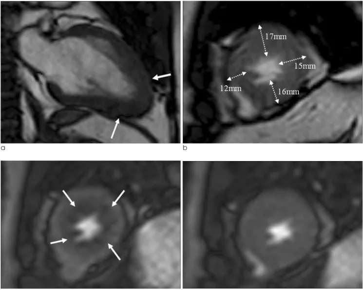
a b
c d
Fig. 1. A 45-year-old male with angina pain who visited to emergency room was confirmed as apical HCM by typical ECG and echocardiographic findings, and he performed CMR for risk stratification.
a. Two-chamber cine image shows apical wall thickening with typical ‘spade of ace’ sign (arrows).
b. Short-axis cine image shows LVH at apical anterior, lateral and inferior wall (NCH-3).
c, d. First-pass stress perfusion (c) and rest perfusion (d) show reversible ring of subendocardial perfusion defect (arrows) at whole apical layer.
(repetition time msec/echo time msec, 3/1.5, flip angle α60�). LGE was additionally performed using an inversion recovery fast gradient echocardiographic pulse sequence (repetition time msec/echo time msec, 4.6/1.5, flip angle α15�, inversion recovery time 200 to 280 msec).
MR image analysis
Myocardial segments were evaluated according to the 17-segment model recommended by the American Heart Association (13). We analyzed the 16 segments at the representative short-axis slices of the basal (segments 1-6), mid-ventricular (segments 7-12), and apical (segments 13-16) regions of the LV, except for apex (segment 17). Therefore, a total of 416 segments were analyzed for 26 patients. Two experienced radiologists evaluated MR images without clinical information or result from other studies. If there was discordant region, two radiologists were finally interpreted with consensus. Image analysis was performed using an independent workstation with dedicated software (Extended MR Workspace 2.6, Philips Healthcare).
Using cine images with SSFP, maximal LV wall thickness was measured on each segment of LV at end diastolic phase. The LVH was defined as more than 15 mm on end-diastolic stage (1). Number of hypertro-
phied segment was defined as the total number of LVH segments. In addition, if the hypertrophied segments were noted at least two adjacent segments, we determined it as number of consecutive hypertro- phied segment (NCH) -2. If the LVH was noted at least 3 adjacent segments, we classified it as NCH -3.
For assessing PD, rest and stress perfusion MR images were displayed side by side on a workstation and were evaluated by manual paging the images for differentiation of low enhancement caused by micro- vascular ischemia from artifact. On MR images, a PD was defined as a clearly visible hypoenhancement in at least one slice that persisted for at least three consecu- tive temporal images of the first-pass stress perfusion MRI (14, 15). According to presence or pattern, we classified the patient with APH into 1) no PD, 2) Presence of PD with sporadic pattern; PD was randomly located, and 3) ring of perfusion defect (ring-PD); ring like subendocardial hypoattenuation at apical layer. LGE was determined using 6 standard deviations (SDs) above the mean signal intensity for the normal nulled myocardium (16). Normal myocardium was defined as a region of myocardium without any apparent LGE at visual inspection. The mean signal intensity and SD were determined by drawing a region of interest in a portion of the normal myocardium.
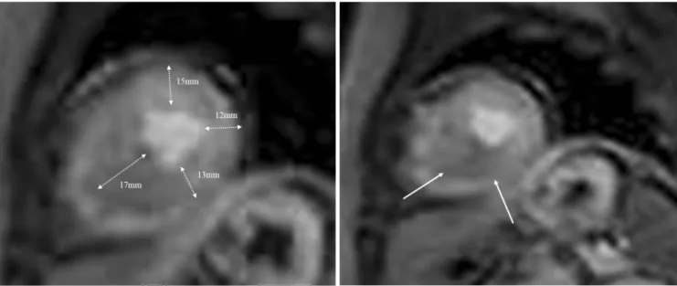
a b
Fig. 2. A 54-year-old female with dyspnea who confirmed as apical HCM by typical ECG and echocardiographic findings.
a. Short-axis cine image shows LVH at apical anterior and septal wall (NCH-2).
b. First-pass stress perfusion show sporadic patterned subendocardial perfusion defect at apical septal wall (arrows).
Statistical analysis
Each categorical variable was expressed as number and percentage of patients. Continuous data was reported as the mean values ± SD. Inter-reader agreement for detection or image measurement of LV wall thickness, PD or LGE on CMR was assessed by using weighted ĸ statistics and interpreted as follows: poor (< 0.20), fair (0.20-0.40), moderate (0.41-0.60), good (0.61-0.80), and excellent agreement (0.81-1.00). For comparison of demographic and imaging features among three PD groups, one way ANOVA analysis was acquired. For independent predictor of the overall PD and ring-PD, we performed univariate logistic regression models. A p- value < 0.05 was considered to indicate statistical signifi- cance.
Demographic and clinical risk factors
Demographics and clinical risk factors in patients with APH were shown in Table 1. The prevalence of male was 80.8% and mean age of the subjects was 57.8 ±9.8 years. Family history of sudden cardiac death or known HCM was 3 patients (11.5%). Fifteen patients (57.7%) complained of symptoms as follows;
chest pain (n=12), dyspnea (n=5), palpitation (n=3) and syncope (n=2). Among them, 7 patients had mixed symptoms such as chest pain and dyspnea, palpitation or syncope. Among the patients with chest pain, 5 patients were atypical and 7 patients were complained typical angina. Eleven patients (42.3%) were asymptomatic, but they performed MRI for risk stratification. The mean EF was 66.8 ±6.6%, LV mass was 199.5 ± 60.9 g.
CMR findings in patients with APH
The mean maximal LV wall thickness was 18.7 ± 3.3 mm (range; 12-24 mm) in 26 patients with APH.
Number of hypertrophied segment (hypertrophied segments more than 15 mm in thickness) in each patient was 4.2 ± 3.2 (range; 1-13) segments.
Eighteen patients (69.2%) showed LGE on delayed enhancement MRI, these LGE were presented as multifocal patchy enhancement in hypertrophied segment of all patients.
Among 26 patients with APH, 20 patients (76.9%)
showed PD and 12 of them (60.0%) represented as ring of subendocardial PD. All PD was reversible pattern, which was defined as nearly normal perfusion on rest-perfusion MR images from hypoattenuated PD on stress-perfusion MR images.
Table 2 was summarized as comparison data of demographic and imaging findings according to three groups which were classified by the presence or pattern of PD. There was no statistically significant difference among the three groups in terms of demographic data including presence of symptom. However, typical angina pain including syncope was frequently noted in ring-PD group, although it was not statistically different
RESULTS
Table 1. Baseline Characteristics of Study Population
Baseline characteristics of the study population (n = 26 ) Age (years)* 57.8±9.8 (range; 43-78)
Male 21 (80.8%)0
BMI* (kg/m2) 025.5±2.3 (range; 21.5-30.5)
Diabetes mellitus 3 (11.5%)
Hypertension 19 (73.1%)
Hypercholesterolemia 8 (30.8%)
Current smoker 8 (30.8%)
Family history of sudden cardiac
death or previous CAD history 3 (11.5%) Medication
Statin 6 (23.1%)
Aspirin 8 (30.8%)
Antithrombotic drug 1 (3.8%)0
Clinical symptom
Asymptomatic, but risk evaluation 11 (42.3%)
Atypical chest pain 5 (19.2%)
Typical angina 7 (23.1%)
Dyspnea 5 (25.0%)
Palpitation 3 (11.5%)
Syncope 2 (7.6%)0
Ejection Fraction (%) 66.8±6.6 (range; 55-83)00 LV mass (g) 199.5±60.9 (range; 114-359) Note.─ Unless otherwise indicated, data are numbers of patients, with percentages in parentheses.
* Data are means ± standard deviations.
�Non-specific symptom; numbness or dizziness (without evidence of syncope), headache etc.
(p=0.152). Maximal LV wall thickness and number of hypertrophied segment were statistically different among the three groups (all, p<0.05), indicating the maximal LV wall thickness and number of hypertro- phied segment were linearly related with PD and ring- PD group.
For evaluation of the characteristics of ring-PD in comparison to no-PD group, we compared the two groups after exclusion of the sporadic-PD group.
Besides LV wall thickness and number of hypertro- phied segment, NCH-3 was significantly related with ring-PD (p < 0.05) (Fig. 1), although NCH-2 was not statistically associated with ring-PD (Fig. 2).
The inter-reader agreements for EF (k=0.86), LV mass (k=0.82) and LV wall thickness (k = 0.85) were
excellent. The agreement with the presence of overall PD and classification of PD pattern were good (k=0.72). The overall agreement of the presence of LGE was excellent (k=0.83).
Predictors for perfusion defect
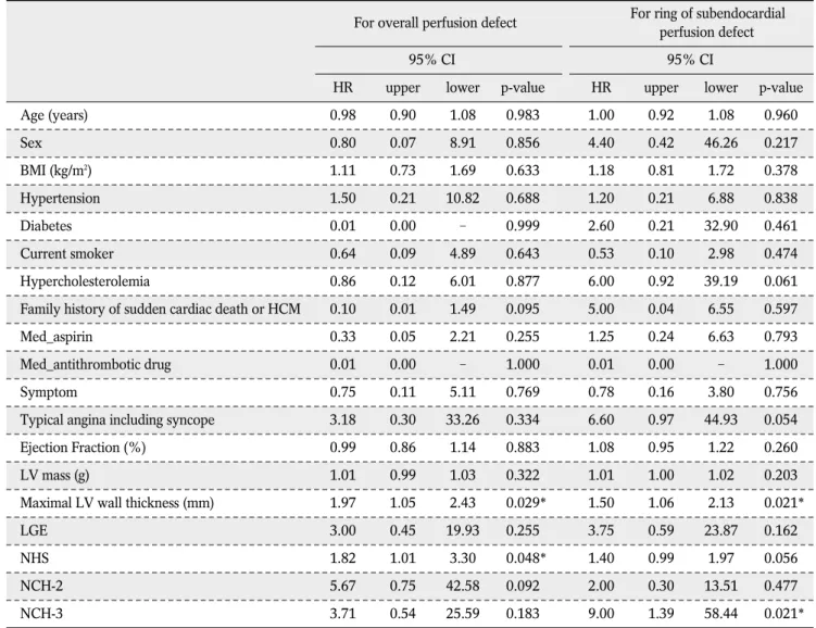
The independent predictors of overall PD and ring- PD in patients with APH were summarized in Table 3.
Using univariate logistic analysis, maximal LV wall thickness (odds ratio [OR], 1.97; 95% confidence interval [CI], 1.05 to 2.43; p=0.029) and number of hypertrophied segment (OR, 1.82; 95% CI, 1.01 to 3.30; p=0.048) were independent predictor for overall PD. The independent predictors for ring-PD were the maximal LV wall thickness (OR, 1.50; 95% CI, 1.06 to
Table 2. Comparison of Demographic and Imaging Findings According to Presence or Pattern of Perfusion Defect
No PD (n=6) Sporadic PD (n=8) Ring-PD (n=12)
Age (years) 59.0 ± 8.1 057.0 ± 12.4 57.7 ± 9.4
Sex 5 (83.3%) 5 (62.5%) 11 (91.7%)
Body mass index (kg/m2) 25.1 ± 1.5 25.2 ± 2.5 25.5 ± 2.3
Hypertension 4 (66.7%) 6 (75.0%) 9 (75.0%)
Diabetes 0 1 (12.5%) 2 (16.7%)
Current smoker 2 (33.3%) 3 (37.5%) 3 (25.0%)
Hypercholesterolemia 3 (50.0%) 0 6 (50.0%)
Family history of sudden cardiac death or HCM 2 (33.3%) 0 1 (8.3%)0
Med_statin 1 (16.7%) 0 5 (41.7%)
Med_aspirin 3 (50.0%) 1 (12.5%) 4 (33.3%)
Med_antithrombotic drug 0 0 1 (8.3%)0
Symptom 2 (33.3%) 4 (26.7%) 9 (75.0%)
Typical angina including syncope 1 (16.7%) 1 (12.5%) 6 (50.0%)
Ejection Fraction (%) 67.2 ± 7.3 64.2 ± 5.3 68.4 ±7.0
LV mass (g) 177.7 ± 61.6 190.7 ± 32.1 216.3 ±73.8
Maximal LV wall thickness (mm) * 15.7 ± 2.6 18.3 ± 2.2 020.4 ± 3.2�
LGE 3 (50.0%) 5 (62.5%) 10 (83.3%)
NHS * 1.8 ± 1.6 3.9 ± 2.4 005.6 ± 3.6�
NCH-2 3 (50.0%) 7 (87.5%) 10 (83.3%)
NCH-3 1 (16.7%) 3 (37.5%) 10 (8.3%)�
Note.─ PD = perfusion defect, LGE = late gadolinium-enhancement, NHS = Number of hypertrophied segment, NCH-2 = more than 2 consecutive hypertrophied segments, NCH-3 = more than 3 consecutive hypertrophied segments
* p < 0.05 by one-way ANOVA analysis for comparison among the three groups
�p < 0.05 for comparison between no PD group and ring-PD group, except for sporadic-PD group
2.13; p=0.021) and NCH-3 (OR, 9.00; 95% CI, 1.39 to 58.44; p=0.021). It means NCH with more than 3 segments is statistically associated with ring-PD.
However, the presence of LGE was not associated with overall PD or ring-PD.
The main findings of the present study of first-pass stress perfusion MR images in patients with APH are the following; 1) PDs were shown in 76.9% of APH, 60% of PD presented as ring of subendocardial PD. 2) LV wall thickness and number of hypertrophied
segment were independent factors for overall PD, and more than three consecutive hypertrophied segment was additional independent factor for ring-PD in APH. However, the presence of LGE was not statisti- cally related with PD or ring-PD in APH.
APH is a common type of HCM in East Asia and it has known to have a relatively favorable prognosis in terms of cardiovascular mortality (17). Although the prevalence of APH has been rare in western countries, its outcome appears to be less benign than in Japan, with atrial fibrillation and myocardial infarction the most frequent complication occurring in up to a third of patients during long term follow up (18).
In APH, severe apical hypertrophy could impose DISCUSSION
Table 3. Independent Predictors for Perfusion Defect
For overall perfusion defect For ring of subendocardial perfusion defect
95% CI 95% CI
HR upper lower p-value HR upper lower p-value
Age (years) 0.98 0.90 1.08 0.983 1.00 0.92 1.08 0.960
Sex 0.80 0.07 8.91 0.856 4.40 0.42 46.26 0.217
BMI (kg/m2) 1.11 0.73 1.69 0.633 1.18 0.81 1.72 0.378
Hypertension 1.50 0.21 10.82 0.688 1.20 0.21 6.88 0.838
Diabetes 0.01 0.00 - 0.999 2.60 0.21 32.90 0.461
Current smoker 0.64 0.09 4.89 0.643 0.53 0.10 2.98 0.474
Hypercholesterolemia 0.86 0.12 6.01 0.877 6.00 0.92 39.19 0.061
Family history of sudden cardiac death or HCM 0.10 0.01 1.49 0.095 5.00 0.04 6.55 0.597
Med_aspirin 0.33 0.05 2.21 0.255 1.25 0.24 6.63 0.793
Med_antithrombotic drug 0.01 0.00 - 1.000 0.01 0.00 - 1.000
Symptom 0.75 0.11 5.11 0.769 0.78 0.16 3.80 0.756
Typical angina including syncope 3.18 0.30 33.26 0.334 6.60 0.97 44.93 0.054
Ejection Fraction (%) 0.99 0.86 1.14 0.883 1.08 0.95 1.22 0.260
LV mass (g) 1.01 0.99 1.03 0.322 1.01 1.00 1.02 0.203
Maximal LV wall thickness (mm) 1.97 1.05 2.43 0.029* 1.50 1.06 2.13 0.021*
LGE 3.00 0.45 19.93 0.255 3.75 0.59 23.87 0.162
NHS 1.82 1.01 3.30 0.048* 1.40 0.99 1.97 0.056
NCH-2 5.67 0.75 42.58 0.092 2.00 0.30 13.51 0.477
NCH-3 3.71 0.54 25.59 0.183 9.00 1.39 58.44 0.021*
Note.─ LGE = late gadolinium-enhancement, NHS = Number of hypertrophied segment, NCH-2 = more than 2 consecutive hypertrophied segments, NCH-3 = more than 3 consecutive hypertrophied segments
* p < 0.05
pressure overload and impair coronary flow in the LV apex, thereby inducing myocardial ischemia (19).
Microvascular dysfunction and structural changes including small intramural coronary arteriole dysplasia with collagen deposition in hypertrophied myocardium has been thought to be a cause of reduced coronary flow reserve in patients with HCM (5). Using several animal studies, a decreased capillary density has been measured in hypertrophied myocardium (20). These findings have led to the clinically silent myocardial ischemia, myocyte death, and replacement scars, which are cause for tachyarrhythmia (21). Many authors reported that reversible perfusion abnormalities are common in HCM (22, 23). According to these reports, about 40% of HCM shows 201 thallium defect despite normal epicardial coronary arteries (22) and about 80% of HCM shows reversible 201 thallium abnormali- ties during exercise stress and most likely identify myocardial ischemia (23). Lee et al. (24) reported 50%
of HCM showed PD, which were predominantly reversible and mostly apical type (69.2%). Our study also showed similar results that 76.9% of APH had PD and most common pattern of PD was ring-PD. The ring of reversible PD during stress-perfusion imaging is also noted as characteristic finding of cardiac syndrome X which is characterized by angina-like chest pain and a positive exercise test, in presence of angiographically smooth coronary arteries (25). Although multiple pathogenic mechanisms have been hypothesized, it was mainly caused by coronary microvascular dysfunction (26). It was similar of reversible PD in APH caused by microvascular ischemia, although the mechanism of microvascular dysfunction is different from cardiac syndrome X. A study by Yamada et al. (27) found reversible PD did not correlate with symptom, but was associated with greater maximal LV wall thickness.
These results were similar with our results of that the independent predictors for PD were maximal LV wall thickness and number of LVH regardless of symptom.
We additionally found the independent factor for ring of PD was at least three adjacent LVH. Although it was not statistically significant, typical angina pain includ- ing syncope occurred in most of patients with ring-PD.
Until now, there was a controversy about whether PD in APH leads to adverse prognosis. Dilsizian et al.
(28) reported the inducible ischemia detected by thallium-201 scan is frequently related to cardiac
arrest and syncope in young patients with HCM, however, Lee et al. (24) reported that reversible thallium defect in the absence of coronary artery disease shows benign prognosis despite the presence of ischemia. Usually, a lot of studies reported that LGE in HCM was correlated with adverse prognosis such as sudden cardiac death, ventricular tachycardia, or heart failure symptom (29). Pathologically, LGE may show areas where replacement fibrosis has occurred following microvascular ischemia and focal necrosis (30). Therefore, PD is presented by earlier structural change of HCM, chronic impairment of myocardial perfusion and repeated inducible ischemia may lead to fibrosis. In addition, PD in APH seems to be different effect for clinical prognosis from the non- APH. Therefore, further comparison study regarding clinical outcomes of PD between APH and non-APH should be carried out.
Our current study has several limitations. First, we assess the PD and LGE by just visual assessment, not quantification. Although the presence of PD or LGE was assessed by two observers with final consensus, the severity of PD or amount of LGE was not graded.
Thus, further quantitative study will be necessary for the exact correlation between PD and LGE. Second, our study did not include follow-up data. As yet there is disagreement regarding the clinical significance of PD, long-term follow-up should be carried out for obtaining more information on this aspect.
In conclusion, about three quarters of the patients with APH showed PD, most of them represented as ring-PD. LVH degree or distribution was related with pattern of PD, however, LGE was not related with PD.
Therefore, the clinical significance of PD in the patients with APH seems to be different from those with non-APH, and further comparison study between the two groups should be carried out.
Acknowledgements
This work was supported by the grant 11-2008-004 from the Seoul National University Bundang Hospital (SNUBH) Research Fund.
References
1. Maron BJ, McKenna WJ, Danielson GK, et al. American college of cardiology/european society of cardiology clinical expert
consensus document on hypertrophic cardiomyopathy. A report of the american college of cardiology foundation task force on clinical expert consensus documents and the european society of cardiology committee for practice guidelines. J Am Coll Cardiol 2003;42:1687-1713
2. Sakamoto T, Tei C, Murayama M, Ichiyasu H, Hada Y. Giant T wave inversion as a manifestation of asymmetrical apical hypertrophy (AAH) of the left ventricle. Echocardiographic and ultrasono-cardiotomographic study. Jpn Heart J 1976;17:611- 629
3. Chun EJ, Choi SI, Jin KN, et al. Hypertrophic cardiomyopathy:
Assessment with MR imaging and multidetector CT.
Radiographics 2010;30:1309-1328
4. Okishige K, Sasano T, Yano K, Azegami K, Suzuki K, Itoh K.
Serious arrhythmias in patients with apical hypertrophic cardiomyopathy. Intern Med 2001;40:396-402
5. Kusukawa J, Suwa M, Nakayama Y, et al. Advanced sequelae of apical hypertrophic cardiomyopathy: report of two cases with wall motion abnormalities. J Cardiol 1988;18:259-269
6. Nishimura RA, Holmes DR Jr. Clinical practice. Hypertrophic obstructive cardiomyopathy. N Engl J Med 2004;350:1320- 1327
7. Spirito P, Bellone P. Natural history of hypertrophic cardiomy- opathy. Br Heart J 1994;72(6 Suppl):S10-12
8. Sipola P, Lauerma K, Husso-Saastamoinen M, et al. First-pass MR imaging in the assessment of perfusion impairment in patients with hypertrophic cardiomyopathy and the Asp175Asn mutation of the alpha-tropomyosin gene. Radiology 2003;226:
129-137
9. Salerno M, Beller GA. Noninvasive assessment of myocardial perfusion. Circ Cardiovasc Imaging 2009;2:412-424
10. Moon JC, Fisher NG, McKenna WJ, Pennell DJ. Detection of apical hypertrophic cardiomyopathy by cardiovascular magnetic resonance in patients with non-diagnostic echocardiography.
Heart 2004;90:645-649
11. Writing Committee M, Yancy CW, Jessup M, et al. 2013 ACCF/AHA guideline for the management of heart failure: a report of the American College of Cardiology Foundation/
American Heart Association Task Force on practice guidelines.
Circulation 2013;128:e240-319
12. Sorajja P, Nishimura RA, Gersh BJ, et al. Outcome of mildly symptomatic or asymptomatic obstructive hypertrophic cardiomyopathya long-term follow-up study. J Am Coll Cardiol 2009;54:234-241
13. Cerqueira MD, Weissman NJ, Dilsizian V, et al. Standardized myocardial segmentation and nomenclature for tomographic imaging of the heart a statement for healthcare professionals from the cardiac imaging committee of the council on clinical cardiology of the american heart association. Circulation 2002;
105:539-542
14. Schwitter J, Wacker CM, van Rossum AC, et al. MR-IMPACT:
comparison of perfusion-cardiac magnetic resonance with single- photon emission computed tomography for the detection of coronary artery disease in a multicentre, multivendor, random- ized trial. Eur Heart J 2008;29:480-489
15. Chung SY, Lee KY, Chun EJ, et al. Comparison of stress perfusion mri and spect for detection of myocardial ischemia in
patients with angiographically proven three-vessel coronary artery disease. AJR Am J Roentgenol 2010;195:356-362 16. Harrigan CJ, Peters DC, Gibson CM, et al. Hypertrophic
cardiomyopathy: quantification of late gadolinium enhancement with contrast-enhanced cardiovascular mr imaging. Radiology 2011;258:128-133
17. Sakamoto T, Amano K, Hada Y, et al. Asymmetric apical hypertrophy: ten years experience. Postgrad Med J 1986;62:
567-570
18. Eriksson MJ, Sonnenberg B, Woo A, et al. Long-term outcome in patients with apical hypertrophic cardiomyopathy. J Am Coll Cardiol 2002;39:638-645
19. Matsubara K, Nakamura T, Kuribayashi T, Azuma A, Nakagawa M. Sustained cavity obliteration and apical aneurysm formation in apical hypertrophic cardiomyopathy. J Am Coll Cardiol 2003;42:288-295
20. Rakusan K, Flanagan MF, Geva T, Southern J, Van Praagh R.
Morphometry of human coronary capillaries during normal growth and the effect of age in left ventricular pressure- overload hypertrophy. Circulation 1992;86:38-46
21. Krams R, Kofflard M, Duncker D, et al. Decreased coronary flow reserve in hypertrophic cardiomyopathy is related to remodeling of the coronary microcirculation. Circulation 1998;
97:230-233
22. von Dohlen TW, Prisant LM, Frank MJ. Significance of positive or negative thallium-201 scintigraphy in hypertrophic cardiomy- opathy. Am J Cardiol 1989;64:498-503
23. Cannon RO 3rd, Dilsizian V, O’Gara PT, et al. Myocardial metabolic, hemodynamic, and electrocardiographic significance of reversible thallium-201 abnormalities in hypertrophic cardiomyopathy. Circulation 1991;83:1660-1667
24. Lee KH, Jang HJ, Lee SC, et al. Myocardial thallium defects in apical hypertrophic cardiomyopathy are associated with a benign prognosis. Int J Cardiovas Imaging 2003;19:381-388 25. Panting JR, Gatehouse PD, Yang G-Z, et al. Abnormal subendo-
cardial perfusion in cardiac syndrome X detected by cardiovas- cular magnetic resonance imaging. N Engl J Med 2002;346:
1948-1953
26. Lanza GA. Cardiac syndrome X: a critical overview and future perspectives. Heart 2007;93:159-166
27. Yamada M, Elliott P, Kaski J, et al. Dipyridamole stress thallium-201 perfusion abnormalities in patients with hypertrophic cardiomyopathy. Relationship to clinical presenta- tion and outcome. Eur Heart J 1998;19:500-507
28. Dilsizian V, Bonow RO, Epstein SE, Fananapazir L. Myocardial ischemia detected by thallium scintigraphy is frequently related to cardiac arrest and syncope in young patients with hypertrophic cardiomyopathy. J Am Coll Cardiol 1993;22:796- 804
29. O’Hanlon R, Grasso A, Roughton M, et al. Prognostic signifi- cance of myocardial fibrosis in hypertrophic cardiomyopathy. J Am Coll Cardiol 2010;56:867-874
30. Cecchi F, Sgalambro A, Baldi M et al. Microvascular dysfunc- tion, myocardial ischemia, and progression to heart failure in patients with hypertrophic cardiomyopathy. J Cardiovasc Transl Res 2009;2:452-461
통신저자 : 전은주, (463-707) 경기도 성남시 분당구 구미동 300, 서울대학교 분당병원 영상의학과
Tel. (031) 787-7618 Fax. (031) 787-4011 E-mail: [email protected]
심첨형 비후성 심근병증에서의 스트레스 부하 관류 자기공명영상 소견:
좌심실 벽 비후 정도와 지연 조영 증강 간의 관련성
1서울대학교 분당병원 영상의학과
2서울대학교 분당병원 순환기내과
유진영1∙전은주1∙김여군1∙최상일1∙최동주2
목적: 심첨형 비후성 심근병증 환자에서 스트레스 부하 관류 자기공명영상을 통한 관류 결손의 빈도와 양상을 평가하 고, 이를 좌심실 비대의 정도와 지연 조영 증강과 비교해 보고자 한다
대상과 방법: 2008년 1월부터 2012년 12월까지 심초음파 및 심전도로 심첨형 비후성 심근병증을 진단받고, 스트 레스 부하 관류, 영화 영상 및 지연 조영 증강 영상을 포함하는 심장 자기공명영상을 시행한 26명의 환자를 대상으로 하였다. 영화 영상에서 이완기 말에 416개 분절의 좌심실 벽 두께를 분석하였고 비후된 분절의 수와 연속하여 비후된 벽 분절의 수를 조사하였다. 또한 모든 환자에서 관류 결손과 지연 조영 증강의 유무를 평가하였다. 자기공명영상에서 관류 결손이 있을 경우, 산발형 혹은 고리형의 2가지 형태로 분류하였다. 단변량 분석을 통해 전체 관류 결손과 고리 형 관류 결손에 대한 독립 변수를 산출하였다.
결과: 심첨형 비후성 심근병증의 76.9%(20명)에서 스트레스 부하 관류 자기공명영상시 관류 결손을 보였으며 이 중 60% (12명)이 고리형 관류 결손을 보였다. 전체 관류 결손에 대한 독립 변수는 최대 좌심실 벽 두께와 비후된 분 절의 수 였고 (p < 0.05), 고리형 관류 결손에 대한 독립 변수는 3개 이상의 연속한 비후된 분절의 수가 추가되었다.
그러나 지연 조영 증강은 관류결손과는 유의한 상관관계가 없었다.
결론: 심첨형 비후성 심근병증 환자의 4분의 3에서(75%) 관류 결손을 보였으며, 대부분이 고리형 관류 결손 형태를 보였다. 좌심실 벽의 비후 정도와 분포는 관류 결손의 형태와 관련이 있었지만 지연 조영 증강과는 유의한 상관성이 없었다. 따라서 심첨형 비후성 심근병증 환자군에서 관류 결손의 임상적 의미는 비심첨형 비후성 심근병증 환자군에 서 보이는 관류결손과는 임상적 의의가 다를 것으로 보이며, 이에 대한 추후의 비교연구가 필요할 것으로 여겨진다.
대한자기공명의과학회지 18:7-16(2014)