93 93
THE EWHA MEDICAL JOURNAL THE EWHA MEDICAL JOURNAL
Magnetic Resonance Imaging of Dermatomyositis with Bilateral Involvement of the Erector Spinae Muscle
Shinjiro Kaieda, Masaki Okamoto, Shiroh Miura, Hiroaki Ida
Division of Respirology, Neurology, and Rheumatology, Department of Medicine, Kurume University School of Medicine, Kurume, Japan
A 58-year-old man had been diagnosed with dermato- myositis and interstitial lung disease in August 2002. Although muscle biopsy was not performed, our patient fulfilled the cri-
teria for dermatomyositis published by Bohan and Peter based on the presence of proximal muscle weakness, elevated serum muscle enzyme levels, electromyographic abnormalities, and
Images and Solution
Ewha Med J 2016;39(3):93-94
http://dx.doi.org/10.12771/emj.2016.39.3.93 pISSN 2234-3180 • eISSN 2234-2591
Received January 11, 2016, Accepted July 1, 2016
Corresponding author Shinjiro Kaieda, Division of Respirology, Neurology, and Rheumatology, Department of Medicine, Kurume University School of Medicine, 67 Asahi-machi, Kurume 830-0011, Japan
Tel: 81-942-31-7560, Fax: 81-942-31-7703, E-mail: [email protected]
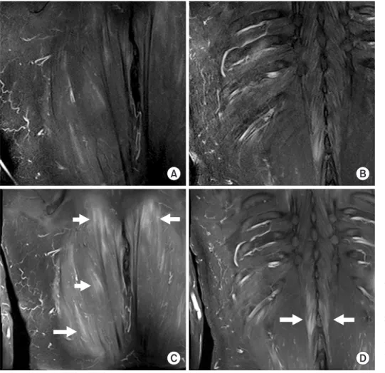
Fig. 1. Magnetic resonance images in a 58-year-old man with dermatomyositis and interstitial lung disease. High signal intensities in the bilateral erector spinae muscles are demonstrated on coronal fat- saturated, T2-weighted magnetic reso- nance images (A and B). Strong contrast enhancement is shown on gadolinium- enhanced T1-weighted images at Th8-L1 (C) and Th12-L1 (D) levels (arrows).
A B
C D
94 THE EWHA MEDICAL JOURNAL Kaieda S, et al
dermatomyositis-specific skin alterations [1]. The presence of anti-PL-7 (anti-threonyl-tRNA synthetase) autoantibodies was confirmed by immunoprecipitation after the initial diagnosis.
Although he remained in a stable condition while receiving 5 mg prednisolone (PSL) and 150 mg cyclosporine A daily for two years, the patient developed sudden onset of back pain and found a subcutaneous mass on his right upper back. He did not have trauma history and myositis ossificans was not suspected.
The subcutaneous mass was approximately 50×50 mm, and mobility was maintained. The subcutaneous tumor surface was smooth, and color change was not observed. Skin rash, proxi- mal muscle weakness, and interstitial lung disease exacerbation were not observed. The serum creatine phosphokinase and se- rum C-reactive protein levels were elevated (1,575 U/L [nor- mal, 45 to 163 U/L] and 0.72 mg/dL [normal <0.14 mg/dL], respectively). Fat-saturated, T2-weighted magnetic resonance images showed high signal intensity in the bilateral erector spi- nae muscles (Fig. 1A, B) and strong contrast enhancement was detected by gadolinium-enhanced T1-weighted images (Fig.
1C, D). Tests for procalcitonin and β-D-glucan were negative.
Increasing the PSL dose to 30 mg provided complete resolution of clinical symptoms in a few days, including the subcutaneous tumor and back pain, and laboratory examination values re- turned to normal.
Whole-body magnetic resonance imaging analysis in patients with adult inflammatory myopathies was performed, and myo- sitis was not found in the erector spinae muscles in 63 patients
with adult inflammatory myopathies, suggesting that involvement of the erector spinae muscles is a rare complication of adult in- flammatory myopathies [2]. Primary erector spinae pyomyositis cases were reported, and abscess was unilateral and localized [3,4]. In the present case, erector spinae muscle myositis was demonstrated to be bilateral, and infectious disease was unlikely to be the cause of erector spinae myositis. Increasing steroid doses in increments completely alleviated his clinical symptoms, including the subcutaneous tumor and back pain, and no relapse occurred after steroid dose reduction. Although skin biopsy was not performed, malignancy was excluded because of good re- sponse to steroid therapy. We suspected that the disease state was related to autoimmunity rather than malignancy and infec- tious diseases. We herein describe a case of dermatomyositis complicated with bilateral erector spinae myositis.
References
1. Bohan A, Peter JB. Polymyositis and dermatomyositis (second of two parts). N Engl J Med 1975;292:403-407.
2. Filli L, Maurer B, Manoliu A, Andreisek G, Guggenberger R.
Whole-body MRI in adult inflammatory myopathies: do we need imaging of the trunk? Eur Radiol 2015;25:3499-3507.
3. Lin MS, Tai CK, Shen CH, Ma TL, Tu DG. Primary erector spinae pyomyositis with an epidural abscess. Spine J 2013;13:1156- 1157.
4. Marshman LA, Bhatia CK, Krishna M, Friesem T. Primary erec- tor spinae pyomyositis causing an epidural abscess: case report and literature review. Spine J 2008;8:548-551.