Korean J Gastroenterol Vol. 72 No. 6, 304-307 https://doi.org/10.4166/kjg.2018.72.6.304 pISSN 1598-9992 eISSN 2233-6869
CASE REPORT
Korean J Gastroenterol, Vol. 72 No. 6, December 2018 www.kjg.or.kr
위장관 출혈을 일으킨 상피하 종양으로 나타난 위 결핵
김태언, 김수진1, 류화성, 김진혁, 정희석, 노지은, 염정아, 박병수2, 김동일2, 김기현2
부산대학교 의과대학 양산부산대학교병원 영상의학과, 소화기내과1, 외과2
Gastric Tuberculosis Presenting as a Subepithelial Mass: A Rare Cause of Gastrointestinal Bleeding
Tae Un Kim, Su Jin Kim1, Hwaseong Ryu, Jin Hyeok Kim, Hee Seok Jeong, Jieun Roh, Jeong A Yeom, Byung Soo Park2, Dong Il Kim2 and Ki Hyun Kim2
Department of Radiology, Division of Gastroenterology, Department of Internal Medicine1, Department of Surgery2, Pusan National University Yangsan Hospital, Pusan National University School of Medicine, Yangsan, Korea
Gastric tuberculosis accounts for approximately 2% of all cases of gastrointestinal tuberculosis. Diagnosis of gastric tuberculosis is challenging because it can present with various clinical, endoscopic, and radiologic features. Tuberculosis manifesting as a gastric subepithelial tumor is exceedingly rare; only several dozen cases have been reported. A 30-year-old male visited emergency room of our hospital with hematemesis and melena. Abdominal CT revealed a 2.5 cm mass in the gastric antrum, and endoscopy revealed a subepithelial mass with a visible vessel at its center on gastric antrum. Primary gastric tuberculosis was diagnosed by surgical wedge resection. We report a rare case of gastric tuberculosis mimicking a subepithelial tumor with acute gastric ulcer bleeding. (Korean J Gastroenterol 2018;72:304-307)
Key Words: Tuberculosis; Endoscopy; Subepithelial; Gastrointestinal hemorrhage
Received June 1, 2018. Revised June 26, 2018. Accepted July 2, 2018.
CC This is an open access article distributed under the terms of the Creative Commons Attribution Non-Commercial License (http://creativecommons.org/licenses/
by-nc/4.0) which permits unrestricted non-commercial use, distribution, and reproduction in any medium, provided the original work is properly cited.
Copyright © 2018. Korean Society of Gastroenterology.
교신저자: 김수진, 50612, 양산시 물금읍 금오로 20, 양산부산대학교병원 소화기내과
Correspondence to: Su Jin Kim, Division of Gastroenterology, Department of Internal Medicine, Pusan National University Yangsan Hospital, 20 Geumo-ro, Mulgeum-eup, Yangsan 50612, Korea. Tel: +82-55-360-1535, Fax: +82-55-360-1536, E-mail: [email protected], ORCID: https://orcid.org/0000-0003-3816-9664
Financial support: None. Conflict of interest: None.
INTRODUCTION
Gastric tuberculosis is extremely rare even in countries with a high prevalence of tuberculosis. Although difficult to de- termine with certainty, gastric tuberculosis constitutes around 2% of gastrointestinal tuberculosis cases.1,2 Its most common symptoms are nonspecific and indistinguishable from those of other ulcerative and obstructive gastric diseases, such as peptic ulcer, neoplasm, and Crohn’s disease.3 The ulcerative and hypertrophic infiltrative types of gastric tuberculosis are most frequently encountered endoscopically,4 and few cases of gastric tuberculosis mimicking a subepithelial tumor have
been reported.5-8 It is important that gastric tuberculosis be differentiated from other tumorous conditions because anti- tubercular treatment is effective against the former but not the latter. We report a case of gastrointestinal bleeding caused by primary gastric tuberculosis presenting as a sub- epithelial tumor that was diagnosed by surgical resection and provide a review of relevant literature.
CASE REPORT
A 30-year-old male visited emergency department of our hospital due to hematemesis and melena, which had devel-
Kim TU, et al. Gastric Tuberculosis Presenting as a Subepithelial Mass 305
Vol. 72 No. 6, December 2018
Fig. 1. (A) Axial contrast-enhanced CT image revealed a relatively well-defined soft tissue mass (arrow) on the anterior wall of antrum and an enlarged lymph node with peripheral rim enhancement and a hypodense center (arrowhead) in the periportal area. (B) Coronal reformatted contrast-enhanced CT scan showing low attenuating masses with thickened walls (arrowheads) in both subphrenic spaces and an exophytic, enhancing mass in gastric antrum (arrow). (C) CT images taken with lung window setting showing airspace consolidation (arrow) in the right lower lobe. CT, computed tomography.
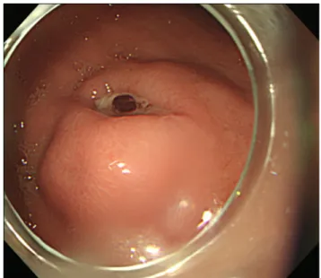
Fig. 2. Gastroduodenal endoscopy revealed an intraluminal protruding mass lesion with overlying ulceration and a visible vessel in the anterior wall of gastric antrum.
oped the previous day. His vital signs were: body temperature 36.4°C, respiration rate 20 breaths/min, pulse rate 120/min, and blood pressure 90/60 mmHg. A physical examination demonstrated anemic conjunctiva but without palpable lymph nodes or a mass on his body. Laboratory investigations re- vealed a low hemoglobin level (9.2 g/dL) and a slightly ele- vated CRP level (3.02 mg/dL). Other laboratory test results were within normal limits.
Contrast-enhanced abdominal CT revealed a 2.5 cm rela- tively well-defined soft tissue mass on the anterior wall of the gastric antrum (Fig. 1A, B), an enlarged lymph node with peripheral rim enhancement and a hypodense center in the periportal area (Fig. 1A), and low attenuating masses with thickened walls in both subphrenic spaces (Fig. 1B). In addi- tion, a CT scan of the chest revealed airspace consolidation in the right lower lobe (Fig. 1C).
At endoscopy, a smooth protruding mass with central ulcer- ation and a visible vessel was observed in the gastric antrum along the greater curvature (Fig. 2). The mass was not soft when pressed with forceps and had a negative rolling sign.
Active bleeding from the mass was not observed. We decided to resect the gastric subepithelial tumor for diagnostic and therapeutic purposes because we could not rule out the possi- bility of malignancy. Exploratory laparotomy revealed a sub- epithelial mass on the greater curvature of the gastric antrum, which was subjected to wedge resection. Whitish miliary nod- ules were observed in peritoneum and omentum and biopsied for histopathologic examination.
Gross inspection of the surgical specimen revealed a well-defined subepithelial mass with central ulceration and
intact surrounding overlying mucosa (Fig. 3A). Histopathologic examination demonstrated chronic granulomatous inflammation with caseating necrosis consistent with tuberculosis (Fig. 3B).
The histopathologic findings of the whitish nodules were sim- ilar to those of the gastric mass.
DISCUSSION
Gastrointestinal tuberculosis is a rare manifestation of tu- berculosis that is most frequently encountered in the ileocecal region. Other sites of involvement include, in descending order of frequency, the ascending colon, jejunum, appendix, duode-
A
A BB CC
306 김태언 등. 상피하 종양으로 나타난 위 결핵
The Korean Journal of Gastroenterology
Fig. 3. (A) The cut surface of the gross specimen showed a pale white, well-circumscribed mass with ulceration in the stomach antrum. (B) Histopathologic examination demonstrated chronic granulomatous inflammation with caseous necrosis surrounded by inflammatory cells (H&E, ×40).
num, stomach, sigmoid colon, and rectum.9 The rarity of gas- tric tuberculosis may be due to the bactericidal activity of gastric acid, resistance of gastric mucosa to tubercular in- fection, scarcity of lymphoid tissue in gastric mucosa, and rapid gastric emptying.10 The incidence of gastric involvement in patients with tuberculosis is approximately 2%.1,2 The mu- cosal layer of the gastrointestinal tract may be infected follow- ing ingestion of contagious material, such as sputum or milk containing Mycobacterium bovis or Mycobacterium tuber- culosis, whereas the most common route of gastric infection is lymph nodal spread via adjacent lymph nodes.6 The ma- jority of cases of gastric tuberculosis are associated with pul- monary tuberculosis and its incidence depends on the se- verity of pulmonary involvement.11
The common clinical presentations of gastric tuberculosis are non-specific, with complaints of epigastric pain, vomiting, and weight loss.6 Morphologically, there are various types of gastric involvement, but ulcerative is the most common and constitutes approximately 80% of cases, because caseous ne- crosis of tubercles in lymphoid tissue of submucosa leads to ulceration of overlying mucosa.4 Ulcers rarely penetrate be- yond submucosa or the muscle layer. Generally, tuberculosis manifesting as gastric ulcer does not perforate or bleed. As the disease progresses, fibrosis and necrosis may lead to transformation to the less common hypertrophic infiltrating type, which in appearance mimics scirrhous gastric cancer.4
Gastric tuberculosis presenting as a subepithelial tumor has only been rarely reported,5-8 and the underlying mecha- nism is unclear. According to Shibagaki et al.,12 granuloma
formation and liquefactive necrosis of caseating lesions may lead to subepithelial mass formation in patients with tuberculosis. In some reports, gastric tuberculosis manifesting as a subepithelial tumor was found to be caused by a tuber- culous lymph node eroding the gastric wall.13,14 Endoscopic ultrasonography is the modality of choice for evaluating gas- tric subepithelial masses as it facilitates the determination of layer of origin, classification, differential diagnosis, and fol- low up. However, endoscopic ultrasonography may depict gas- tric tuberculosis as hypoechoic lesions within the muscularis propria that are indistinguishable from gastrointestinal stro- mal tumors (the most common gastric mesenchymal tu- mors).6 Given its rarity, varied clinical presentations, and lack of specific endoscopic or radiological features, the correct di- agnosis of gastric tuberculosis is difficult.
Histopathologic confirmation of tuberculosis remains a challenge. The presence of acid-fast bacilli in endoscopic bi- opsies or surgical specimens leads to a definitive diagnosis of gastric tuberculosis, but acid-fast bacilli are observed in only one-third of cases. However, in the absence of acid-fast bacilli, the presence of caseating granuloma may be consid- ered diagnostic. According to the criteria proposed by Paustian and Marshall,15 the presence of at least one of the following findings is indicative of intestinal tuberculosis; caseating gran- uloma, acid-fast bacilli, or positive bacterial cultures.
Although it is difficult to diagnose, some have suggested the possibility of tuberculosis should be considered when an ulcer exhibits no therapeutic response or a tuberculous lesion is present at another site.16 In our case, pulmonary and abdomi-
A
A BB
Kim TU, et al. Gastric Tuberculosis Presenting as a Subepithelial Mass 307
Vol. 72 No. 6, December 2018
nal lesions were presumed to be tuberculosis. Antitubercular agents are the mainstay of treatment. Surgery is frequently required for complications such as pyloric obstruction, perfo- ration, bleeding, and fistula17 and should be followed by treat- ment with an antituberculosis drug regimen.
In conclusion, although gastric tuberculosis can manifest in various ways, its presentation as a subepithelial tumor, as in our case, is rare. However, physicians should be aware of the possibility of gastric tuberculosis in patients with tuber- culosis in other organ(s), especially in lungs. We report an unusual case of gastric tuberculosis mimicking subepithelial tumor on endoscopic and imaging studies that was ultimately diagnosed histopathologically following surgical resection.
REFERENCES
1. Singh V, Jain AK, Agrawal AK, et al. Clinicopathological profile of abdominal tuberculosis. Br J Clin Pract 1995;49:22-24.
2. Subei I, Attar B, Schmitt G, Levendoglu H. Primary gastric tuber- culosis: a case report and literature review. Am J Gastroenterol 1987;82:769-772.
3. Petroianni A, Mugnaini L, Laurendi G, et al. Abdominal tuber- culosis mimicking Crohn's disease: a difficult diagnosis. Report of a case. Panminerva Med 2002;44:155-158.
4. Talukdar R, Khanna S, Saikia N, Vij JC. Gastric tuberculosis pre- senting as linitis plastica: a case report and review of the literature. Eur J Gastroenterol Hepatol 2006;18:299-303.
5. Rana SS, Bhasin DK, Srinivasan R, Singh K. Gastric outlet ob- struction caused by tuberculosis and diagnosed by endoscopic ultrasound-guided fine needle aspiration. Endoscopy 2011;43 Suppl 2 UCTN:E117-E118.
6. Kim SH, Park JH, Kang KH, et al. Gastric tuberculosis presenting
as a submucosal tumor. Gastrointest Endosc 2005;61:319-322.
7. Gupta V, Goel MM, Noushif M, Rai P, Gupta P, Chandra A. Primary gastric tuberculosis mimicking gastrointestinal stromal tumor.
Am J Gastroenterol 2012;107:1269-1270.
8. Ardengh JC, Vaiciunas S, Kemp R, Venco F, Lima-Filho ER, dos Santos JS. Upper endoscopy versus endosonography in differ- ential diagnosis of gastrointestinal bulging. Arq Gastroenterol 2011;48:236-241.
9. Kruijshaar ME, Abubakar I. Increase in extrapulmonary tuber- culosis in England and Wales 1999-2006. Thorax 2009;64:
1090-1095.
10. Tromba JL, Inglese R, Rieders B, Todaro R. Primary gastric tuber- culosis presenting as pyloric outlet obstruction. Am J Gastroenterol 1991;86:1820-1822.
11. Mitchell RS, Bristol LJ. Intestinal tuberculosis: an analysis of 346 cases diagnosed by routine intestinal radiography on 5,529 ad- missions for pulmonary tuberculosis, 1924-49. Am J Med Sci 1954;227:241-249.
12. Shibagaki K, Miyaike J, Onji M, et al. Submucosal tumor-like le- sion originating from colon tuberculosis: a case report and re- view of the literature. Clin J Gastroenterol 2015;8:207-211.
13. Kim DY, Bang S, Park BK, et al. Tuberculous mesenteric lympha- denitis involving the gastric wall: case report. Gastrointest Endosc 2005;62:799-802.
14. Lee TH, Cho JY, Bok GH, Cho WY, Jin SY. Intra-abdominal tuber- culous lymphadenitis diagnosed using an endoscopic ultra- sonography-guided Procore needle biopsy. Clin Endosc 2013;
46:77-80.
15. Paustian FF, Marshall JB. Intestinal tuberculosis. In: Berk JE, ed.
Gastroenteroscopy. Volume 3. 4th ed. Philadelphia: WB Saunders, 1985:2018-2036.
16. Gaines W, Steinbach HL, Lowenhaupt E. Tuberculosis of the stomach. Radiology 1952;58:808-819.
17. Rao YG, Pande GK, Sahni P, Chattopadhyay TK. Gastroduodenal tuberculosis management guidelines, based on a large experi- ence and a review of the literature. Can J Surg 2004;47:364-368.