대한소화기학회지 2006;48:63-66
□ IMAGE OF THE MONTH □
증례: 74세 남자환자가 3주 전부터 시작된 복부 불편감과 복부 팽만감을 주소로 입원하였다. 60년 전 결핵을 진단받 고 약물치료를 받았으며 50갑년의 흡연자였다. 2개월 동안 2 kg의 체중감소가 있었고 2주 동안 지속된 변비를 호소하 였다. 복부는 팽만되었고 고음의 증가된 기계음이 청진되었 으며 압통과 반발통이 관찰되었다. 활력징후는 혈압 130/80 mmHg, 맥박 90회/분, 체온 36.5oC, 호흡수 20회였다. 검사실 소견에서 백혈구 13,800/mm3, 혈색소 12.8 g/dL, 혈소판은 496,000/mm3였으며 생화학검사에서 적혈구침강속도 50 mm/
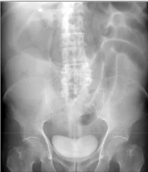
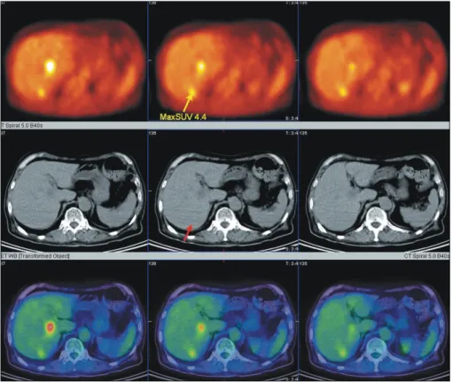
hr, C-반응단백 4.0 mg/dL, AST/ALT 49/74 IU/L, LDH/ALP 499/375 IU/L였다. 단순 복부촬영에서 기계적 폐쇄에 의한 장 폐쇄 소견을 보였고(Fig. 1), 외부에서 시행한 복부 CT에 서는 S결장과 직장의 접합부위에 종괴가 관찰되었으며 근 위부 대장의 확장소견이 관찰되고, 간의 4구역에 2 cm 크기 의 전이가 의심되는 병변이 관찰되었다(Fig. 2). 응급 S결장 경을 시행하였는데 항문피부선 7 cm 상방에 직장암에 의한 완전 폐쇄를 보여 응급으로 스텐트를 삽입하였다(Fig. 3). 수 술 전 복부 CT에서 보이는 간 병변 이외의 전이 여부를 확 인하기 위하여 F-18 FDG 양전자방출단층촬영을 시행하였 다. 양전자방출단층촬영 결과 복부 CT에서 보였던 간의 4구 역 이외에 6구역에서도 섭취 증가가 관찰되었다(Fig. 4). 이 후 lower anterior resection과 전이 간암에 대해 수술실에서 고주파 소작술을 시행하였다.
환자는 수술 후 합병증 없이 외래에서 추적관찰을 하였으 며 수술 후 11개월째 수술 시 83 ng/mL에서 1.8 ng/mL까지 감소하였던 암종배아항원(carcinoembryonic antigen) 수치가
양전자방출단층촬영술을 이용한 직장암 환자의 간내 다발성 전이 진단과 수술 후 재발 진단
을지의과대학교 내과학교실
전대원․최정호․박영숙
Detection of Liver Metastases and Recurrence Using PET-CT Scan in a Patient with Rectal Cancer
Dae Won Jun, M.D., Jeoung Ho Choi, M.D., and Young Sook Park, M.D.Department of Internal Medincine, Eulji University College of Medicine, Eulji Medical Center, Seoul, Korea
ꠏꠏꠏꠏꠏꠏꠏꠏꠏꠏꠏꠏꠏꠏꠏꠏꠏꠏꠏꠏꠏꠏꠏꠏꠏꠏꠏꠏꠏꠏꠏꠏꠏꠏ
연락처: 박영숙, 139-711, 서울시 노원구 하계 1동 280-1 을지의과대학교 노원을지병원 소화기내과 Tel: (02) 970-8207, Fax: (02) 970-8621 E-mail: [email protected]
Fig. 1. Simple abdominal X-ray shows small and large bowel dilatation. This finding suggested the possible distal large bowel obstruction.
ꠏꠏꠏꠏꠏꠏꠏꠏꠏꠏꠏꠏꠏꠏꠏꠏꠏꠏꠏꠏꠏꠏꠏꠏꠏꠏꠏꠏꠏꠏꠏꠏꠏꠏ Correspondence to: Young Sook Park, M.D.
Department of Internal Medicine, Eulji University School of Medi- cine, 280-1, Hagye 1-dong, Nowon-gu, Seoul 139-711, Korea Tel: +82-2-970-8207, Fax: +82-2-970-8621
E-mail: [email protected]
64 대한소화기학회지: 제48권 제2호, 2006
13 ng/mL으로 다시 증가하였다. 추적 복부 CT에서 특이소 견이 없었으나 F-18 FDG 양전자방출단층촬영에서 천골 앞 (presacral region)에서 의미 있는 섭취증가가 관찰되어 국소 재발로 판단하고 2차 수술을 시행하였다(Fig. 5). 조직검사 에서 국소재발로 확인되어 환자는 인공항문조성술을 시행 받고 퇴원하였으며 현재 재발 증후 없이 외래에서 추적관찰 중이다.
진단: 간내 다발성 전이를 동반한 직장암
대장 직장암은 미국에서 세 번째로 흔한 암종이며,1 국내 에서도 국립암센터의 2002년 암등록사업 결과에 따르면 대 장암은 간암에 이어 네 번째로 발생빈도가 높았다.2 이러한 수치는 1984년도 통계와 비교하였을 때 무려 533%나 증가 된 것이다. 대장 직장암은 간 전이가 많아 수술 전 이에 대
Fig. 2. Abdomen CT scan of the liver. A 20 mm sized single, low attenuated mass with peripheral enhancement is shown at S4 of liver.
Fig. 4. F-18 FDG PET findings.
FDG uptake is seen at S4 and newly developed lesion is seen at S6. Hot uptake at S6 region with SUV 4.4 is not noticed in pre- vious CT scan.
Fig. 3. Sigmoidoscopic finding. Huge ulcerofungating mass replaced nearly entire colonic lumen and spontaneous bleeding is noticed on the tumor surface.
전대원 외 2인. 양전자방출단층촬영술을 이용한 직장암 환자의 간내 다발성 전이 진단과 수술 후 재발 진단 65
한 평가가 매우 중요하다. 일반적으로 대장 직장암의 수술 전 병기 평가를 위하여 복부 CT 촬영이 이용되고 있으며 최근 양전자방출단층촬영 검사의 활용빈도가 높아지면서 대장 직장암 환자에서 수술 전 병기결정과 재발의 조기 진 단 등의 목적으로 사용빈도가 높아지고 있다.
양전자방출단층촬영 검사는 암세포에서 포도당 이용이 증가한다는 원리를 이용하여 암세포를 영상화하여 악성종 양의 병기 결정과 재발의 조기 진단에 사용하는 검사다. 그 러나 대장암의 조기 진단과 선별검사로서의 역할은 매우 제 한적이다. 선행 연구들에서 양전자방출단층촬영은 대장암 의 진단에 있어서 90-100%의 높은 예민도를 나타내나 염증 병소나 양성 용종에서도 섭취가 증가되기 때문에 특이도가 50% 내외로 낮아 대장암의 조기 진단과 선별검사로서의 역 할은 제한적이다.3,4 따라서 실제 용도에서 양전자방출단층 촬영 검사의 역할은 간 전이를 포함한 원격 전이의 평가와 수술 후 재발 병소의 평가에 더 많이 이용되고 있다. 현재까 지 연구는 주로 대장 직장암의 수술 후 재발의 조기 발견에 초점이 맞추어져 진행되어 왔다. 이는 대장 직장암 수술 후 천골 앞의 종괴가 발견되는 경우가 많은데 이러한 종괴가 재발에 의한 것인지 수술 후 발생되는 육아종, 반흔 등에 의 한 것인지 복부 CT나 자기공명장치와 같은 영상방법으로는 감별이 어려우며,5 대장내시경을 이용한 조직검사에서도 위 음성으로 나타나는 경우가 많아 진단이 어려운 경우가 많 다.6 이러한 경우 양전자방출단층촬영은 복부 CT에 비하여 정확도가 90-100%로 높아 수술 후 재발이 의심되는 환자에 게 매우 유용한 검사로 입증되어 있다.7-9
그러나 수술 전 병기 결정에서 양전자방출단층촬영이 반 드시 필요한지에 대해서는 아직 논란의 여지가 있다. 초기 의 연구들은 양전자방출단층촬영이 대장 직장암 환자의 수
술 전 병기 결정에서 매우 고무적인 결과를 발표하였으
며,10-12 Wiering 등13은 메타 분석을 통하여 양전자방출단층
촬영의 민감도와 특이도를 각각 88.0%와 96.1%, 그리고 간 전이를 진단하는 민감도와 특이도를 91.5%, 95.4%로 보고하 여 복부 CT의 전반적인 민감도와 특이도(60.9%, 91.1%)보다 높았으며 간내 전이를 평가하는 데도 우수하였다. 그러나 이후 Furukawa 등14이 처음으로 다채널 복부 CT (multi- detector CT)를 이용하여 대장암 환자 44명을 대상으로 양전 자방출단층촬영과의 수술 전 유용성에 대한 무작위 대조연 구를 시행하였으며 두 평가방법에 유의한 차이가 없었다.
그러나 아직 이에 대한 무작위 대조실험이 부족하며 보다 많은 연구가 필요하다.
결론으로, 양전자방출단층촬영 검사가 현재까지 연구결 과 복부 CT 등 기존 영상 검사나 침습적인 진단 검사를 대 체할 수 있는 검사라기보다는 상호보완적인 검사라 생각한 다. 고가이며 아직 임상에서 보편적으로 사용될 수 없는 장 비라는 제한점이 있지만, 이번 환자에서와 같이 수술 전 병 기 판정과 재발의 조기 발견을 통하여 생존율과 삶의 질 향 상을 가져올 수 있으며 앞으로 대장 직장암 환자에서 양전 자방출단층촬영 검사의 역할에 대하여 보다 많은 연구가 필 요하다고 생각한다.
참고문헌
1. Jemal A, Murray T, Ward E, et al. Cancer statistics, 2005. CA Cancer J Clin 2005;55:10-30.
2. National Cancer Center. Cancer Resistry and Statistics in KO- REA/Cancer Statictics 2002, http://www.ncc.re.kr, 2004.
3. Rohren EM, Provenzale JM, Barboriak DP, Coleman RE. Sc- reening for cerebral metastases with FDG PET in patients undergoing whole-body staging of non-central nervous system malignancy. Radiology 2003;226:181-187.
4. Gupta NC, Falk PM, Frank AL, Thorson AM, Frick MP, Bo- wman B. Pre-operative staging of colorectal carcinoma using positron emission tomography. Nebr Med J 1993;78:30-35.
5. Imbriaco M, Akhurst T, Hilton S, et al. Whole-body FDG- PET in patients with recurrent colorectal carcinoma. A com- parative study with CT. Clin Positron Imaging 2000;3:107- 114.
6. Grabbe E, Winkler R. Local recurrence after sphincter-saving resection for rectal and rectosigmoid carcinoma. Value of va- rious diagnostic methods. Radiology 1985;155:305-310.
7. Kanyari Z, Orosz L, Juhasz B, et al. The role of positron emission tomography (PET) in the detection of local recurr- ence and metastases of colorectal cancer. Magy Seb 2005;58:
179-183.
Fig. 5. Post-operative PET-CT. High metabolic regions were noticed at the presacral area and rectal wall with SUV 7.9 and 4.1, respectively. These findings were compatible with local recurrence.
66 The Korean Journal of Gastroenterology: Vol. 48, No. 2, 2006
8. Tanaka T, Kawai Y, Kanai M, Taki Y, Nakamoto Y, Takaba- yashi A. Usefulness of FDG-positron emission tomography in diagnosing peritoneal recurrence of colorectal cancer. Am J Surg 2002;184:433-436.
9. Takeuchi O, Saito N, Koda K, Sarashina H, Nakajima N. Cli- nical assessment of positron emission tomography for the diagnosis of local recurrence in colorectal cancer. Br J Surg 1999;86:932-937.
10. Delbeke D, Martin WH. PET and PET-CT for evaluation of colorectal carcinoma. Semin Nucl Med 2004;34:209-223.
11. Arulampalam TH, Francis DL, Visvikis D, Taylor I, Ell PJ.
FDG-PET for the pre-operative evaluation of colorectal liver metastases. Eur J Surg Oncol 2004;30:286-291.
12. Bohm B, Voth M, Geoghegan J, et al. Impact of positron emission tomography on strategy in liver resection for pri- mary and secondary liver tumors. J Cancer Res Clin Oncol 2004;130:266-272.
13. Wiering B, Krabbe PF, Jager GJ, Oyen WJ, Ruers TJ. The impact of fluor-18-deoxyglucose-positron emission tomogr- aphy in the management of colorectal liver metastases. Can- cer 2005;104:2658-2670.
14. Furukawa H, Ikuma H, Seki A, et al. PET scanning is not superior to whole-body multi-detector helical computed tomo- graphy in the preoperative staging of colorectal cancer. Gut 2006;55:1007-1011.