http://dx.doi.org/10.4078/jrd.2012.19.3.132
132
<Received:January 4, 2012, Revised:(1st: March 15, 2012, 2nd: May 22, 2012, 3rd: May 25, 2012) Accepted:May 29, 2012>
Corresponding to:Sung-Hwan Park, Division of Rheumatology, Department of Internal Medicine, School of Medicine, The Catholic University of Korea, Seoul St Mary’s Hospital, 505, Banpo-dong, Seocho-gu, Seoul 137-040, Korea. E-mail:
[email protected] pISSN: 2093-940X, eISSN: 2233-4718
Copyright ⓒ 2012 by The Korean College of Rheumatology
This is a Free Access article, which permits unrestricted non-commerical use, distribution, and reproduction in any medium, provided the original work is properly cited.
Gender Differences in Clinical Features and Anti-TNF Agent Use in Korean Ankylosing Spondylitis Patients
Chang Hoon Lee1, Myeung Su Lee1, Kwi Young Kang2, Su Jin Mun3, Ji Min Kim4, Ho Seung Yun3, Seung Gi Kwak3, Ji Hyeon Ju3, Kyung Su Park3, Ho-Youn Kim3, Sung-Hwan Park3
Department of Internal Medicine, School of Medicine, Wonkwang University1, Iksan, Chungbuk National University College of Medicine2, Cheongju, The Catholic University of Korea School of Medicine3, Seoul, Division of Rheumatology, Department of Internal Medicine, Pusan National University Yangsan Hospital,
Pusan National University School of Medicine4, Yangsan, Korea
Objective. The aim of this study was to assess the gender dif- ferences in the clinical presentation and treatment patterns between Korean women and men with ankylosing spondylitis (AS).
Methods. We retrospectively analyzed the data from extensive clinical assessments of 721 patients (162 women and 559 men) with AS, who were diagnosed at Seoul St. Mary’s Hospital, between January 2000 and September 2009. Clinical data, re- garding the disease onset, disease duration, clinical pre- sentations, status of human leukocyte antigen (HLA)-B27, and bone mineral density, were determined using a dual-energy X-ray absorptiometry (DEXA). Finally, we analyzed the medi- cal treatments prescribed for these patients.
Results. The ratio of men to women was 3.45:1. Compared to men, women were older at the time of diagnosis, had short- er disease durations, and were diagnosed in earlier stages of the disease. More women had a history of uveitis at diagnosis
than men. Back pain was the main presenting symptom, and its prevalence was the same in both genders. Fewer women showed cervical and thoracic axial involvement than men.
Initially, more women had wrist and hand pain than men;
however, at some point, peripheral arthritis development was equally likely in both genders. Women experienced shoulder pain, during the disease course, more often thanmen. On the other hand, men presented with knee and hip pain more often than women. Sulfasalazine and anti-TNF agents were more often prescribed to women.
Conclusion. The presentation and progression of AS showed a difference between women and men. Because of these differ- ences, AS should be considered when a women presents with peripheral arthritis or uveitis in the early stage of the disease.
Key Words. Differences, Clinical presentation, Women, Ankylosing spondylitis, Men
Introduction
Ankylosing spondylitis (AS) is a chronic systemic in- flammatory disorder. It is a seronegative spondyloarthropathy characterized by sacroiliitis and spondylitis. Although AS has historically been observed primarily in men, recent studies have shown that a significant proportion of patients with AS are women, with the ratio of men to women approaching 2- 3:1 (1-3). The incorrect assumption that AS affects men al-
most exclusively has persisted throughout the first half of the twentieth century. Axial involvement with back pain are com- mon features of AS, but a definitive diagnosis can be delayed because of the current lack of specific diagnostic indices. The differences in clinical presentation and disease course between men and women may also contribute to a delay in diagnosis.
The aim of this study was to compare the disease features of AS in women and men in order to better characterize any
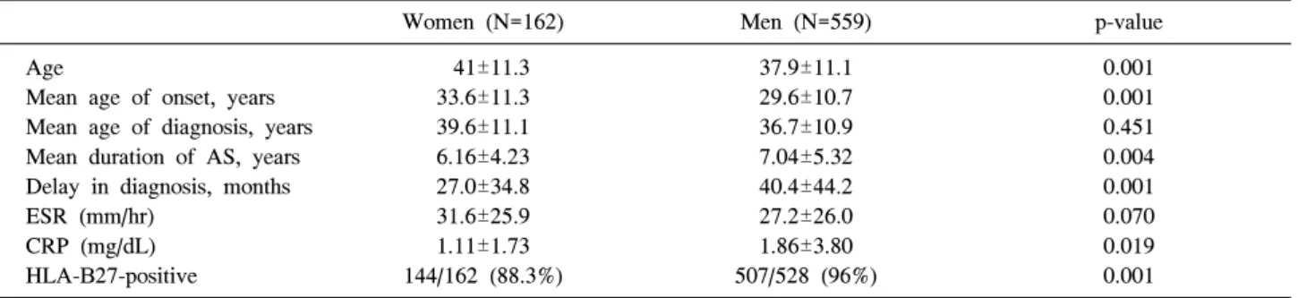
Table 1. Clinical features compared between women and men with ankylosing spondylitis
Women (N=162) Men (N=559) p-value
Age
Mean age of onset, years Mean age of diagnosis, years Mean duration of AS, years Delay in diagnosis, months ESR (mm/hr)
CRP (mg/dL) HLA-B27-positive
41±11.3 33.6±11.3 39.6±11.1 6.16±4.23 27.0±34.8 31.6±25.9 1.11±1.73 144/162 (88.3%)
37.9±11.1 29.6±10.7 36.7±10.9 7.04±5.32 40.4±44.2 27.2±26.0 1.86±3.80 507/528 (96%)
0.001 0.001 0.451 0.004 0.001 0.070 0.019 0.001 Data are shown in mean±SD or %. AS: ankylosing spondylitis, HLA: human leukocyte antigen
gender differences in clinical features, medications, and bone mineral density, which would help elucidate the potential in- fluence of gender on the severity of AS in patients.
Materials and Methods
We retrospectively analyzed the medical records of AS pa- tients who visited the Rheumatology Department at Seoul St.
Mary’s Hospital between January 2000 and August 2009 to identify patients who met the European Spondyloarthropathy Study Group Criteria (4) with radiographic sacroiliitis or Modified New York Criteria for AS (5). Patients who were thought to have degenerative arthritis and patients with a his- tory of inflammatory bowel disease or psoriasis were excluded. A total of 162 women and 559 men met these criteria. The patient data that we analyzed addressed 2 aspects of the disease: (1) disease presentation at the time of diagnosis and (2) disease course.
Disease onset, presentation, duration, and delay in diagnosis In order to determine the delay in diagnosis, we determined patient age at the time of disease onset and diagnosis; the de- lay was calculated by deducting the age at disease onset from the age at disease diagnosis. Disease duration was obtained by deducting the date of disease onset from the date of investigation.
Clinical features
We analyzed clinical features of AS, including first presenting symptom, initial site of pain, and history of uveitis at the time of disease onset. Data regarding history of peripheral arthritis, uveitis, and enthesitis and the site of axial involvement and pe- ripheral arthritis were collected during the course of the disease.
We also assessed human leukocyte antigen (HLA)-B27 status, erythrocyte sedimentation rate (ESR), C-reactive protein (CRP) level, familial history of AS in first-degree relatives and X-ray pictures of the sacroiliac joints and spine. Bone mineral density
of L-spine and femur were measured using dual-energy X-ray absorptiometry (DEXA). Osteoporosis was defined as a T-score less than or equal to -2.5 at the spine or hip. We also inves- tigated the patients’ medication history, including the use of nonsteroidal anti-inflammatory drugs (NSAIDs), methotrexate, sulfasalazine, and anti–tumor necrosis factor (anti-TNF) agents.
Statistical analysis
Statistical analyses were performed using the Statistical Package for Social Sciences version 12.0. One-way analysis of variance and independent t tests were used to compare age at diagnosis, disease duration, and delay in diagnosis between men and women. Chi-square tests were used to compare the status of HLA-B27, the site of the first presenting symptom, a history of uveitis, site of peripheral arthritis and axial involve- ment, osteoporosis, and drug use. The comparison of T-scores in bone mineral density was adjusted for age (p<0.05).
Results Baseline characteristics
In total, 721 patients (162 women, 559 men) were included in this study (Table 1). The ratio of male to female patients was 3.45:1. The mean age of women at the time of diagnosis was greater than that of men (39.6±11.1 years vs. 36.7±10.9, p=0.451). However, there tended to be a shorter delay in diag- nosis (27.0±34.8 months [women] vs. 40.4±44.2 months [men], p<0.001) and a shorter disease duration in case of women (6.16±4.23 years [women] vs. 7.04±5.32 years [men], p=0.004).
Furthermore, the number of first-degree relatives with AS in cases of women was greater than that in cases of men.
Compared to men, women had lower CRP levels (1.11±1.73 mg/dL [women] vs. 1.86±3.80 mg/dL [men], p<0.019) at the first visit time and were less likely to be HLA-B27 positive (88.3% [women] vs. 96% [men], p<0.001). There ware no sig- nificant differences in the rates of positive familial history be- tween the genders.
Table 2. Clinical manifestations at diagnosis
Women (N=162) Men (N=559) p-value Back pain
Peripheral arthritis Enthesitis Uveitis*
133 (82.1%) 71 (43.8%) 20 (12.3%) 34 (20.9%)
457 (81.8%) 219 (39.1%) 85 (15.2%) 71 (12.7%)
0.966 0.288 0.364 0.008
*including previous history of uveitis at diagnosis
Table 3. Site of peripheral arthritis at diagnosis
Women (N=162) Men (N=559) p-value Shoulder
Wrist and hand Hip
Knee
Ankle and foot
11 (6.8%) 21 (12.9%) 22 (13.5%) 32 (19.7%) 12 (7.4%)
28 (5.0%) 29 (5.9%) 74 (13.2%) 101 (18.0%) 51 (9.1%)
0.378
<0.001 0.910 0.496 0.496
Table 4. Clinical manifestations during the disease course
Women (N=162) Men (N=559) p-value Back pain
Peripheral arthritis Enthesitis Uveitis
142 (87.6%) 110 (67.1%) 33 (20.3%) 51 (31.4%)
506 (90.5%) 399 (71.3%) 122 (21.8%) 134 (23.9%)
0.287 0.393 0.692 0.054
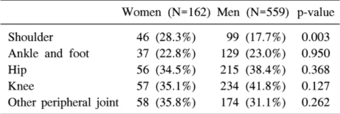
Table 5. Site of peripheral arthritis during the disease course
Women (N=162) Men (N=559) p-value Shoulder
Ankle and foot Hip
Knee
Other peripheral joint
46 (28.3%) 37 (22.8%) 56 (34.5%) 57 (35.1%) 58 (35.8%)
99 (17.7%) 129 (23.0%) 215 (38.4%) 234 (41.8%) 174 (31.1%)
0.003 0.950 0.368 0.127 0.262
Table 6. Axial involvement & osteoporosis in women and men
Women (N=162) Men (N=559) p-value Cervical spine
Thoracic spine Lumbar spine BMD T<−2.5*
(L-spine or femur)
23 (14.1%) 8 (4.9%) 124 (76.4%) 19/72 (26.3%)
147 (26.2%) 69 (12.3%) 431 (77.1%) 39/251 (15.5%)
0.001 0.007 0.882 0.053
BMD: bone mineral density. *adjusted for age
Table 7. Comparison of drug use in patients with AS
Women (N=161) Men (N=556) p-value NSAIDs
Sulfasalazine Methotrexate Anti-TNF agents
149 (92.5%) 117 (72.7%) 61 (37.9%) 33 (20.4%)
499 (89.7%) 450 (80.9%) 258 (46.4%) 167 (30.0%)
0.289 0.023 0.056 0.010 NSAIDs: Non-steroidal anti-inflammatory drugs
Clinical manifestations at diagnosis
Back pain was the most common symptom at the time of diagnosis for both women and men (82.1% and 81.8%, re- spectively). More women had a history of uveitis (20.9% vs.
12.7%, p=0.008) and peripheral arthritis (39.1% vs. 43.8%) than men; on the other hand, fewer women had enthesitis at diagnosis than men (12.3% vs. 15.2%). However, the above-mentioned differences between male and female pa- tients with regard to enthesitis and peripheral arthritis were not statistically significant (Table 2).
Regarding the site of peripheral arthritis, more women than men complained of wrist and hand pain at diagnosis (12.9%
vs. 5.9%, p<0.001), but there were no significant differences in the rates of pain in the knee, hip, ankle, foot and shoulder between men and women (Table 3).
Additional manifestations of AS
As the disease progressed, patients typically experienced addi- tional symptoms. Back pain was the most common symptom in both women and men. Although fewer women than men complained of back pain (87.6% vs. 90.5%, respectively), en- thesitis (20.3% vs. 21.8%), and peripheral arthritis (67.1% vs.
71.3%), there were no statistically significant differences in the
prevalence of these symptoms during disease progression be- tween men and women. The symptoms shown by AS patients during disease progression have been shown in Table 4.
As shown in Table 5, more women than men experienced shoulder pain (28.3% vs. 17.7%, p=0.003) during the disease progression, but fewer women complained of pain in the knee (35.1% vs. 41.8%, p=0.127) and hip (34.5% vs. 38.4%, p=0.368).
Axial involvement and osteoporosis
In terms of axial involvement, compared to men, women showed lesser C-spine (14.1% vs. 26.2%, p=0.001) and T-spine (4.9% vs. 12.3%, p=0.007) involvement. We measured bone mineral density of the L-spine and femur with DEXA. More women than men had osteoporosis (26% vs. 15%), but the dif- ference was not statistically significant (Table 6).
Medication
Women were less likely to be treated with anti-TNF agents or sulfasalazine than were men (p=0.010). More women than men used NSAIDs, and fewer women than men used metho- trexate (MTX); however, the differences were not statistically
significant (Table 7).
Discussion
Previous studies have shown distinct differences in the prev- alence and clinical manifestation of AS between women and men (1,2). However, to make better diagnostic and treatment decisions for AS patients, recognition of broad differences in these factors between genders is needed. Characterizing these differences will provide insightful ideas for spondyloarthritis research.
This study evaluated the differences between women and men with AS at disease onset (age of diagnosis, delay in diag- nosis, first symptom at the time of presentation, familial his- tory, status of HLA-B27, and acute phase reactant), clinical manifestation during disease progression, and management of AS in Korea. We retrospectively investigated the medical re- cords at the Rheumatology Department of Seoul St. Mary’s Hospital.
The earliest studies, performed in the 1940s, suggested the prevalence ratio of AS between men and women to be 9-10:1 (6,7). However, these numbers were affected by selection bias;
therefore, it is likely that the ratio has been overestimated in favor of men. Following the discovery of HLA-B27 and the development of standardized diagnostics (10-12), reports pub- lished in the 1970s have placed the ratio lower, at approx- imately 2-3:1 between men and women. The findings of our study are similar to those of these previously published studies and indicate the ratio of men to women to be 3.45:1. As Hill et al. (3) stated in 1976, “The precise ratio is less important than the fact that AS does occur in women and should be in- cluded in the differential diagnosis of back pain”.
There is an opinion that the diagnosis of AS may be more delayed in women than in men and that the disease may be underdiagnosed or even overlooked in women. Certainly, pa- tient-selection methods and diagnostic criteria can affect the delay in diagnosis, but the point appears to be that AS pre- senting in women is often overlooked for a longer period of time than in men (8-10). In a study conducted in 1976, Hill et al. (3) examined the delay in AS diagnosis and reported that the median delay in diagnosis was 10 years for women versus 3 years for men. A later study by Calin et al. (11) in 1988 suggested that the delay in diagnosis was greater in women than in men (9∼14 vs. 5∼7 years). However, our study revealed that the delay to diagnosis in women was shorter than that in men (25.9 vs. 41.6 months). Several fac- tors could contribute to this shift. For example, given the high- er incidence of uveitis prior to diagnosis among women in our study, it is likely that more women who were initially diag-
nosed with uveitis in ophthalmology were subsequently eval- uated for AS in rheumatology.
Uveitis is reported to be the most common extra-articular manifestation of AS. In 2008, Zeboulon et al. (12) reported that the prevalence of uveitis was 33.2% in a large population of patients with AS (N=12,768) and that the prevalence was higher in women than in men (odds ratio=1.3). Similar to these findings, we found that more women had uveitis than men (odds ratio=1.8) in our study. As the disease progressed, this difference became less pronounced.
The major known genetic factor for AS susceptibility is HLA-B27, and the prevalence of HLA-B27 among women with AS is equivalent to that in men with AS (3). In 1994, Bae reported that 96% of women and 94% of men with AS in Korea had the gene but that the difference in prevalence did not reach statistical significance (13). However, our find- ings indicated that the prevalence of HLA-B27 in women with AS was lower than that in men with AS (88.3% vs. 96.0%) and that the difference in prevalence was statistically significant. Therefore, it is possible that, in Korean patients with AS, the prevalence of HLA-B27 in women is in fact lower. The presence of patients with back pain and negative HLA-B27 test results referred from other hospitals as fi- bromyalgia patients but were later diagnosed with AS may have contributed to the lower prevalence of HLA-B27 in the female patients in our study.
Regarding acute phase reactants, there was no significant dif- ference in ESR but women had lower CRP level. However, because CRP of both genders was within normal range, we think there was no significant difference in acute phase reactants.
In a study from Mexico, it has been reported that women have fewer severe symptoms of the disease, thus resulting in less disability (14).
Since the 1950s, several studies have consistently shown that, among patients with AS, more women had cervical and peripheral joint pain than men (1,2). We also reported that more women had peripheral arthritis at the time of diagnosis.
Furthermore, with respect to the site of peripheral arthritis, more women complained of wrist and hand pain than men.
One possibility that could account for this discrepancy is that women with AS were initially being misdiagnosed and treated for seronegative rheumatoid arthritis instead of AS. The short- er delay in diagnosis of women reported here may be attrib- uted to their greater prevalence of a history of uveitis at diagnosis.
The findings of our study were consistent with those of pre- vious studies, in that back pain was reported as the predom-
inant first symptom by 82.1% of the women and 81.8% of the men, but the difference was not statistically significant.
However, the previously reported results for axial involvement (1,2,14-18) differed from the results of our study, in that cer- vical and thoracic vertebral involvement occurred less fre- quently in the women in our study.
During the disease progression stage, the primary symptom reported by AS patients was back pain (reported by approx- imately 90% of both women and men). More women had shoulder pain than men, but the prevalence of knee, hip, and ankle and foot pain was lower than that in men. However, in terms of overall peripheral arthritis and enthesitis, we ob- served no significant differences between genders.
Regarding osteoporosis, Deborah et al reported that women with AS had reduced bone mineral density at the hip than the age- and sex-matched controls. Although there are generally more women with osteoporosis than men in elderly pop- ulations, we found that the rate of women with low bone min- eral density was no different from (age-adjusted) men with AS. Such data have not been reported in previous comparative studies between women and men with AS (1,2).
Finally, we examined the medications prescribed to patients with AS. In a study from the Prospective Study of Outcomes in Ankylosing Spondylitis (PSOAS) cohort (19), women were treated more often with MTX or sulfasalazine, perhaps due to a higher incidence of peripheral arthritis. In our study, women were less likely than men to be prescribed anti-TNF agents or sulfasalazine. Because anti-TNF drugs are predom- inantly used in the management of severe AS while sulfasala- zine is used for peripheral arthritis, our data suggest that wom- en with AS may have less severe symptoms and less severe peripheral arthritis than men. However, since we observed no significant gender differences in lower back pain or peripheral arthritis, the difference in prescription patterns of anti-TNF agents and sulfasalazine remains unclear.
The limitations of this study are as follows. First, as a retro- spective review of medical records, some data of the patient were not perfect. This prevented us from analyzing the in- dependent predictors of AS susceptibility. Second, there are likely recall biases in the data owing to the fact that some pa- tients were referred for evaluation long after the onset of their symptoms. Third, because the disease activity of patients could not be analyzed, we could not adjust for this effect. Forth, pe- ripheral arthritis was defined as the presence of swelling and/or restricted range of motion in at least one peripheral joint, and/or history of intra-articular steroid injection by a rheumatologist, and/or the presence of inflammation on bone scan or sonog- raphy, and/or addition of medication with elevated ESR or
CRP, so there are likely selection biases in the data.
Conclusion
The male to female ratio of AS patients in our study was 3.45:1. Our results revealed that the predominant initial symptom in both women and men was back pain, but women more often had a history of uveitis and wrist and hand pain.
During the course of disease, women were more likely to de- velop shoulder pain but less likely to have cervical and thora- cic spine involvement. There was no difference in the overall prevalence of uveitis and osteoporosis between genders as the disease progressed. AS should always be included in the dif- ferential diagnosis when a female patient presents with wrist and hand pain along with uveitis, because these symptoms may indicate diagnostic clues for female patients with AS.
Acknowledgements
This study was supported by a grant from Wonkwang University in 2012.
References
1. Will R, Edmunds L, Elswood J, Calin A. Is there sexual inequality in ankylosing spondylitis? A study of 498 women and 1202 men. J Rheumatol 1990;17:1649-52.
2. Gran JT, Ostensen M, Husby G. A clinical comparison between males and females with ankylosing spondylitis.
J Rheumatol 1985;12:126-9.
3. Hill HF, Hill AG, Bodmer JG. Clinical diagnosis of anky- losing spondylitis in women and relation to presence of HLA-B27. Ann Rheum Dis 1976;35:267-70.
4. Dougados M, van der Linden S, Juhlin R, Huitfeldt B, Amor B, Calin A, et al. The European Spondylarthropa- thy Study Group preliminary criteria for the classification of spondylarthropathy. Arthritis Rheum 1991;34:1218-27.
5. Goie The HS, Steven MM, van der Linden SM, Cats A.
Evaluation of diagnostic criteria for ankylosing spondyli- tis: a comparison of the Rome, New York and modified New York criteria in patients with a positive clinical his- tory screening test for ankylosing spondylitis. Br J Rheumatol 1985;24:242-9.
6. Polley HF, Slocumb CH. Rheumatoid spondylitis: a study of 1,035 cases. Ann Intern Med 1947;26:240-9.
7. West HF. Aetiology of Ankylosing Spondylitis. Ann Rheum Dis 1949;8:143-8.
8. Boyer GS, Templin DW, Bowler A, Lawrence RC, Heyse SP, Everett DF, et al. Spondyloarthropathy in the com- munity: differences in severity and disease expression in Alaskan Eskimo men and women. J Rheumatol 2000;27:
170-6.
9. Gran JT, Husby G. Ankylosing spondylitis in women.
Semin Arthritis Rheum 1990;19:303-12.
10. Brophy S, Taylor G, Blake D, Calin A. The interrelation- ship between sex, susceptibility factors, and outcome in
ankylosing spondylitis and its associated disorders includ- ing inflammatory bowel disease, psoriasis, and iritis. J Rheumatol 2003;30:2054-8.
11. Calin A, Elswood J, Rigg S, Skevington SM. Ankylosing spondylitis--an analytical review of 1500 patients: the changing pattern of disease. J Rheumatol 1988;15:
1234-8.
12. Zeboulon N, Dougados M, Gossec L. Prevalence and characteristics of uveitis in the spondyloarthropathies: a systematic literature review. Ann Rheum Dis 2008;67:
955-9.
13. Bae SC. Clinical features of ankylosing spondylitis. J Korean Rheum Assoc 1994;1:13-8.
14. Jiménez-Balderas FJ, Mintz G. Ankylosing spondylitis:
clinical course in women and men. J Rheumatol 1993;
20:2069-72.
15. Maldonado-Cocco JA, Babini S, Garcia-Morteo O.
Clinical features of ankylosing spondylitis in women and men and its relationship with age of onset. J Rheumatol 1985;12:179-80.
16. Marks SH, Barnett M, Calin A. Ankylosing spondylitis in women and men: a case-control study. J Rheumatol 1983;10:624-8.
17. Resnick D, Dwosh IL, Goergen TG, Shapiro RF, Utsinger PD, Wiesner KB, et al. Clinical and radiographic abnor- malities in ankylosing spondylitis: a comparison of men and women. Radiology 1976;119:293-7.
18. McBryde AM Jr, McCollum DE. Ankylosing spondylitis in women. The disease and its prognosis. N C Med J 1973;34:34-7.
19. Lee W, Reveille JD, Davis JC Jr, Learch TJ, Ward MM, Weisman MH. Are there gender differences in severity of ankylosing spondylitis? Results from the PSOAS cohort.
Ann Rheum Dis 2007;66:633-8.