INTRODUCTION
External-beam radiation therapy (EBRT) with intracavitary high-dose-rate (HDR) brachytherapy is the standard treatment modality for advanced cervical cancer. Although long-term survival rates in patients with cervical cancer treated with definitive radiotherapy (RT) have improved considerably in re- cent years, the incidence of late gastrointestinal complications has also increased. The rectum is a major organ at risk during treatment planning because of its close proximity to the pel- vic organs and relative immobility [1]; radiation proctitis has been reported in 5% to 11% of patients treated for gyneco- logical cancer [2]. Sigmoid complications such as obstructive sigmoiditis and perforation are rarely reported, although some
previous studies have estimated they occur in 0.6-6.4% of cas- es [3,4]. Herein we report two uncommon cases of radiation sigmoiditis mimicking sigmoid colon cancer after EBRT with intracavitary HDR brachytherapy for uterine cervical cancer with dosimetric consideration. We also discuss the importance of radiation dose to the sigmoid colon in three-dimensional image-based brachytherapy.
CASE REPORTS 1. Case 1
A 79-year-old woman received definitive RT for International Federation of Gynecology and Obstetrics (FIGO) stage IIIB cer- vical cancer. EBRT was performed at a total dose of 50.4 Gy in 1.8 Gy fractions for five days each week using a 15-MV photon.
Intracavitary HDR brachytherapy (microSelectron-HDR) of 24 Gy in six fractions was conducted twice each week using a tandem and colpostat. Following the completion of RT, the cervical cancer completely disappeared. After one year, the
Case Report
Radiation sigmoiditis mimicking sigmoid colon cancer after radiation therapy for cervical cancer:
the implications of three-dimensional image-based brachytherapy planning
Hyebin Lee, Seung Jae Huh, Dongryul Oh, Bae Kwon Jeong, Sang Gyu Ju
Department of Radiation Oncology, Samsung Medical Center, Sungkyunkwan University School of Medicine, Seoul, Korea
Received Sep 20, 2011, Revised Oct 26, 2011, Accepted Oct 26, 2011 Correspondence to Seung Jae Huh
Department of Radiation Oncology, Samsung Medical Center, Sungkyunkwan University School of Medicine, 81 Ilwon-ro, Gangnam-gu, 137-710, Seoul, Korea.
Tel: 82-2-3410-2613, Fax: 82-2-3410-2619, E-mail: [email protected]
pISSN 2005-0380 eISSN 2005-0399
Copyright © 2012. Asian Society of Gynecologic Oncology, Korean Society of Gynecologic Oncology This is an Open Access article distributed under the terms of the Creative Commons Attribution Non-Commercial License (http://creativecommons.org/licenses/by-nc/3.0/) which permits unrestricted non-commercial use, distribution, and
reproduction in any medium, provided the original work is properly cited.
www.ejgo.org
External-beam radiation therapy with intracavitary high-dose-rate brachytherapy is the standard treatment modality for advanced cervical cancer; however, late gastrointestinal complications are a major concern after radiotherapy. While radiation proctitis is a well-known side effect and radiation oncologists make an effort to reduce it, the sigmoid colon is often neglected as an organ at risk. Herein, we report two cases of radiation sigmoiditis mimicking sigmoid colon cancer after external-beam radiation therapy with intracavitary high-dose-rate brachytherapy for uterine cervical cancer with dosimetric consideration.
Keywords: Radiation complication, Radiation therapy, Sigmoiditis, Uterine cervical carcinoma
J Gynecol Oncol Vol. 23, No. 3:197-200 http://dx.doi.org/10.3802/jgo.2012.23.3.197
Hyebin Lee, et al.
http://dx.doi.org/10.3802/jgo.2012.23.3.197 198 www.ejgo.org
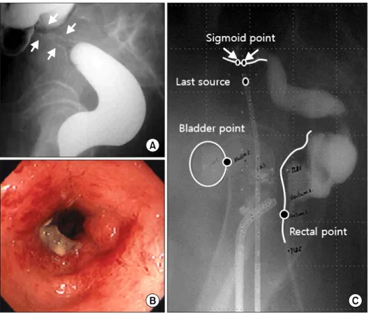
patient returned to the hospital complaining of intermittent rectal bleeding. A barium study revealed a 3-cm-long severe narrowing of the sigmoid wall, which was suspicious for sig- moid colon cancer (Fig. 1A). The patient was referred to our hospital. Sigmoidoscopy revealed luminal narrowing with a friable hyperemic nodular mucosal change 15 cm from the anal verge (Fig. 1B). Biopsies were performed, and the results showed focal chronic active inflammation. The patient was diagnosed as having radiation sigmoiditis and was managed conservatively.
2. Case 2
A 58-year-old woman received definitive RT with concur- rent chemotherapy for FIGO stage IIIA cervical cancer. EBRT was performed at a total dose of 50.4 Gy in 1.8 Gy fractions for five days each week using a 15-MV photon. Intracavitary HDR brachytherapy (microSelectron-HDR) of 24 Gy in six frac- tions was conducted twice each week using a tandem and colpostat. Six cycles of weekly cisplatin (30 mg/m2) were given during RT. The patient has been followed with no evidence of recurrence. After five years, the patient visited our emergency
Fig. 1. Case 1: Lateral view (A) of a barium enema study shows a stricture (between arrows) of the sigmoid colon and endo- scopy shows luminal narrowing and friable hyperemic nodular mucosal changes (B). A close sigmoid-to-tandem distance may cause a high sigmoid dose, which could lead to the development of sigmoiditis (C).
Fig. 2. Case 2: Endoscopy revealed luminal narrowing, hemorrhage and hyperemic mucosal nodularities with mucosal edema of the sigmoid colon (A). A CT scan of the abdomen and pelvis showed diffuse submucosal thickening of the sigmoid wall (arrow) infiltrating into the adjacent mesenteric fat, but no evidence of malignancy was seen (B).
Radiation sigmoiditis mimicking sigmoid colon cancer
J Gynecol Oncol Vol. 23, No. 3:197-200 www.ejgo.org 199
department with complaints of poor oral intake, lower ab- dominal pain and no stool passage over ten days. Sigmoidos- copy revealed an approximately 5-cm-long section of diffuse luminal narrowing in the sigmoid colon and hyperemic mu- cosal nodularities with mucosal edema (Fig. 2A). The endosco- pist suspected primary sigmoid colon cancer or local invasion of recurrent cervical cancer. Pathologic examination reported diffuse active inflammation with inflamed granulation tissue.
A computed tomography (CT) scan also revealed thickening of the wall of the rectosigmoid colon probably caused by ra- diation or inflammation; however no evidence of any cancer recurrence was found (Fig. 2B). She has been managed con- servatively with laxatives and has been clinically followed with improved symptoms.
3. Dosimetric analysis
We were only able to restore the old simulation film in case 1. The simulation film of the HDR brachytherapy is shown in Fig. 1C. The radiation dose was prescribed at point A in 4 Gy fractions. We estimated an irradiated dose of brachytherapy of 518.7 to 642.3 cGy at the sigmoid colon point, which was 29.5% to 60.5% higher than prescribed dose at point A per fraction.
DISCUSSION
Acute and late GI complications are a major concern during and after RT. Radiation sigmoiditis is not well-known com- pared to other GI complications such as proctitis and enteritis because it has been rarely reported. Galland and Spencer [5].
reported 23 cases of sigmoid complications among 80 cases of radiation colitis induced by pelvic RT. Ramirez et al. [4] re- ported that a total of 35 patients developed sigmoid perfora- tion among over 5,000 patients who were treated with RT for stage IB-IIIB cervical cancer.
In this report, we described two cases of late sigmoid com- plication after pelvic RT. The sigmoiditis occurred one year after RT completion for one patient and five years after RT completion for the other patient. Both cases showed obstruc- tive symptoms, which make clinicians suspicious of sigmoid colon cancer or recurrent cancer. During the follow-up of cancer patients who received RT previously, clinicians need to keep the possibility of RT complications in mind.
Holloway et al. [6] reported that the sigmoid-to-tandem dis- tance is significantly related to sigmoid dose using 3D imag- ing techniques. Concordantly, the simulation films of case 1 showed close proximity between the sigmoid colon and the tandem, which suggests the possibility of a high sigmoid dose
during brachytherapy (Fig. 1C). The estimated dose at the sigmoid colon point during brachytherapy was 518.7 to 642.3 cGy, which was 29.5% to 60.5% higher than prescribed dose at point A (400 cGy) per fraction.
The International Commission on Radiation Units (ICRU) 38 did not describe this hot spot in the sigmoid colon. The rec- tal and bladder reference dose points are only standard for reporting normal tissue doses with brachytherapy. Indeed, these points are based on plain films and two-dimensional (2D) brachytherapy planning. As EBRT is moving from con- ventional to conformal planning, the incorporation of three- dimensional (3D) sectional imaging such as CT and MRI into brachytherapy planning is under active investigation [6,7].
Recently, there has been a proposal to establish image-guided intracavitary brachytherapy guidelines for cervical carcinoma.
In the Group European de Curietherapies and European So- ciety for Therapeutic Radiology and Oncology (GEC-ESTRO) recommendations, the rectum and bladder, as well as the sig- moid colon, are highlighted as organs at risk [8]. Many studies have reported a sigmoid dose in 3D image-based planning for the treatment of cervical carcinoma with HDR brachytherapy [6,7]. Kim et al. [7] reported that the sigmoid dose was higher than the rectal dose in 8 of 13 (61%) patients and its volume was discontinuously located as multiple ‘‘hot spots’’ in 9 of 13 (69%) patients on dosimetric analysis of 3D image-based planning of intracavitary brachytherapy for cervical cancer.
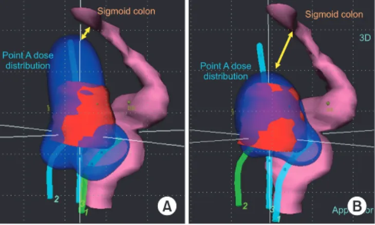
Currently at our institution, 3D image-based brachytherapy is implemented to decrease the radiation dose to organs at risk, including the sigmoid colon (Fig. 3).
3D image-based brachytherapy planning can be helpful, es- pecially for patients who have risk factors. Patients who have
Fig. 3. Treatment planning with three dimensional (3D) image- based brachytherapy. The reconstruction image of dose distribution (blue) using traditional planning based on point A (A) shows a closer proximity to the sigmoid colon compared to 3D planning (B).
applicator, sky blue; clinical target volume, red; rectum and sigmoid colon, purple.
Hyebin Lee, et al.
http://dx.doi.org/10.3802/jgo.2012.23.3.197 200 www.ejgo.org
a history of abdominal surgery, pelvic inflammatory disease, or diverticulosis are at a greater risk of developing radiation- induced GI complications because such conditions may cre- ate adhesions to the uterus, which prevent the loops of the bowel from moving during RT [9].
In conclusion, radiation sigmoiditis is characterized by ob- structive symptoms and its radiological findings can mimic sigmoid colon cancer. Radiation oncologists should acknowl- edge the sigmoid colon as well as rectum and bladder as organs at risk. 3D image-based brachytherapy could be an op- timal modality to reduce the radiation-toxicity associated with treatment for uterine cervical cancer.
CONFLICT OF INTEREST
No potential conflict of interest relevant to this article was reported.
REFERENCES
1. Perez CA, Grigsby PW, Lockett MA, Chao KS, Williamson J.
Radiation therapy morbidity in carcinoma of the uterine cervix: dosimetric and clinical correlation. Int J Radiat Oncol Biol Phys 1999;44:855-66.
2. Perez CA, Breaux S, Bedwinek JM, Madoc-Jones H, Camel HM, Purdy JA, et al. Radiation therapy alone in the treatment of carcinoma of the uterine cervix. II. Analysis of complications. Cancer 1984;54:235-46.
3. Syed AM, Puthawala AA, Abdelaziz NN, el-Naggar M,
Disaia P, Berman M, et al. Long-term results of low-dose- rate interstitial-intracavitary brachytherapy in the treat- ment of carcinoma of the cervix. Int J Radiat Oncol Biol Phys 2002;54:67-78.
4. Ramirez PT, Levenback C, Burke TW, Eifel P, Wolf JK, Gershenson DM. Sigmoid perforation following radiation therapy in patients with cervical cancer. Gynecol Oncol 2001;82:150-5.
5. Galland RB, Spencer J. The natural history of clinically esta blished radiation enteritis. Lancet 1985;1:1257-8.
6. Holloway CL, Racine ML, Cormack RA, O'Farrell DA, Viswa- n athan AN. Sigmoid dose using 3D imaging in cervical- cancer brachytherapy. Radiother Oncol 2009;93:307-10.
7. Kim RY, Shen S, Duan J. Image-based three-dimensional treat ment planning of intracavitary brachytherapy for cancer of the cervix: dose-volume histograms of the bladder, rectum, sigmoid colon, and small bowel. Brachytherapy 2007;6:187-94.
8. Potter R, Haie-Meder C, Van Limbergen E, Barillot I, De Brabandere M, Dimopoulos J, et al. Recommendations from gynaecological (GYN) GEC ESTRO working group (II): concepts and terms in 3D image-based treatment planning in cervix cancer brachytherapy-3D dose volume parameters and aspects of 3D image-based anatomy, radiation physics, radiobiology. Radiother Oncol 2006;78:
67-77.
9. Lanciano RM, Martz K, Montana GS, Hanks GE. Influence of age, prior abdominal surgery, fraction size, and dose on complications after radiation therapy for squamous cell cancer of the uterine cervix: a patterns of care study.
Cancer 1992;69:2124-30.