Copyright © 2015 The Korean Society for Bone and Mineral Research
This is an Open Access article distributed under the terms of the Creative Commons Attribution Non-Commercial Li- cense (http://creativecommons.org/licenses/by-nc/3.0/) which permits unrestricted non-commercial use, distribu- tion, and reproduction in any medium, provided the original work is properly cited.
pISSN 2287-6375 eISSN 2287-7029
Change of Bone Mineral Density and Biochemical Markers of Bone Turnover in Patients on
Suppressive Levothyroxine Therapy for Differentiated Thyroid Carcinoma
Chei Won Kim1, Seokbo Hong1, Se Hwan Oh1, Jung Jin Lee1,2, Joo Young Han1, Seongbin Hong1, So Hun Kim1, Moonsuk Nam1, Yong Seong Kim1
1Department of Endocrinology, Inha University School of Medicine, Incheon;
2Department of Internal Medicine, Hallym General Hospital, Incheon, Korea
Untreated hyperthyroidism and high-dose thyroid hormone are associated with osteo- porosis, and increased bone mineral density (BMD) has been demonstrated in post- menopausal females with hypoparathyroidism. Studies on the effect of suppressive le- vothyroxine (LT4) therapy on BMD and bone metabolism after total thyroidectomy in patients with differentiated thyroid carcinoma have presented conflicting results, and few studies in relation to the status of hypoparathyroidism have been studied. One hun- dred postmenopausal women and 24 premenopausal women on LT4 suppression ther- apy were included in this study. BMD of lumbar spine and femur and bone turnover markers were measured at the baseline and during the follow-up period up to 18 months using dual energy X-ray absorptiometry. Biochemical marker of bone resorption was measured by urine deoxypyridinoline and bone formation by serum osteocalcin.
The age ranged from 36 to 64 years old. Thyroid stimulating hormone (TSH) was sup- pressed during the study. The results showed that BMD of femur and lumbar spine were not significantly changed in both pre- and postmenopausal women except femur neck in postmenopausal women without hypoparathyroidism. Patients with hypoparathy- roidism had higher BMD gain than those without hypoparathyroidism in total hip (1.25 vs. -1.18%, P=0.015). Biochemical markers of bone turnover, serum osteocalcin, and urine deoxypyridinoline did not show significant change. In conclusion, patients with well differentiated thyroid carcinoma are not at a great risk of bone loss after LT4 sup- pressive therapy. The state of hypoparathyroidism is associated with increased BMD, particularly in postmenopausal women.
Key Words: Bone density, Hypoparathyroidism, Postmenopause, Thyroid neoplasms, Thyroxine
INTRODUCTION
The incidence of differentiated thyroid cancer (DTC) is rapidly increasing, althou- gh mortality is stable.[1] Suppression of thyroid stimulating hormone (TSH) is re- quired in an effort to decrease the risk of tumor recurrence.[2] Because hyperthy- Corresponding author
Seongbin Hong
Department of Endocrinology, Inha University School of Medicine, 27 Inhang-ro, Jung-gu, Incheon 22332, Korea
Tel: +82-32-890-2819 Fax: +82-32-890-6578 E-mail: [email protected] Received: August 10, 2015 Revised: August 31, 2015 Accepted: August 31, 2015
No potential conflict of interest relevant to this article was reported.
Original Article
roidism accelerates bone turnover and shortens the normal bone remodeling cycle, it was expected that suppressive levothyroxine (LT4) therapy might decrease the bone min- eral density (BMD).[3]
Although many studies in patients with suppressive LT4 therapy for DTC have been reported, there was no uniform answer to this suspicion. While the majority of reports con- cluded that suppressive LT4 therapy has no deteriorating effect on BMD,[4-8] some studies reported opposite con- clusions in which LT4 suppressive therapy showed a slight- ly negative effect on bone metabolism.[9-13] Several of these studies evaluated the influence of menopausal sta- tus and LT4 dose as well.[9-11]
Most studies excluded patients with hypoparathyroid- ism as considering a confounding variable. No study on the effects of thyroid hormone on BMD and biochemical marker of bone turnover according to the presence of hy- poparathyroidism and estrogen status has been reported.
Even though it occurs in 1 to 10% of patients permanently after thyroidectomy, transient hypoparathyroidism is more common.[14]
Therefore, the aim of our study is to evaluate the effect of suppressive therapy with LT4 on BMD in relation to post- operative hypoparathyroidism in Korean female patients who underwent total thyroidectomy due to DTC.
METHODS
1. PatientsThe research utilized patients’ information between Jan- uary, 2003 and April, 2015, based on digitalized medical records of Inha University hospital. Of all patients who had undergone total or near total thyroidectomy during the period, only those who checked BMD at post-operation and repeated BMD exam from 12 to 18 month from the baseline exam were included. Only female patients were selected, and their states of menopause at the time of the first BMD were confirmed. Postmenopausal women were defined as women with amenorrhea for the 12 months fol- lowing the final menstrual period and who did not receive hormone replacement therapy (HRT). Patients who under- went thyroid lobectomy or had a history of treatment of osteoporosis using bisphosphonate, selective estrogen re- ceptor modulators, calcitonin, and intermittent parathyroid hormone (PTH) were excluded. Patients who had been
taking medication of calcium supplementation and/or ac- tive form of vitamin D were included in the list of study. Fif- ty postmenopausal, 6 premenopausal women who had undergone total thyroidectomy for diagnosed thyroid can- cer and were diagnosed postoperative hypoparathyroid- ism were selected. Fifty postmenopausal and 18 postmeno- pausal women who had intact parathyroid function, and matched based on age and body weight, with the women with hypoparathyroidism were included in the study. A to- tal of 112 patients (90%) had been previously treated with iodine-131 ablation therapy according to the indication.
All included patients had been taking LT4 in order to main- tain the serum TSH concentration between 0.1 and 0.5 mU/L.
The occurrence of hypoparathyroidism after surgery was defined as re-occurrence of hypocalcemia after cessation of active form of vitamin D or dose de-escalation and also refractory hypocalcemia to medication. Serum PTH was confirmed in all patients.
2. Methods
BMD of the lumbar spine (lumbar vertebrae, L1–L4), to- tal hip, and femoral neck was measured at baseline (post- operative) and during the follow-up period (from 12 to 18 months) using dual energy X-ray absorptiometry (DXA;
Hologic QDR-4500, Hologic, Inc., Bedford, MA, USA). The coefficient of variation for BMD measurement at our center is 1.41% in the lumbar spine and 1.89% in the femur. All serum tests including TSH, free T4, intact PTH (iPTH) and bone markers were measured at the time when the first BMD was performed and the latter one was performed.
Biochemical marker of bone resorption was measured by urine deoxypyridinoline (mmol/cr) and bone formation by serum osteocalcin (radioimmunoassay [RIA], ng/mL). iPTH was determined by immunoradiometric assay (IRMA; Ni- cholos Institute Diagonostics, San Juan Capistrano, CA, USA).
Dose of LT4 was titrated by the level of TSH according to the guideline of American Association of Thyroid.[2]
3. Statistical analysis
All results were expressed as mean±standard deviation (SD) unless indicated. Statistical analyses were performed using SPSS version 12.1 (SPSS Inc., Chicago, IL, USA). The characteristics of the participants were compared accord- ing to group using independent samples Student’s t-tests or Mann–Whitney U test for continuous measures and χ2
tests for categorical measures. The BMD at baseline and follow-up were compared using paired t test. Logistic re- gression was used for categorical variables. Observations were considered significant if two-sided P-values were <
0.05.
RESULTS
1. General characteristics of the study population
Of 124 subjects in the study population, 24 women were premenopausal status and 100 women were postmeno- pausal at the time of the first BMD. Thus subjects were sub- divided into 4 different subgroups by menopausal state and presence of hypoparathyroidism. The baseline charac-
teristics of each subgroup are shown in Table 1. Age ranged from 36 to 64 at the time of the earlier BMD. As expected, baseline characteristics of patients with hypoparathyroid- ism were not different except the serum concentration of PTH compared to those without hypoparathyroidism in both premenopausal and postmenopausal groups respec- tively.
2. Postoperative changes in BMD
The value of BMD at the early postoperative period (base- line) was compared with later one. In all patients, follow- up BMD of lumbar spine and femur neck decreased from baseline exam significantly (P<0.001 and P=0.018, respec- tively) (Table 2). However, when same analysis was done in 4 groups according to menopausal status and hypopara-
Table 1. The baseline characteristics of study population
Premenopausal women Postmenopausal women
HypoP (-)
n=18 HypoP (+)
n=6 Pa) HypoP (-)
n=50 HypoP (+)
n=50 Pa)
Age (yr) 43±7 45±2 0.207 56±8 54±7 0.102
Height (cm) 158.3±6.0 155.6±3.8 0.321 155.6±4.9 155.6±5.4 0.983
Weight (kg) 60.3±10.1 58.8±7.9 0.741 62.1±8.2 58.5±8.8 0.065
LT4 dose (μg/day) 169.7±63.2 170.8±29.2 0.968 147.6±36.8 137.8±30.8 0.151
Calcium (mg/dL) 9.1±0.5 8.7±0.8 0.212 9.1±0.6 8.8±0.8 0.062
Phosphorus (mg/dL) 3.7±0.6 4.4±0.8 0.097 4.0±0.8 4.4±0.8 0.061
Free T4 (ng/dL) 1.48±0.66 1.32±0.98 0.006 1.66±0.44 1.56±0.65 0.380
TSH (mIU/L) 0.18±0.25 0.18±0.29 0.980 0.30±0.77 0.22±0.50 0.566
Osteocalcin (ng/mL) 2.99±1.88 2.11±1.55 0.706 5.86±3.07 7.69±4.78 0.408
uDPD (mmol/cr) 7.63±4.81 3.10±1.66 0.462 10.40±6.39 7.27±3.84 0.356
PTH (pg/mL) 31.54±9.94 4.34±4.26 0.006 33.84±16.88 22.03±15.80 0.015
a)P-values were obtained using the χ2 test for categorical variables and the Mann-Whitney U test for continuous variables and represent the significanc- es of differences between patients with and without hypoparathyroidism.
HypoP, hypoparathyroidism; LT4, levothyroxine; PTH, parathyroid hormone; TSH, thyroid stimulating hormone; uDPD, urine deoxypyridinoline.
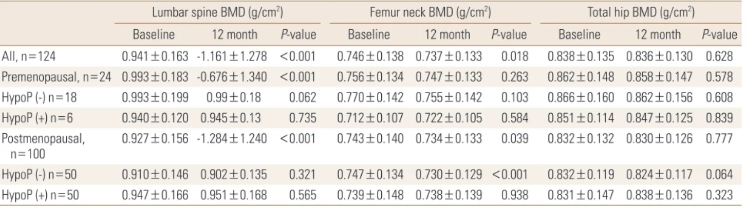
Table 2. Bone mineral density changes in patients with thyroid cancer
Lumbar spine BMD (g/cm2) Femur neck BMD (g/cm2) Total hip BMD (g/cm2) Baseline 12 month P-value Baseline 12 month P-value Baseline 12 month P-value All, n=124 0.941±0.163 -1.161±1.278 <0.001 0.746±0.138 0.737±0.133 0.018 0.838±0.135 0.836±0.130 0.628 Premenopausal, n=24 0.993±0.183 -0.676±1.340 <0.001 0.756±0.134 0.747±0.133 0.263 0.862±0.148 0.858±0.147 0.578 HypoP (-) n=18 0.993±0.199 0.99±0.18 0.062 0.770±0.142 0.755±0.142 0.103 0.866±0.160 0.862±0.156 0.608 HypoP (+) n=6 0.940±0.120 0.945±0.13 0.735 0.712±0.107 0.722±0.105 0.584 0.851±0.114 0.847±0.125 0.839 Postmenopausal,
n=100 0.927±0.156 -1.284±1.240 <0.001 0.743±0.140 0.734±0.133 0.039 0.832±0.132 0.830±0.126 0.777 HypoP (-) n=50 0.910±0.146 0.902±0.135 0.321 0.747±0.134 0.730±0.129 <0.001 0.832±0.119 0.824±0.117 0.064 HypoP (+) n=50 0.947±0.166 0.951±0.168 0.565 0.739±0.148 0.738±0.139 0.938 0.831±0.147 0.838±0.136 0.323 P-values were obtained using paired t-test.
BMD, bone mineral density; HypoP, hypoparathyroidism.
thyroidism, there were no significant differences between levels at baseline and follow-up period on all three sites, except femur neck in postmenopausal women without hy- poparathyroidism (P<0.001).
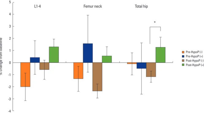
The mean change of BMD after thyroidectomy was cal- culated as percent change between levels at baseline and follow-up (Fig. 1). In women with hypoparathyroidism, Per- cent change of BMD of hip in patients with hypoparathy- roidism increased compared with those in women without hypoparathyroidism in the postmenopausal group (1.25
vs. -1.18%, P=0.015). Percent change of lumbar spine BMD of patient without hypoparathyroidism was not different significantly compared with those with hypoparathyroid- ism in both pre- and postmenopausal women.
3. Changes of serum level of PTH and calcium Since hypoparathyroidism is usually accompanied by hypocalcemia and low level of PTH, hypocalcemia would have been expected in patients with hypoparathyroidism.
As expected, there were significant differences in the fol- Table 3. Variables of bone metabolism and bone turnover markers at follow up
Premenopausal women Postmenopausal women
HypoP (-)
n=18 HypoP (+)
n=6 Pa) HypoP (-)
n=50 HypoP (+)
n=50 Pa)
Calcium (mg/dL) 9.08±0.93 8.75±0.61 0.139 9.15±0.50 8.71±0.80 0.002
Phosphorus (mg/dL) 4.02±0.50 4.10±0.69 0.757 3.94±0.50 4.35±0.67 0.001
Free T4 (ng/dL) 1.74±0.94 1.40±0.81 0.424 1.59±0.70 1.70±0.39 0.267
TSH (mIU/L) 0.10±0.07 0.34±0.66 0.407 0.19±0.33 0.20±0.31 0.825
Osteocalcin (ng/mL) 10.26±8.42 3.36±1.52 0.285 13.19±4.21 8.04±4.74 0.033
uDPD (mmol/cr) 6.53±1.50 4.60±1.67 0.381 10.16±7.10 8.52±5.54 0.209
PTH (pg/mL) 35.00±6.85 4.54±0.37 0.001 32.47±11.0 16.7±10.0 0.001
a)P-values were obtained using the Mann-Whitney U test for continuous variables and represent the significances of differences between patients with and without hypoparathyroidism.
HypoP, hypoparathyroidism; TSH, thyroid stimulating hormone; uDPD, urine deoxypyridinoline; PTH, parathyroid hormone.
Fig. 1. Bone mineral density changes (%) according to menopausal status and presence of hypoparathyroidism *P<0.05. Pre-HypoP (-), premeno- pausal patients without hypoparathyroidism; Pre-HypoP (+), premenopausal patients with hypoparathyroidism; Post-HypoP (-), postmenopausal patients without hypoparathyroidism; Post-HypoP (-), postmenopausal patients with hypoparathyroidism.
5 4 3 2 1 0 -1 -2 -3 -4
% change from baseline
L1-4 Femur neck Total hip
*
Pre-HypoP (-) Pre-HypoP (+) Post-HypoP (-) Post-HypoP (+)
low up level of serum calcium and phosphorus (P=0.001, P=0.001, respectively), not only the level of PTH in relation to hypoparathyroidism. However level of serum TSH showed no significant difference between patients with and with- out hypoparathyroidism in pre-, postmenopausal women (P=0.407, P=0.825, respectively). Baseline or follow up TSH, PTH or age could not predict BMD change in all groups on multiple regression study. .
4. Changes in biochemical markers of bone turnover
The levels of serum osteocalcin and urine deoxypyridin- oline were not significantly changed. Follow up serum os- teocalcin level was higher in patients without hypothyroid- ism compared to those with hypoparathyroidism in post- menopausal women (Table 3). For all patients enrolled in this study, osteocalcin and urine deoxypyridinoline were not associated with BMD change in lumbar and femur.
DISCUSSION
Our study showed no deleterious effect of suppressive LT4 treatment in women with DTC, regardless of their es- trogen status, and a protective effect was found in women with post-operative hypoparathyroidism. Even recommen- dations for management of thyroid carcinoma have no definite suggestion with regard to monitoring TSH level to prevent accelerated bone turnover.[15] Thyroid hormone excess increases osteoclastic and osteoblastic activities, both in vitro studies.[16] Although the majority of investi- gations showed that a carefully monitored LT4 suppressive therapy is not associated with bone loss in premenopausal women with differentiated thyroid cancer, change of BMD in postmenopausal women was still conflicting.
LT4 suppressive therapy was associated with bone loss in postmenopausal women,[6,8] which could be prevent- ed by either calcium supplementation or intranasal calci- tonin.[10] LT4 suppressive therapy accelerates bone loss, particularly in postmenopausal women and exclusively during the early post-thyroidectomy period.[11] On the other hand, Reverter et al.[7] reported that long-term sup- pressive LT4 treatment did not affect skeleton in women regardless of estrogen status.
However, most previous studies did not include patients with hypoparathyroidism, because hypoparathyroidism is
regarded as a confounding factor. PTH plays an important role in regulation of bone metabolism. It regulates calcium homeostasis by affecting intestinal calcium absorption, re- nal calcium excretion, and the rate of bone resorption.[17]
In our study, percent change of BMD in patients with hy- poparathyroidism increased significantly in total hip com- pared to patients with normal parathyroid function. Chan et al.[18] reported that the state of hypoparathyroidism ei- ther idiopathic or post-thyroidectomy is associated with increased BMD, most notably at the spine. However that study included 6 patients with post-thyroidectomy hypo- parathyroidism and 8 with idiopathic hypoparathyroidism.
In addition, their age ranged from 23 to 57 years old and menopause was not considered. In another study, BMD was increased in postmenopausal females at the lumbar spine and the proximal femur with hypoparathyroidism af- ter thyroidectomy, but it was not statistically significant and a cross- sectional study.[19] Increase of BMD may ei- ther be attributable to hypoparathyroidism or to treatment with active form of vitamin D and calcium supplementa- tion.[20,21] An increase in bone mineralization secondary to suppressed bone turnover can increase BMD. Accelerat- ed bone loss can be attenuated in patients after menopause, indicating a reduced remodeling rate with calcium and vi- tamin D.[22] Although high dose calcium and vitamin D supplement means severe hypoparathyroidism status, dose of this correlated with BMD. Vitamin D receptors have been found in osteoblasts and in normal subjects, vitamin D stim- ulates both the number and activity of osteoblasts, also in- creased BMD.[23,24]
Previous studies have focused on menopausal state and endogenous and drug induced hyperthyroidism. Meta-anal- yses have shown a significant reduction in BMD only in post- menopausal women on long-term LT4 suppressive therapy that was more marked on cortical bone than on trabecular bone.[25] The study group included cancer and hypothy- roidism, and studied BMD alone. In addition, changes of BMD could be different according to the replacement or suppressive treatment.
Bone marker study did not show significant change dur- ing the follow up period, and did not show correlation with percent change of BMD. Serum osteocalcin increased dur- ing treatment with calcium and vitamin D in patients with hypoparathyroidism.[26]
Currently there are no studies concerning the effect of
LT4 on BMD and bone turnover marker according to meno- pausal and parathyroid status. Therefore our study is the first study considering the effect of thyroid hormone on BMD and biological bone turnover marker in relation to parathyroid state and estrogen status.
The limitations of our study include small sample size and the fact that all participants were enrolled at a single center. Because our study was retrograde, a thyroid hor- monal status during the follow-up period was not strictly controlled. The second limitation is that the follow up peri- od was not long enough to evaluate the long-term effect of LT4 suppression on bone metabolism. Further studies are necessary to clarify the long term effect of TSH suppres- sion in multiple centers. Also, baseline serum vitamin D lev- el was not measured, which could have differently affected BMD changes.
In summary, we have shown little deleterious effect of LT4 suppressive treatment, suggesting that patients with LT4 suppressive therapy are not at great risk of bone loss.
In addition, in patients with hypoparathyroidism, it showed an improved BMD which might be explained in part by sup- plementation of calcium and active vitamin D metabolites.
Further series of intervening studies to prove this opinion is required.
REFERENCES
1. Brito JP, Davies L. Is there really an increased incidence of thyroid cancer? Curr Opin Endocrinol Diabetes Obes 2014;
21:405-8.
2. Cooper DS, Doherty GM, Haugen BR, et al. Revised Ameri- can Thyroid Association management guidelines for pa- tients with thyroid nodules and differentiated thyroid can- cer. Thyroid 2009;19:1167-214.
3. Mosekilde L, Eriksen EF, Charles P. Effects of thyroid hor- mones on bone and mineral metabolism. Endocrinol Metab Clin North Am 1990;19:35-63.
4. Lee MY, Park JH, Bae KS, et al. Bone mineral density and bone turnover markers in patients on long-term suppres- sive levothyroxine therapy for differentiated thyroid can- cer. Ann Surg Treat Res 2014;86:55-60.
5. Heijckmann AC, Huijberts MS, Geusens P, et al. Hip bone mineral density, bone turnover and risk of fracture in pa- tients on long-term suppressive L-thyroxine therapy for differentiated thyroid carcinoma. Eur J Endocrinol 2005;
153:23-9.
6. Larijani B, Gharibdoost F, Pajouhi M, et al. Effects of levo- thyroxine suppressive therapy on bone mineral density in premenopausal women. J Clin Pharm Ther 2004;29:1-5.
7. Reverter JL, Holgado S, Alonso N, et al. Lack of deleterious effect on bone mineral density of long-term thyroxine sup- pressive therapy for differentiated thyroid carcinoma. En- docr Relat Cancer 2005;12:973-81.
8. Marcocci C, Golia F, Bruno-Bossio G, et al. Carefully moni- tored levothyroxine suppressive therapy is not associated with bone loss in premenopausal women. J Clin Endocri- nol Metab 1994;78:818-23.
9. Jódar E, Begoña López M, Garcia L, et al. Bone changes in pre- and postmenopausal women with thyroid cancer on levothyroxine therapy: evolution of axial and appendicu- lar bone mass. Osteoporos Int 1998;8:311-6.
10. Kung AW, Yeung SS. Prevention of bone loss induced by thyroxine suppressive therapy in postmenopausal wom- en: the effect of calcium and calcitonin. J Clin Endocrinol Metab 1996;81:1232-6.
11. Kim MK, Yun KJ, Kim MH, et al. The effects of thyrotropin- suppressing therapy on bone metabolism in patients with well-differentiated thyroid carcinoma. Bone 2015;71:101-5.
12. Mohammadi B, Haghpanah V, Tavangar SM, et al. Model- ing the effect of levothyroxine therapy on bone mass den- sity in postmenopausal women: a different approach leads to new inference. Theor Biol Med Model 2007;4:23.
13. Mazokopakis EE, Starakis IK, Papadomanolaki MG, et al.
Changes of bone mineral density in pre-menopausal wom- en with differentiated thyroid cancer receiving L-thyroxine suppressive therapy. Curr Med Res Opin 2006;22:1369-73.
14. Youngwirth L, Benavidez J, Sippel R, et al. Parathyroid hor- mone deficiency after total thyroidectomy: incidence and time. J Surg Res 2010;163:69-71.
15. Cooper DS, Specker B, Ho M, et al. Thyrotropin suppres- sion and disease progression in patients with differentiat- ed thyroid cancer: results from the National Thyroid Can- cer Treatment Cooperative Registry. Thyroid 1998;8:737- 44.
16. Lee MS, Kim SY, Lee MC, et al. Negative correlation between the change in bone mineral density and serum osteocal- cin in patients with hyperthyroidism. J Clin Endocrinol Me- tab 1990;70:766-70.
17. De Sanctis V, Soliman A, Fiscina B. Hypoparathyroidism:
from diagnosis to treatment. Curr Opin Endocrinol Diabe-
tes Obes 2012;19:435-42.
18. Chan FK, Tiu SC, Choi KL, et al. Increased bone mineral den- sity in patients with chronic hypoparathyroidism. J Clin Endocrinol Metab 2003;88:3155-9.
19. Duan Y, De Luca V, Seeman E. Parathyroid hormone defi- ciency and excess: similar effects on trabecular bone but differing effects on cortical bone. J Clin Endocrinol Metab 1999;84:718-22.
20. Dawson-Hughes B, Dallal GE, Krall EA, et al. A controlled trial of the effect of calcium supplementation on bone den- sity in postmenopausal women. N Engl J Med 1990;323:
878-83.
21. Reid IR, Ames RW, Evans MC, et al. Effect of calcium sup- plementation on bone loss in postmenopausal women. N Engl J Med 1993;328:460-4.
22. Chapuy MC, Arlot ME, Duboeuf F, et al. Vitamin D3 and
calcium to prevent hip fractures in the elderly women. N Engl J Med 1992;327:1637-42.
23. Haussler MR, Haussler CA, Jurutka PW, et al. The vitamin D hormone and its nuclear receptor: molecular actions and disease states. J Endocrinol 1997;154 Suppl:S57-73.
24. Cranney A, Weiler HA, O'Donnell S, et al. Summary of evi- dence-based review on vitamin D efficacy and safety in relation to bone health. Am J Clin Nutr 2008;88:513s-9s.
25. Uzzan B, Campos J, Cucherat M, et al. Effects on bone mass of long term treatment with thyroid hormones: a meta- analysis. J Clin Endocrinol Metab 1996;81:4278-89.
26. Hawkins F, Escobar-Jiménez F, Jódar E, et al. Bone mineral density in hypoparathyroid women on LT4 suppressive therapy. Effect of calcium and 1,25(OH)2 vitamin D3 treat- ment. J Musculoskelet Neuronal Interact 2003;3:71-6.