http://dx.doi.org/10.3988/jcn.2012.8.2.146 J Clin Neurol 2012;8:146-150
Introduction
Sleep deprivation (SD) is known to cause profound impair- ments in executive function and vigilant attention.1,2 SD is reported to be associated with increases in blood pressure and stress hormone levels, and a decrease in parasympathetic tone.3,4 Stress hormones are regarded as factors that can in- terfere with cognitive function, including memory5; it has been shown that the increased levels of adrenal steroids in- duced by psychological stress blocks hippocampal long-term potentiation.6 The present study was designed to investigate
whether 24 h of SD negatively affects the attention and working memory and increases the serum concentrations of stress hormones, glucose, and inflammatory markers.
Methods
Subjects and study protocol
Six normal healthy men (mean age 25.3 years, range 23-27 years; mean duration of education 13.5 years, range 14-18 years) participated in the study. All subjects were good sleep- ers and non habitual nappers with no excessive daytime sleep- iness, and they had low anxiety levels. Sleep quality and qu- antity were assessed by questionnaires and actigraphy re- cordings. The subjects performed a cognitive test [the Conti- nuous Performance Test (CPT)] in the morning after a nor-
Adverse Effects of 24 Hours of Sleep Deprivation on Cognition and Stress Hormones
Eun Yeon Joo,* Cindy W Yoon,* Dae Lim Koo, Daeyoung Kim, Seung Bong Hong
Department of Neurology, Samsung Medical Center, Sungkyunkwan University School of Medicine, Seoul, Korea
Received September 26, 2011 Revised January 2, 2012 Accepted January 2, 2012 Correspondence Seung Bong Hong, MD Department of Neurology, Samsung Medical Center, Sungkyunkwan University School of Medicine, 50 Irwon-dong, Gangnam-gu, Seoul 135-710, Korea Tel +82-2-3410-3592 Fax +82-2-3410-0052 E-mail [email protected]
*Two first authors contributed equally to write the manuscript in this study.
Background and PurposezzThe present study was designed to investigate whether 24 h of SD negatively affects the attention and working memory and increases the serum concentrations of stress hormones, glucose, and inflammatory markers.
MethodszzThe acute effects of sleep deprivation (SD) on cognition and the stress hormones were evaluated in six healthy volunteers (all men, age 23-27 years). All were good sleepers, had no history of medical or neuropsychiatric diseases, and were not taking any kind of medication.
All of the volunteers were subjected to the Continuous Performance Test (CPT) for attention and working memory of cognition and blood tests both before and after 24 h of SD. Electroencepha- lographic monitoring was performed during the study to confirm the wakefulness of the sub- jects.
ResultszzSD significantly elevated the serum concentrations of stress hormones (cortisol, epi- nephrine, and norepinephrine), but serum levels of glucose and inflammatory markers were not changed compared to baseline. For easier steps of the CPT the subjects performed well in giving correct responses after SD; the correct response scores decreased only at the most difficult step of the CPT. However, the subjects performed consistently poor for the error responses at all steps after SD. There was no correlation between the CPT scores and stress hormone levels.
ConclusionszzThe 24 h of SD significantly heightened the levels of stress hormones and low- ered attention and working memory. The acute SD condition seems to render the subject more
susceptible to making errors. J Clin Neurol 2012;8:146-150
Key Wordszz sleep deprivation, attention, working memory, stress hormone.
Open Access
cc This is an Open Access article distributed under the terms of the Cre- ative Commons Attribution Non-Commercial License (http://creative- commons.org/licenses/by-nc/3.0) which permits unrestricted non-com- mercial use, distribution, and reproduction in any medium, provided the ori- ginal work is properly cited.
mal night of sleep and then again after 24 h of SD. Serum concentrations of stress hormones (cortisol, epinephrine, and norepinephrine), glucose, and inflammatory markers such as erythrocyte sedimentation rate (ESR) and C-reactive protein (CRP) were measured at the same time that the tests were performed.
The CPT is a psychological test that measures a subject’s sustained and selective attention and impulsivity.7 All tasks were presented on a computer screen, and the entire testing procedure was fully computerized. Commercial software (STIM2, Neuroscan, Compumedics, Charlotte, NC, USA) was used to present the stimuli on a 17-inch LCD monitor. A sequential series of single letters was presented visually at a rate of one item per 500 msec. The subjects were asked to respond every time the letter “X”, “Y”, or “Z” appeared among a string of random letters (level 1). At a higher step (level 2), the subjects were required to respond as quickly as possible only when the stimulus appeared in a particular con- text (e.g., the letter “X” or “Y” only when it followed the let- ter “Y” or “X”), and not press the button if “X” or “Y” ap- peared following the same letter. At the highest step (level 3), the number of response stimuli was increased from two to three (the letters “X”, “Y”, and “Z”). At each level, the scores of correct responses and error responses were calcu- lated automatically.
Subjects completed the Stanford Sleepiness Scale (SSS)8 and a visual analog scale (VAS) for fatigue every 2 h in order to measure subjective alertness and fatigue. Electroencepha- lographic video monitoring was conducted to confirm the wakefulness of the subjects objectively. Blood pressure and pulse rate were also measured every 2 h. Subjects were served four meals a day (at 08:00, 13:00, 19:00, and 01:00 h) with no differences in the amount or composition of food.
All subjects gave their written informed consent to partici- pate before the commencement of the study. The Institutional Review Board at Samsung Medical Center authorized the in- formed consent form and the study protocol.
Statistical analysis
The parameters (the scores of correct and error responses) obtained from the CPT and the serum concentrations of cor- tisol, epinephrine, norepinephrine, glucose, ESR, and CRP were compared before and after SD. The Wilcoxon signed- rank test was used for the values with a nonparametric distri- bution, and multiple comparisons were performed using Bonferroni’s correction. The level of statistical significance was set at p<0.05. All statistical analyses were conducted us- ing commercially available software (SPSS for Windows, version 17.0, SPSS, Chicago, IL, USA).
Results
Subjective fatigue and alertness
A higher VAS score indicated greater fatigue. The mean VAS scores for fatigue increased with time for all subjects. The mean SSS scores significantly increased from 1.2 to 6.3 at 09:00 h the next morning (p<0.001). Mean blood pressure (systolic/diastolic) and pulse rate did not differ between be- fore and after 24 h of SD.
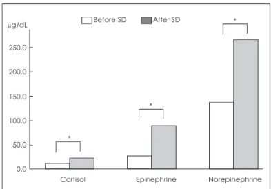
Stress hormones and inflammatory markers The mean serum concentrations of all stress hormones such as cortisol, epinephrine, and norepinephrine were signifi- cantly elevated in the morning after SD (Fig. 1). The refer- ence ranges used at Samsung Medical Center are as follows:
cortisol, 5.9-26.1 µg/dL; epinephrine <60 µg/dL during sitting;
and norepinephrine, 120-680 µg/dL during sitting. However, mean serum concentrations of glucose and inflammatory markers (ESR and CRP) were not changed after SD. Reference ranges are as follows: ESR, 0-22 mm/h; CRP, 0-0.3 mg/dL.
Continuous Performance Test
The mean score of correct responses was significantly de- creased at the highest (most difficult) step after 24 h of SD;
there were no statistical differences at easier steps. In con- trast, the mean scores of error responses were significantly increased at all steps after 24 h of SD (Fig. 2). No significant correlations were found between CPT scores and the serum concentrations of stress hormones, glucose, and inflammato- ry markers. Detailed data of patients before and after SD are listed and compared in Table 1.
Fig. 1. Comparison of serum concentrations of stress hormones before and after 24 h of SD. Mean serum concentrations of corti- sol, epinephrine, and norepinephrine were significantly increased after SD. *p<0.05 (Wilcoxon signed-rank test with multiple compari- sons). SD: sleep deprivation.
0.0 50.0 100.0 150.0 200.0 250.0
Cortisol Epinephrine Norepinephrine
*
*
µg/dL Before SD After SD *
Discussion
The hypothesis underlying this study was that acute SD for 24 h negatively influences daytime function and increases hormone levels in healthy volunteers. As expected, the fa-
tigue level increased and alertness decreased over time in our subjects. After a total SD of 24 h, subjects recorded a VAS score of 95 (extremely fatigued), and an SSS alertness score of 6 (foggy, losing interest in remaining awake, and slowed- down state). The CPT used in this study measures a person’s
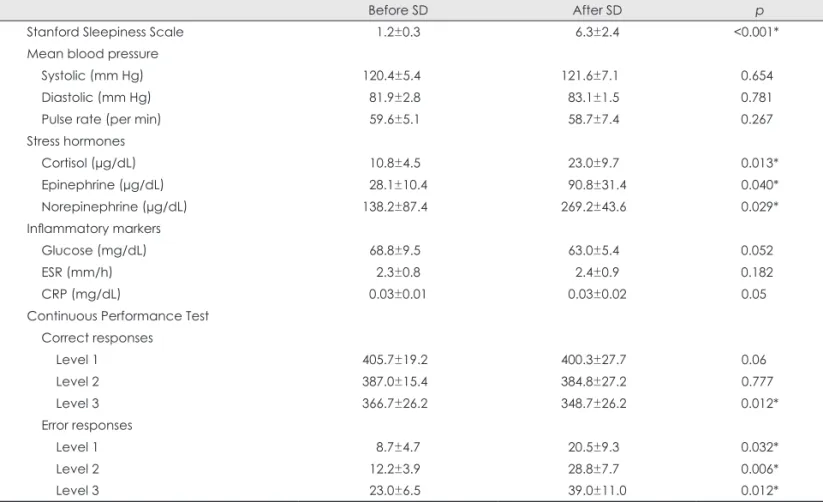
Table 1. Comparison of data before and after sleep deprivation (SD) in patients
Before SD After SD p
Stanford Sleepiness Scale 1.2±0.3 6.3±2.4 <0.001*
Mean blood pressure
Systolic (mm Hg) 120.4±5.4 121.6±7.1 0.654
Diastolic (mm Hg) 81.9±2.8 83.1±1.5 0.781
Pulse rate (per min) 59.6±5.1 58.7±7.4 0.267
Stress hormones
Cortisol (µg/dL) 10.8±4.5 23.0±9.7 0.013*
Epinephrine (µg/dL) 28.1±10.4 90.8±31.4 0.040*
Norepinephrine (µg/dL) 138.2±87.4 269.2±43.6 0.029*
Inflammatory markers
Glucose (mg/dL) 68.8±9.5 63.0±5.4 0.052
ESR (mm/h) 2.3±0.8 2.4±0.9 0.182
CRP (mg/dL) 0.03±0.01 0.03±0.02 0.05
Continuous Performance Test Correct responses
Level 1 405.7±19.2 400.3±27.7 0.06
Level 2 387.0±15.4 384.8±27.2 0.777
Level 3 366.7±26.2 348.7±26.2 0.012*
Error responses
Level 1 8.7±4.7 20.5±9.3 0.032*
Level 2 12.2±3.9 28.8±7.7 0.006*
Level 3 23.0±6.5 39.0±11.0 0.012*
Data are mean±standard deviation values.
*p<0.05 (Wilcoxon signed-rank test with multiple comparisons using Bonferroni’s correction).
Fig. 2. Comparison of the Continuous Performance Test results before and after 24 h of sleep deprivation (SD). The mean scores of correct (A) and error (B) responses are shown before and after SD according to the test level (i.e., 1-3). The mean score of correct responses was signifi- cantly reduced after SD only at the most difficult test level. However, the mean scores of error responses were significantly increased at all test levels. *p<0.05 (Wilcoxon signed-rank test with multiple comparisons).
0.0 5.0 10.0 15.0 20.0 25.0 30.0 35.0 40.0 45.0
Level 1 Level 2
*
*
*
Level 3
Error (n)
B A
310 320 330 340 350 360 370 380 390 400 410 420
Level 1 Level 2
*
Level 3
Correct (n)
Before SD After SD Before SD After SD
sustained and selective attention and working memory. Our results showed that at easier steps (levels 1 and 2), the mean scores of correct responses were not significantly decreased after SD, but they were significantly reduced (decreased by 2.2%) at the most difficult step (level 3). The mean scores of error responses were significantly increased at all levels (in- creased by 69.5-136%) after SD. These findings show that attention and working memory are impaired after acute SD of 24 h, with the performance decrements being more dra- matic for more complex tasks. Interestingly, subjects exhibit- ed a relatively good performance in correct responses at eas- ier steps after SD, but they made errors frequently even at easier steps as well as most difficult level. The findings sug- gest that the SD condition results in the subject being more susceptible to making an error.
Regardless of the task, cognitive performance progressive- ly deteriorated for increasing time spent on the task; this is a classic “fatigue” effect that is exacerbated by sleep loss.9 Previous findings that performance on even brief cognitive tasks that measure attention and working memory was sensi- tive to SD2,10,11 support our results.
This study showed that acute (24 h) SD induces increases in the serum concentrations of stress hormones, and suggests that SD itself is the stress-eliciting situation. Although the in- creased concentrations of hormones after SD were still with- in normal ranges, statistically heightened levels of hormones after SD may indicate the important effect of sleep on stress hormones. Some studies have shown that stress hormones may interfere with memory and other cognitive functions. A significant relationship between pre-retrieval stress or high cortisol levels and impaired memory retrieval has been re- ported consistently.12,13 Acute elevations of exogenous gluco- corticosteroids were shown to impair working memory with- out affecting declarative memory.13,14 A significant associa- tion was found between insomnia duration and left dor- somedial prefrontal damage in patients with focal brain lesions.15 The gray-matter volume in the left orbitofrontal cortex is reduced in chronic insomnia, and was found to be strongly correlated with the subjective severity of insomnia.16 Although the aim of the present experiment was to examine 24 h SD, significant deficits in attention and working memo- ry after SD suggested that acute SD causes a deterioration in prefrontal function.
While cross-sectional and longitudinal studies point to a U-shaped relationship between self-reported sleep duration and the risk of type 2 diabetes mellitus,17,18 it was recently re- ported that prebreakfast concentrations of blood glucose and other inflammatory markers were not changed after restrict- ing sleep (to <4.5 h/day) for 2 nights.19 We also found un- changed serum concentrations of glucose after SD. Whether
or not acute SD alters glucose metabolism therefore remains uncertain. Additional measurements of other hormones such as insulin and glucagon would be helpful to clarify this issue.
Insufficient sleep and poor sleep quality are risk factors for inflammation-related conditions.19-21 Total SD for 40 h caused significant increases in E-selectin, intercellular adhe- sion molecule 1, interleukin-1b, and interleukin-1Ra, and de- creases in CRP and interleukin-6, suggesting that sleep loss triggers a stress response that includes stimulation of both pro- and anti-inflammatory proteins.22 In contrast, we did not find any differences in the serum concentrations of ESR and CRP, which are nonspecific measures of inflammation. It is not clear whether the difference in these results is attribut- able to the duration of sleep deprivation or the specificity of inflammatory markers.
While this study has not revealed a direct relationship be- tween stress hormone levels and cognitive function, we did find that acute SD for 24 h significantly heightened the levels of stress hormones and caused a deterioration of cognitive function. The acute SD condition appears to render the sub- ject more susceptible to making an error.
Conflicts of Interest
The authors have no financial conflicts of interest.
Acknowledgements
This study was supported by a Grant of the Korean Health Technology R&D Project, Ministry for Health, Welfare & Family Affairs, Republic of Korea (No. A110097), and by the Global Frontier R&D Program on
<Human-centered Interaction for Coexistence> funded by the National Research Foundation of Korea grant funded by the Korean Government (MEST) (NRF-M1AXA003-2011-0031688).
REFERENCES
1. Nilsson JP, Söderström M, Karlsson AU, Lekander M, Akerstedt T, Lindroth NE, et al. Less effective executive functioning after one night’s sleep deprivation. J Sleep Res 2005;14:1-6.
2. Adam M, Rétey JV, Khatami R, Landolt HP. Age-related changes in the time course of vigilant attention during 40 hours without sleep in men. Sleep 2006;29:55-57.
3. Leproult R, Copinschi G, Buxton O, Van Cauter E. Sleep loss results in an elevation of cortisol levels the next evening. Sleep 1997;20:865- 4. Spiegel K, Leproult R, Van Cauter E. Impact of sleep debt on meta-870.
bolic and endocrine function. Lancet 1999;354:1435-1439.
5. Ryu SY, Kwon MJ, Lee SB, Yang DW, Kim TW, Song IU, et al.
Measurement of precuneal and hippocampal volumes using magnetic resonance volumetry in Alzheimer’s disease. J Clin Neurol 2010;6:
196-203.
6. Diamond DM, Fleshner M, Rose GM. Psychological stress repeated- ly blocks hippocampal primed burst potentiation in behaving rats.
Behav Brain Res 1994;62:1-9.
7. Conners CK; MHS Staff. Conners’ Continuous Performance Test II:
Computer Program for Windows Technical Guide and Software Manual. North Tonawanda, NY: Multi-Health Systems, 2000.
8. Hoddes E, Zarcone V, Smythe H, Phillips R, Dement WC. Quantifi- cation of sleepiness: a new approach. Psychophysiology 1973;10:
431-436.
9. Wilkinson RT. Sleep deprivation: performance tests for partial and selective sleep deprivation. Prog Clin Psychol 1968;8:28-43.
10. Whitney P, Hinson JM. Measurement of cognition in studies of sleep deprivation. Prog Brain Res 2010;185:37-48.
11. Hagewoud R, Whitcomb SN, Heeringa AN, Havekes R, Koolhaas JM, Meerlo P. A time for learning and a time for sleep: the effect of sleep deprivation on contextual fear conditioning at different times of the day. Sleep 2010;33:1315-1322.
12. de Quervain DJ, Henke K, Aerni A, Treyer V, McGaugh JL, Berthold T, et al. Glucocorticoid-induced impairment of declarative memory retrieval is associated with reduced blood flow in the medial tempo- ral lobe. Eur J Neurosci 2003;17:1296-1302.
13. Lupien SJ, Gillin CJ, Hauger RL. Working memory is more sensitive than declarative memory to the acute effects of corticosteroids: a dose-response study in humans. Behav Neurosci 1999;113:420-430.
14. Wolf OT, Convit A, McHugh PF, Kandil E, Thorn EL, De Santi S, et al. Cortisol differentially affects memory in young and elderly men.
Behav Neurosci 2001;115:1002-1011.
15. Koenigs M, Holliday J, Solomon J, Grafman J. Left dorsomedial frontal brain damage is associated with insomnia. J Neurosci 2010;30:
16041-16043.
16. Altena E, Vrenken H, Van Der Werf YD, van den Heuvel OA, Van Someren EJ. Reduced orbitofrontal and parietal gray matter in chron- ic insomnia: a voxel-based morphometric study. Biol Psychiatry 2010;67:182-185.
17. Gangwisch JE, Heymsfield SB, Boden-Albala B, Buijs RM, Kreier F, Pickering TG, et al. Sleep duration as a risk factor for diabetes inci- dence in a large U.S. sample. Sleep 2007;30:1667-1673.
18. Mallon L, Broman JE, Hetta J. High incidence of diabetes in men with sleep complaints or short sleep duration: a 12-year follow-up study of a middle-aged population. Diabetes Care 2005;28:2762- 2767.
19. Schmid SM, Hallschmid M, Jauch-Chara K, Wilms B, Lehnert H, Born J, et al. Disturbed glucoregulatory response to food intake after moderate sleep restriction. Sleep 2011;34:371-377.
20. Mullington JM, Haack M, Toth M, Serrador JM, Meier-Ewert HK.
Cardiovascular, inflammatory, and metabolic consequences of sleep deprivation. Prog Cardiovasc Dis 2009;51:294-302.
21. Knutson KL, Spiegel K, Penev P, Van Cauter E. The metabolic con- sequences of sleep deprivation. Sleep Med Rev 2007;11:163-178.
22. Frey DJ, Fleshner M, Wright KP Jr. The effects of 40 hours of total sleep deprivation on inflammatory markers in healthy young adults.
Brain Behav Immun 2007;21:1050-1057.