http://dx.doi.org/10.3988/jcn.2015.11.1.80 J Clin Neurol 2015;11(1):80-86
Low-Density-Lipoprotein Particle Size Predicts a Poor Outcome in Patients with Atherothrombotic Stroke
Tae-Jin Song,a,b Hyun-Ji Cho,c Yoonkyung Chang,a Minjung Youn,a Min-Jeong Shin,d Inho Jo,e Ji Hoe Heo,b Yong-Jae Kima
aDepartments of Neurology and eMolecular Medicine, Ewha Womans University School of Medicine, Seoul, Korea
bDepartment of Neurology, Yonsei University College of Medicine, Seoul, Korea
cDepartment of Neurology, Incheon St. Mary’s Hospital, The Catholic University of Korea, Incheon, Korea
dDepartment of Food and Nutrition and Institute of Health Sciences, Korea University, Seoul, Korea
Received June 20, 2014 Revised September 19, 2014 Accepted September 24, 2014 Correspondence Yong-Jae Kim, MD, PhD Department of Neurology, Ewha Womans University School of Medicine, 1071 Anyangcheon-ro, Yangcheon-gu, Seoul 158-710, Korea Tel +82-2-2650-2677 Fax +82-2-2650-5958 E-mail [email protected]
Background and PurposezzLow-density lipoprotein (LDL) particle size is considered to be one of the more important cardiovascular risk factors, and small LDL particles are known to have atherogenic potential. The aim of this study was to determine whether LDL particle size is associated with stroke severity and functional outcome in patients with atherothrombotic stroke.
MethodszzBetween January 2009 and May 2011, 248 patients with first-episode cerebral in- farction who were admitted to our hospital within 7 days after symptom onset were prospec- tively enrolled. LDL particle size was measured using the nondenaturing polyacrylamide gradi- ent gel electrophoresis assay. Stroke severity was assessed by applying the National Institutes of Health Stroke Scale (NIHSS) at admission. Functional outcome was investigated at 3 months after the index stroke using the modified Rankin Scale (mRS), and poor functional out- come was defined as an mRS score of ≥3.
ResultszzThe LDL particle size in the 248 patients was 25.9±0.9 nm (mean±SD). LDL parti- cle size was inversely correlated with the degree of cerebral artery stenosis (p=0.010). Multino- mial multivariate logistic analysis revealed that after adjustment for age, sex, and variables with p<0.1 in univariate analysis, LDL particle size was independently and inversely associated with stroke severity (NIHSS score ≥5; reference, NIHSS score 0–2; odds ratio=0.38, p=0.028) and poor functional outcome (odds ratio=0.44, p=0.038).
ConclusionszzThe results of this study demonstrate that small LDL particles are independent- ly correlated with stroke outcomes. LDL particle size is thus a potential biomarker for the prog- nosis of atherothrombotic stroke. J Clin Neurol 2015;11(1):80-86 Key Wordszz low-density-lipoprotein particle size, lipoprotein, stroke severity,
stroke outcome, atherosclerosis.
Open Access
cc This is an Open Access article distributed under the terms of the Cre- ative Commons Attribution Non-Commercial License (http://creative- commons.org/licenses/by-nc/3.0) which permits unrestricted non-com- mercial use, distribution, and reproduction in any medium, provided the ori- ginal work is properly cited.
Introduction
Hypercholesterolemia is closely associated with an increased risk of cardiovascular disease, including stroke, due to ath-
erosclerosis of blood vessels. Total cholesterol is made up of low-density lipoprotein (LDL), very-low-density lipoprotein, triglycerides (TG), and high-density lipoprotein (HDL). Ath- erosclerosis is generally characterized by elevated plasma concentrations of TG or LDL and a decreased HDL concen- tration.1 It has recently been shown that certain LDL, subclas- sified according to particle size, are associated with athero- sclerotic risk.2 LDL particle size is negatively associated with plasma TG levels, and the combination of small LDL particles
and increased plasma TG levels is considered to be the athero- genic lipoprotein phenotype.3 Small LDL particles are more easily taken up by arterial tissue4 and are more susceptible to oxidative stress than are large LDL particles.5 Therefore, small LDL particles are considered to be associated with the pro- gression of atherosclerosis6 and have been accepted by the National Cholesterol Education Program Adult Treatment Pan- el III as one of the emerging cardiovascular risk factors.7
Clinical studies have emphasized the role of small LDL par- ticles in the development of early atherosclerosis in meno- pausal women,8 coronary heart disease including acute myo- cardial infarction or coronary vasospasm,9-12 or peripheral arterial disease, regardless of the presence of diabetes melli- tus.13 In stroke patients, small LDL particles are known to be associated with short-term mortality after acute ischemic stroke.14 However, there is as yet little information available on the relationship between LDL particle size and functional outcome after stroke, and including stroke severity. Therefore, in this study, the association between LDL particle and stroke severity and functional outcomes was investigated in patients with atherothrombotic stroke.
Methods
Subjects
Patients diagnosed with first-episode ischemic stroke and ad- mitted to Ewha Womans University Mokdong Hospital with- in 7 days after symptom onset were prospectively enrolled in this study between January 2009 and June 2011. Blood sam- ples to be used for LDL particle analysis were obtained from all enrolled patients. Patient information was collected, and data were evaluated including past medical, medication, and familial history, brain imaging studies (CT and/or MRI), vas- cular imaging studies (digital subtraction angiography, CT an- giography, or MR angiography), chest X-ray, 12-lead electro- cardiography, electrocardiography monitoring during a median time period of 3 days at a stroke intensive care unit, transtho- racic echocardiography, and routine blood tests.15 Patients were excluded if they did not agree to provide blood samples for this study. Of the 427 initially eligible patients, 2 who received incomplete vascular imaging, 40 who had transient ischemic attacks with negative diffusion-weighted images, and 10 with rare causes of stroke, such as moyamoya disease, arterial dis- section, or venous thrombosis, were excluded. Moreover, pa- tients with a moderate or high risk of cardiac sources of embo- lism (n=127) based on the Trial of Org 10172 in Acute Stroke Treatment classification system16 were excluded because acute cardioembolic occlusion of the artery could be mistaken for true arterial occlusion despite the absence of stenosis. Ulti- mately, the data of 248 patients were analyzed in this study,
which was approved by the Institutional Review Board of Ewha Womans University Mokdong Hospital.
Measurement of the degree of stenosis, stroke severity, and functional outcome
The degree of arterial stenosis was measured according to the method used in the North American Symptomatic Carotid Endarterectomy Trial17 for extracranial arteries, or based on the method used in the Warfarin-Aspirin Symptomatic Intracrani- al Disease study18 for intracranial arteries. Vascular images were evaluated by two independent vascular neurologists (T.- J.S. and H.-J.C.) who were blinded to the clinical information.
Interobserver agreement on the presence of more than 50%
stenosis and/or occlusion was excellent (κ=0.96), and cerebral artery stenosis was classified into three groups: ≥50% stenosis (one or more vessels with ≥50% stenosis), <50% stenosis (one or more vessels with <50% stenosis), and no atherosclerosis (i.e., no stenosis). The severity of neurologic deficits was de- termined using the National Institutes of Health Stroke Scale (NIHSS) at admission.19 Functional outcomes were also as- sessed using the modified Rankin Scale (mRS) at 3 months af- ter the index stroke.
Measurement of LDL particle size and lipid profile
Blood samples were collected after fasting for >12 h for lipid profiling into plain, ethylenediaminetetraacetic acid-treated tubes within 24 hours after admission. The blood was centri- fuged to separate the plasma or serum from the whole blood, and then stored at -70°C until analysis. Nondenaturing poly- acrylamide gradient gel electrophoresis with lipid staining of the plasma was performed to determine the peak LDL parti- cle diameter, as described elsewhere.20 Briefly, the whole plasma and the plasma fraction with a density of <1.063 kg/L (prepared by ultracentrifugation) were separated by electro- phoresis using gradient gel (PAA 2/16, Pharmacia, Uppsala, Sweden). Gels were stained and then scanned with a scanning densitometer (Transidyne RFT, Ann Arbor, MI, USA), and the peak particle diameters of the main LDL subclasses were calculated from calibration curves using standards of known size. Serum concentrations of cholesterol, LDL, and HDL were measured with commercially available kits (Choong- wae, Seoul, Korea) using enzymatic methods.21 The serum TG level was analyzed using a total glycerol test kit (Roche, Basel, Switzerland). All measurements were performed on an Hitachi 747 autoanalyzer (Hitachi, Tokyo, Japan). Plasma levels of lipoprotein (a) were measured using an enzyme- linked immunosorbent assay, according to the manufactur- er’s instructions (R&D Systems, Minneapolis, MN, USA).
Each sample was measured in duplicate and the mean of the
two values was used in the analysis.21 Risk factors
Hypertension was defined as a resting systolic blood pressure of ≥140 mm Hg or a diastolic blood pressure of ≥90 mm Hg on repeated measurements, or receiving treatment with antihy- pertensive medications. Diabetes mellitus was diagnosed if the patient had a fasting blood glucose level of ≥7.0 mmol/L or was being treated with oral hypoglycemic agents or insulin.
Hyperlipidemia was diagnosed if the patient had an LDL-cho- lesterol level of ≥4.1 mmol/L, a total cholesterol level of ≥6.2 mmol/L, or if the patient was being treated with lipid-lowering agents after being diagnosed with hyperlipidemia. Patients were defined as smokers if they were current smokers or if they had stopped smoking within 1 year before the stroke event.
The presence of coronary heart disease was determined when a patient had a history of unstable angina, myocardial infarc- tion, or angiographically confirmed coronary artery disease. A positive family history was considered as a history of coronary heart disease or stroke, regardless of their type (ischemic or hemorrhagic). If the cause of disease was unknown in a family member, the family history in the subject was defined as neg- ative.
Statistical analysis
Statistical analyses were performed using the Windows SPSS software package (version 18.0, SPSS Inc., Chicago, IL, USA).
Continuous variables are expressed as mean±SD values, or as medians and interquartile ranges (IQRs). Categorical variables are expressed as frequencies and percentages. Independent t- test, Mann-Whitney U test, one-way analysis of variance with Bonferroni corrected post-hoc analyses, and the Kruskal-Wal- lis test were used to compare continuous values. Categorical variables were compared using the chi-square test or Fisher’s exact test. Since the continuous and ordinal variables of this study did not exhibit normality according to the Kolmogorov- Smirnov test (except HDL and total cholesterol), even though logarithmically transformed, NIHSS score was trichotomized based on their tertiles for uni- and multivariate analyses. The other continuous or ordinal variables were dichotomized ac- cording to their median values. Univariate and multivariate multinomial logistic regression analysis was used for stroke severity (NIHSS score with the first tertile as the reference group). Univariate and multivariate binary logistic regression analyses were also performed to determine the predictive fac- tors for functional outcome. Functional outcome was dichoto- mized into good (mRS score of <3) or poor (mRS score of
≥3). The cutoff for statistical significance was set at p<0.05 (two-tailed).
Results
The demographic data of study subjects are given in Supple- mentary Table 1 (in the online-only Data Supplement). The patients were aged 62±11 years, and 64.9% (161/248) were male. The LDL particle size was 25.9± 0.9 nm and the median NIHSS score was 3 (IQR=2–5). A history of diabetes mellitus, hemoglobin A1C and fasting glucose stroke was more fre- quent or elevated in the ≥50% stenosis group than in the no- atherosclerosis group. Smoking was more frequent in the no- atherosclerosis and ≥50% stenosis groups than in the <50%
stenosis group. Twenty-one patients had taken lipid-lowering agents prior to the index stroke (a statin only in 19 patients, and a statin and fenofibrate in 2). The LDL particles were smaller in the ≥50% stenosis group (24.5±0.8 nm) than in the no-atherosclerosis (25.9±0.8 nm) and <50% stenosis (25.2±0.7 nm) groups (Supplementary Table 1).
Association between clinical variables and LDL particle size
LDL particle size was inversely correlated with age (r=-0.171, p=0.013), serum TG concentration (r=-0.155, p=0.024), TG/
HDL ratio (r=-0.139, p=0.044), high-sensitivity C-reactive protein [high-sensitivity C-reactive protein (hs-CRP; r=-0.139, p=0.044)], NIHSS score (r=-0.228, p=0.012), and mRS score (r=-0.190, p=0.005). Furthermore, LDL particle size had mar- ginally significant negative associations with hemoglobin (r=
-0.118, p=0.086), white blood cell count (r=-0.122, p=0.077), hemoglobin A1C (r=-0.115, p=0.095), and fasting glucose level (r=-0.122, p=0.075). There was no statistically significant correlation between LDL particle size and any of the other variables.
Association between LDL particle size and stroke outcomes (severity and functional outcome at 3 months)
The relationship between LDL particle size and initial stroke severity was investigated after trichotomizing NIHSS scores.
The LDL particles were smaller in the third tertile (NIHSS score ≥5) than in the second (NIHSS score 3 or 4) and first (NIHSS score 0–2) tertiles (24.8±0.7, 25.7±0.8, and 26.1±0.8 nm, respectively; p=0.004) (Table 1). Moreover, 194 (78.2%) of the 248 patients had a good functional outcome, and the LDL particle size in these patients was 26.1±0.8 nm. The LDL particles were smaller (25.5±0.7 nm, p=0.003) in the remain- ing 54 (21.8%) patients with a poor outcome than in patients with a good functional outcome (Table 2).
Regarding stroke severity, after adjusting for factors includ- ing age, sex, and variables with p<0.1 in univariate analysis (white blood cell count, total cholesterol, LDL cholesterol, hs-
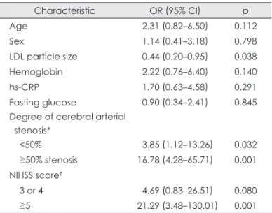
CRP, and fasting glucose), LDL particle size was indepen- dently and inversely associated with stroke severity [NIHSS score ≥5, reference NIHSS score 0–2; odds ratio (OR)=0.38, p=0.028] (Table 3). Furthermore, LDL particle size was inde- pendently and inversely associated with poor functional out- come at discharge (mRS score ≥3; OR=0.44, p=0.038) after adjusting for age, sex, and variables with p<0.1 in univariate analysis (hemoglobin, hs-CRP, fasting glucose, degree of cere- bral arterial stenosis, and NIHSS score) (Table 4).
Discussion
The findings of this study show that small LDL particles were associated with stroke severity and poor functional outcome in the studied patients, even after adjusting for NIHSS score, which is a strong predictive factor for stroke outcome. There are a few previous reports on this relationship. One study of 200 patients with acute ischemic stroke found that small LDL particles were significantly associated with in-hospital mortal- ity.14 Furthermore, in patients with coronary artery disease, and especially in those with acute myocardial infarction, the Table 1. Comparison of the demographic and clinical data according to stroke severity
Characteristic Stroke severity (NIHSS score)
0–2 (n=144) 3 or 4 (n=81) ≥5 (n=23) p
Demographic data
Gender, male 98 (68.1) 49 (60.5) 14 (60.9) 0.476
Age (years) 61±12 63±12 65±10 0.070
Risk factors
Hypertension 62 (43.1) 31 (38.3) 9 (39.1) 0.767
Diabetes mellitus 50 (34.7) 21 (25.9) 6 (26.1) 0.339
Hyperlipidemia 18 (13.2) 17 (21.0) 2 (8.7) 0.194
Smoking 27 (18.8) 15 (18.5) 6 (26.1) 0.691
Familial history 6 (4.2) 3 (3.7) 2 (8.7) 0.574
Coronary heart disease 15 (10.4) 12 (14.8) 6 (26.1) 0.108
Medications
Prestroke lipid-lowering agents 12 (8.3) 8 (9.9) 1 (4.3) 0.700
Laboratory data
LDL particle size (nm) 26.1±0.8 25.7±0.8 24.8±0.7 0.004
Hemoglobin (g/L) 13±1 13±2 12±2 0.234
White blood cell count (×1012/L) 7±2 7±2 9±7 0.017
Total cholesterol (mg/dL) 179±34 187±31 169±40 0.041
HDL (mg/dL) 44±11 44±13 44±12 0.813
LDL (mg/dL) 111±32 122±28 100±34 0.013
Lipoprotein (a) (mg/dL) 23±21 23±20 18±11 0.438
TG (mg/dL) 124±64 107±56 122±82 0.282
Hemoglobin A1C (%) 6±1 6±1 7±2 0.285
hs-CRP 1±1 1±3 1±2 0.061
Fasting glucose (mg/dL) 104±33 109±43 127±57 0.031
TG/HDL ratio 3±2 3±2 3±4 0.443
Total cholesterol/HDL ratio 4±1 4±1 4±1 0.164
LDL/HDL ratio 2±0 2±1 2±1 0.014
Clinical data
Relevant cerebral artery stenosis 0.524
No atherosclerosis* 79 (54.9) 48 (59.3) 9 (39.1)
<50% stenosis† 35 (24.3) 19 (23.5) 7 (30.4)
≥50% stenosis‡ 30 (20.8) 14 (17.3) 7 (30.4)
Body mass index 23±2 23±2 23±2 0.203
The data are presented as n (%) or mean±SD values.
*No stenosis, †One or more vessels with <50% stenosis, ‡One or more vessels with 50% stenosis.
HDL: high-density lipoprotein, hs-CRP: high-sensitivity C-reactive protein, LDL: low-density lipoprotein, NIHSS: National Institutes of Health Stroke Scale, TG: triglycerides.
LDL particles were reportedly relatively small, and this ten- dency persisted during hospitalization.11 Consistent with this previous study in patients with acute myocardial infarction, the LDL particle size measured in the present study is also likely to have been affected by the acute-phase reaction of the lipolysis during acute ischemic stroke.
In addition, we found that small LDL particles were corre- lated with atherogenic molecules such as TG and hs-CRP.
These data are relatively consistent with the results of a study that evaluated carotid artery stenosis using a hospital-based,
cross-sectional design22 and of a study that measured the rela- tive LDL particle sizes in patients with metabolic syndrome, insulin resistance, or coronary heart disease.3 Since both serum TG and hs-CRP are closely associated with atherosclerosis via the inflammatory pathway,1 the present findings suggest that small LDL particles play an important role in atherogenicity or the inflammatory reaction in atherosclerosis.
The presence of an association between LDL particle size and stroke severity and poor functional outcomes may be at- tributable to the following pleiotropic roles of small LDL par- Table 2. Comparison of clinical variables between patients with a good and poor functional outcomes at 3 months
Characteristic Functional outcome (mRS score)
0–2 (n=194) 3–6 (n=54) p
Demographic data
Gender, male 130 (67.0) 31 (57.4) 0.191
Age (years) 61±12 64±11 0.152
Risk factors
Hypertension 80 (41.2) 22 (40.7) 0.948
Diabetes mellitus 60 (30.9) 17 (31.5) 0.938
Hyperlipidemia 29 (14.9) 9 (16.7) 0.757
Smoking 40 (20.6) 8 (14.8) 0.340
Familial history 7 (3.6) 4 (7.4) 0.230
Coronary heart disease 23 (11.9) 10 (18.5) 0.202
Medications
Prestroke lipid-lowering agents 19 (9.8) 2 (3.7) 0.155
Laboratory data
LDL particle size (nm) 26.1±0.8 25.5±0.7 0.003
Hemoglobin (g/L) 13±1 12±2 0.013
White blood cell count (×1012/L) 7±2 7±4 0.279
Total cholesterol (mg/dL) 179±34 187±31 0.761
HDL (mg/dL) 44±11 45±13 0.539
LDL (mg/dL) 113±32 115±30 0.750
Lipoprotein (a) (mg/dL) 22±19 25±23 0.421
TG (mg/dL) 119±64 114±63 0.613
Hemoglobin A1C (%) 6±1 7±2 0.074
hs-CRP 0±2 2±3 0.004
Fasting glucose (mg/dL) 105±37 119±46 0.048
TG/HDL ratio 3±2 3±2 0.856
Total cholesterol/HDL ratio 4±1 4±1 0.781
LDL/HDL ratio 2±1 2±1 0.930
Clinical data
Relevant cerebral artery stenosis 0.003
No atherosclerosis* 116 (59.8) 20 (37.0)
<50%† 46 (23.7) 15 (27.8)
≥50% stenosis‡ 32 (16.5) 19 (35.2)
Body mass index 23±2 24±2 0.343
NIHSS [median (interquartile range)] 1 [0–2] 3 [3–4] 0.001
The data are presented as n (%), mean±SD values or median (interquatile ranges).
*No stenosis, †One or more vessels with <50% stenosis, ‡One or more vessels with 50% stenosis.
HDL: high-density lipoprotein, hs-CRP: high-sensitivity C-reactive protein, LDL: low-density lipoprotein, mRS: modified Rankin Scale, TG:
triglycerides.
ticles. First, small LDL particles are more atherogenic than their larger counterparts.22 It is known that small LDL particles are more easily taken up by arterial tissue, suggesting greater transendothelial transport, and they exhibit increased binding with polyanionic proteoglycans, which play a deterministic role in atherosclerosis.2 Since progressive cerebral atheroscle- rosis is associated with a poor stroke outcome,23 the present results regarding the association between small LDL particles and stroke prognosis may be valid. Second, small LDL parti- cles have a lower affinity for LDL receptors than do larger LDL particles.24 The lower affinity of small LDL particles for LDL receptors was reported to result in a longer retention time and consequently increased susceptibility to oxidative modifi- cations.25 This altered balance in the antioxidant/oxidant mech-
anism may cause increased vascular thrombogenesis as well as severe atherogenesis.26 Moreover, increased thrombogene- sis may contribute to atherothrombosis and poor stroke out- come. Finally, small LDL particles are involved in the inflam- matory cytokine signaling pathways. The excess infiltration in the arterial intima and local enzymatic modification of small LDL particles may trigger the expression of inflammatory cy- tokines and vascular cell adhesion molecules. These cascades could ultimately lead to the formation of destabilizing lipid- dominant cores and rupture prone fibrous caps on plaque.27 This vulnerable atherosclerotic plaque is an important deter- minant for thrombosis-related stroke with poor outcome, thus explaining the present finding of a relationship between stroke outcome and LDL particle size. Small LDL particles have also been reported to be inversely correlated with tumor necrosis factor α and interleukin-1β.28 These inflammatory cytokines are associated with activation of nuclear factor-κB, which is triggered by hypoxia, reactive oxygen species, and several in- flammatory mediators, and is responsible for neuronal cell death and neurovascular unit injuries.29 Therefore, the pres- ence of small LDL particles could reflect the severity or out- come of ischemic brain injury.
This study was subject to some limitations. Blood samples were not drawn from a sample of the normal population for comparison. However, the objective of this study was to deter- mine the relationship between LDL particle size and prognosis in stroke patients. Furthermore, such a comparative study be- tween normal controls and stroke patients has already been performed,14 and it was found that LDL particles were smaller in stroke patients. Another limitation of the present study is that blood samples were obtained from acute stroke patients at admission, and LDL particle size was not serially assessed during the stroke time course. However, the patients’ lipid and lipoprotein levels remained stable during the 4-week follow- up.30
In conclusion, small LDL particles are associated with ini- tial stroke severity and are an independent predictor of poor functional outcome. Therefore, LDL particle size is a poten- tial biomarker for the prognosis of atherothrombotic stroke.
Supplementary Materials
The online-only Data Supplement is available with this article at http://dx.doi.org/10.3988/jcn.2015.11.1.80.
Conflicts of Interest
The authors have no financial conflicts of interest.
Acknowledgements
This work was supported by a grant from Ewha Womans University School of Medicine.
Table 3. Multinomial multivariate logistic regression regarding the association between LDL particle size and stroke severity (NIHSS score)†
Stroke severity (NIHSS score)
3 or 4 ≥5
Unadjusted model 0.69 (0.38–1.24) 0.36 (0.19–0.70)*
Adjusted model‡§ 0.69 (0.38–1.27) 0.33 (0.17–0.65)*
Adjusted model‡ǁ 0.72 (0.38–1.35) 0.31 (0.15–0.63)*
Adjusted model‡¶ 0.76 (0.40–1.44) 0.38 (0.19–0.78)*
*Statistically significant at p<0.05, †Reference NIHSS score, 0–2,
‡Entered variables, §Age, sex, and LDL particle size, ǁAge, sex, and variables regarding lipid profile [LDL particle size, total cholesterol, HDL, LDL cholesterol, lipoprotein (a), and TG],
¶Age, sex, and variables with p<0.1 in univariate analysis (LDL particle size, white blood cell count, total cholesterol, LDL cho- lesterol, hs-CRP, and fasting glucose).
HDL: high-density lipoprotein, hs-CRP: high-sensitivity C-reac- tive protein, LDL: low-density lipoprotein, NIHSS: National Insti- tutes of Health Stroke Scale, TG: triglycerides.
Table 4. Multivariate binary logistic regression regarding the de- terminant factors associated with poor functional outcome
Characteristic OR (95% CI) p
Age 2.31 (0.82–6.50) 0.112
Sex 1.14 (0.41–3.18) 0.798
LDL particle size 0.44 (0.20–0.95) 0.038
Hemoglobin 2.22 (0.76–6.40) 0.140
hs-CRP 1.70 (0.63–4.58) 0.291
Fasting glucose 0.90 (0.34–2.41) 0.845 Degree of cerebral arterial
stenosis*
<50% 3.85 (1.12–13.26) 0.032
≥50% stenosis 16.78 (4.28–65.71) 0.001 NIHSS score†
3 or 4 4.69 (0.83–26.51) 0.080
≥5 21.29 (3.48–130.01) 0.001
*Reference: no relevant artery atherosclerosis, †Reference: NI- HSS score, 0–2.
hs-CRP: high-sensitivity C-reactive protein, LDL: low-density li- poprotein, NIHSS: National Institutes of Health Stroke Scale, OR:
odds ratio, 95% CI: 95% confidence interval.
REFERENCES
1. Tousoulis D, Kampoli AM, Papageorgiou N, Androulakis E, Antonia- des C, Toutouzas K, et al. Pathophysiology of atherosclerosis: the role of inflammation. Curr Pharm Des 2011;17:4089-4110.
2. Rizzo M, Berneis K. Should we measure routinely the LDL peak par- ticle size? Int J Cardiol 2006;107:166-170.
3. Berneis KK, Krauss RM. Metabolic origins and clinical significance of LDL heterogeneity. J Lipid Res 2002;43:1363-1379.
4. Björnheden T, Babyi A, Bondjers G, Wiklund O. Accumulation of li- poprotein fractions and subfractions in the arterial wall, determined in an in vitro perfusion system. Atherosclerosis 1996;123:43-56.
5. Tribble DL, Rizzo M, Chait A, Lewis DM, Blanche PJ, Krauss RM.
Enhanced oxidative susceptibility and reduced antioxidant content of metabolic precursors of small, dense low-density lipoproteins. Am J Med 2001;110:103-110.
6. Berneis K, Shames DM, Blanche PJ, La Belle M, Rizzo M, Krauss RM. Plasma clearance of human low-density lipoprotein in human apolipoprotein B transgenic mice is related to particle diameter. Me- tabolism 2004;53:483-487.
7. National Cholesterol Education Program (NCEP) Expert Panel on De- tection, Evaluation, and Treatment of High Blood Cholesterol in Adults (Adult Treatment Panel III). Third Report of the National Cho- lesterol Education Program (NCEP) Expert Panel on Detection, Eval- uation, and Treatment of High Blood Cholesterol in Adults (Adult Treatment Panel III) final report. Circulation 2002;106:3143-3421.
8. Gentile M, Panico S, Mattiello A, Ubaldi S, Iannuzzo G, De Michele M, et al. Association between small dense LDL and early atherosclero- sis in a sample of menopausal women. Clin Chim Acta 2013;426:1-5.
9. Rizzo M, Pernice V, Frasheri A, Di Lorenzo G, Rini GB, Spinas GA, et al. Small, dense low-density lipoproteins (LDL) are predictors of cardio- and cerebro-vascular events in subjects with the metabolic syn- drome. Clin Endocrinol (Oxf) 2009;70:870-875.
10. Cromwell WC, Otvos JD, Keyes MJ, Pencina MJ, Sullivan L, Vasan RS, et al. LDL Particle Number and Risk of Future Cardiovascular Disease in the Framingham Offspring Study - Implications for LDL Management. J Clin Lipidol 2007;1:583-592.
11. Barbagallo CM, Rizzo M, Cefalu’ AB, Noto D, Scimeca A, Castello A, et al. Changes in plasma lipids and low-density lipoprotein peak parti- cle size during and after acute myocardial infarction. Am J Cardiol 2002;89:460-462.
12. Miwa K. Low density lipoprotein particles are small in patients with coronary vasospasm. Int J Cardiol 2003;87:193-201.
13. O’Neal DN, Lewicki J, Ansari MZ, Matthews PG, Best JD. Lipid lev- els and peripheral vascular disease in diabetic and non-diabetic sub- jects. Atherosclerosis 1998;136:1-8.
14. Zeljkovic A, Vekic J, Spasojevic-Kalimanovska V, Jelic-Ivanovic Z, Bogavac-Stanojevic N, Gulan B, et al. LDL and HDL subclasses in acute ischemic stroke: prediction of risk and short-term mortality. Ath- erosclerosis 2010;210:548-554.
15. Gorelick PB. Primary and comprehensive stroke centers: history, val- ue and certification criteria. J Stroke 2013;15:78-89.
16. Adams HP Jr, Bendixen BH, Kappelle LJ, Biller J, Love BB, Gordon
DL, et al. Classification of subtype of acute ischemic stroke. Defini- tions for use in a multicenter clinical trial. TOAST. Trial of Org 10172 in Acute Stroke Treatment. Stroke 1993;24:35-41.
17. North American Symptomatic Carotid Endarterectomy Trial Collabo- rators. Beneficial effect of carotid endarterectomy in symptomatic pa- tients with high-grade carotid stenosis. N Engl J Med 1991;325:445- 18. Warfarin-Aspirin Symptomatic Intracranial Disease (WASID) Trial In-453.
vestigators. Design, progress and challenges of a double-blind trial of warfarin versus aspirin for symptomatic intracranial arterial stenosis.
Neuroepidemiology 2003;22:106-117.
19. Oh MS, Yu KH, Lee JH, Jung S, Ko IS, Shin JH, et al. Validity and re- liability of a Korean version of the national institutes of health stroke scale. J Clin Neurol 2012;8:177-183.
20. Kim OY, Chung HK, Shin MJ. Higher levels of serum triglyceride and dietary carbohydrate intake are associated with smaller LDL particle size in healthy Korean women. Nutr Res Pract 2012;6:120-125.
21. Kim YJ, Lee SM, Cho HJ, Do HJ, Hong CH, Shin MJ, et al. Plasma levels of apolipoprotein E and risk of intracranial artery stenosis in acute ischemic stroke patients. Ann Nutr Metab 2013;62:26-31.
22. Shoji T, Hatsuda S, Tsuchikura S, Shinohara K, Kimoto E, Koyama H, et al. Small dense low-density lipoprotein cholesterol concentration and carotid atherosclerosis. Atherosclerosis 2009;202:582-588.
23. Lei C, Wu B, Liu M, Chen Y. Risk factors and clinical outcomes asso- ciated with intracranial and extracranial atherosclerotic stenosis acute ischemic stroke. J Stroke Cerebrovasc Dis 2014;23:1112-1117.
24. Chen GC, Liu W, Duchateau P, Allaart J, Hamilton RL, Mendel CM, et al. Conformational differences in human apolipoprotein B-100 among subspecies of low density lipoproteins (LDL). Association of altered proteolytic accessibility with decreased receptor binding of LDL subspecies from hypertriglyceridemic subjects. J Biol Chem 1994;269:29121-29128.
25. Tan KC, Ai VH, Chow WS, Chau MT, Leong L, Lam KS. Influence of low density lipoprotein (LDL) subfraction profile and LDL oxidation on endothelium-dependent and independent vasodilation in patients with type 2 diabetes. J Clin Endocrinol Metab 1999;84:3212-3216.
26. Banfi C, Camera M, Giandomenico G, Toschi V, Arpaia M, Mussoni L, et al. Vascular thrombogenicity induced by progressive LDL oxidation:
protection by antioxidants. Thromb Haemost 2003;89:544-553.
27. Hansson GK. Inflammation, atherosclerosis, and coronary artery dis- ease. N Engl J Med 2005;352:1685-1695.
28. Chae JS, Kim OY, Paik JK, Kang R, Seo WJ, Jeong TS, et al. Associ- ation of Lp-PLA(2) activity and LDL size with interleukin-6, an in- flammatory cytokine and oxidized LDL, a marker of oxidative stress, in women with metabolic syndrome. Atherosclerosis 2011;218:499- 29. Ridder DA, Schwaninger M. NF-kappaB signaling in cerebral isch-506.
emia. Neuroscience 2009;158:995-1006.
30. Kargman DE, Tuck C, Berglund L, Lin IF, Mukherjee RS, Thompson EV, et al. Lipid and lipoprotein levels remain stable in acute ischemic stroke: the Northern Manhattan Stroke Study. Atherosclerosis 1998;
139:391-399.
Characteristics
Degree of stenosis
Total p value No atherosclerosis,
n=136
0<stenosis<50%, n=61
≥50% stenosis, n=51 Demographic data
Gender, male, n (%) 92 (67.6) 37 (60.7) 32 (62.7) 161 (64.9) 0.595
Age, years, mean±SD 61±11 63±14 63±10 62±11 0.405
Risk factors, n (%)
Hypertension 56 (41.2) 20 (32.8) 26 (51.0) 102 (41.1) 0.150
Diabetes mellitus 40 (29.4) 14 (23.0) 23 (45.1) 77 (31.0) 0.034
Hyperlipidemia 20 (14.7) 7 (11.5) 11 (21.6) 38 (15.3) 0.322
Smoking 32 (23.5) 5 (8.2) 11 (21.6) 48 (19.4) 0.038
Familial history 3 (2.2) 4 (6.6) 4 (7.8) 11 (4.4) 0.162
Coronary heart disease 13 (9.6) 10 (16.4) 10 (19.6) 33 (13.3) 0.141
Medications, n (%)
Pre-stroke lipid lowering agents 15 (11.0) 4 (6.6) 2 (3.9) 21 (8.5) 0.247
Laboratory data, mean±SD
LDL particle size, nm 25.9±0.8 25.2±0.7 24.5±0.8 25.9±0.9 0.010
Hemoglobin, g/L 13±2 13±1 12±2 13±2 0.098
White blood cell count, ×1012/L 6±2 7±2 8±4 7±3 0.104
Total cholesterol, mg/dL 177±34 189±28 181±37 181±34 0.085
High density lipoprotein, mg/dL 44±11 46±13 42±11 44±12 0.375
Low density lipoprotein, mg/dL 110±33 119±29 117±31 114±31 0.142
Lipoprotein (a), mg/dL 21±18 22 ±17 29±24 23±20 0.098
Triglyceride, mg/dL 116±67 120±59 122±62 118±64 0.497
Hemoglobin A1C, % 6±1 6±1 7±2 7±1 0.003
High sensitivity C-reactive protein 1±2 1±2 1±3 1±2 0.123
Fasting glucose, mg/dL 104±32 101±39 126±51 108±39 0.009
TG/HDL ratio 2±2 3±2 3±2 3±2 0.432
Total cholesterol/HDL ratio 4±1 4±1 4±2 4±1 0.325
LDL/HDL ratio 2±1 2±1 3±1 3±1 0.108
Clinical data
Body mass index, mean±SD 24±2 23±2 23±2 24±1 0.025
Initial NIHSS score, median (interquartile range)
3 (1–5) 3 (1–5) 3 (2–5) 3 (2–5) 0.782
mRS score at 3 month, median (interquartile range)
3 (1–5) 3 (1–5) 3 (2–5) 3 (2–5) 0.003
The data are presented as n (%), mean±SD values or median (interquatile ranges).
HDL: high-density lipoprotein, LDL: low-density lipoprotein, mRS: modified Rankin scale, NIHSS: National Institute of Health Stroke Scale, n: number, SD: standard deviation, TG: triglyceride.