Calcium overload is essential for the acceleration of
staurosporine-induced cell death following neuronal
differentiation in PC12 cells
Su Ryeon Seo
1,3and Jeong Taeg Seo
2,3 1Department of Molecular Bioscience School of Bioscience and Biotechnology Kangwon National University
Chuncheon 200-701, Korea
2
Department of Oral Biology, BK 21 Project Yonsei University College of Dentistry Seoul 120-752, Korea
3
Corresponding authors: Tel, 82-33-250-8541;
Fax, 82-33-241-4627; E-mail, [email protected] (S.R. Seo) Tel, 82-2-2228-3054; Fax, 82-2-364-1085;
E-mail, [email protected] (J.T. Seo) DOI 10.3858/emm.2009.41.4.030 Accepted 3 December 2008
Abbreviations: NGF, nerve growth factor; PARP, poly ADP-ribose polymerase
Abstract
Differentiation of neuronal cells has been shown to
ac-celerate stress-induced cell death, but the underlying
mechanisms are not completely understood. Here, we
find that early and sustained increase in cytosolic
([Ca
2+]
c) and mitochondrial Ca
2+levels ([Ca
2+]
m) is
es-sential for the increased sensitivity to staurosporine-
induced cell death following neuronal differentiation in
PC12 cells. Consistently, pretreatment of
differ-entiated PC12 cells with the intracellular Ca
2+-chelator
EGTA-AM diminished staurosporine-induced PARP
cleavage and cell death. Furthermore, Ca
2+overload
and enhanced vulnerability to staurosporine in
differ-entiated cells were prevented by Bcl-X
Loverexpr-ession. Our data reveal a new regulatory role for
differ-entiation-dependent alteration of Ca
2+signaling in cell
death in response to staurosporine.
Keywords: bcl-X protein; calcium; cell death; cell
dif-ferentiation; PC12 cells; staurosporine
Introduction
Previously, it has been reported that PC12 cells
differentiated into sympathetic neurons in response to nerve growth factor (NGF) are more sensitive to apoptotic stimuli, such as TNF-α and ethanol, than undifferentiated PC12 cells (Oberdoerster et al., 1999; Zhang et al., 2007). On the other hand, however, a carbonyl stressor (methylglyoxal) was shown to induce apoptosis more robustly in undi-fferentiated PC12 cells (Okouchi et al., 2005). These different reports suggest that mechanisms involved in death pathways are divided between neurotoxic factors and may be significantly influen-ced by cellular phenotypes.
In many cell types, alteration of intracellular Ca2+
homeostasis plays a pivotal role in initiating apop-tosis (Park et al., 2002; Demaurex et al., 2003). Analysis of brain tissue from Alzheimer Disease (AD) patient showed that alteration of Ca2+ ho-moeostasis is associated with the neurofibrillary tangle-bearing neurons (Murray et al., 1992). Nu-merous findings have also suggested that pertur-bation of Ca2+ signaling contributes to many age- related neurodegenerative disorders, including: Par-kinson's Disease (PD), Huntington’s Disease (HD), ischemic stroke, and amyotrophic lateral sclerosis (Beal, 1998; Rodnitzky, 1999; Simpson, et al., 2002). Increases in cytosolic ([Ca2+]
c) or mitochondrial
Ca2+ concentrations ([Ca2+]m) have been shown to
mediate cell death in various cell types. For example, exposure of PC12 cells to staurosporine causes cytosolic and mitochondrial Ca2+ overload, which is an essential event for initiation of cell death (Kruman et al., 1998). Staurosporine, a broad spectrum protein kinase inhibitor, has been used to induce cell death in a wide range of cell types (Kabir et al., 2002; Witasp et al., 2005; Wang et al., 2007). Although the exact mechanism responsible for staurosporine-induced cell death is unknown, ac-tivation of caspases triggered by cytochrome c release from mitochondria into cytosol is required (Johansson et al., 2003).
We investigated if neuronal differentiation of PC12 cells by NGF accelerates staurosporine-induced cell death and if an increase in [Ca2+]
c is a
con-tributing factor to the increased sensitivity to stau-rosporine-induced cell death following neuronal differentiation. Our data suggest that early and su-stained increase in [Ca2+]c is responsible for
re-lease of mitochondrial cytochrome c, caspase-3 activation, DNA fragmentation, and cell death in
270 Exp. Mol. Med. Vol. 41(4), 269-276, 2009
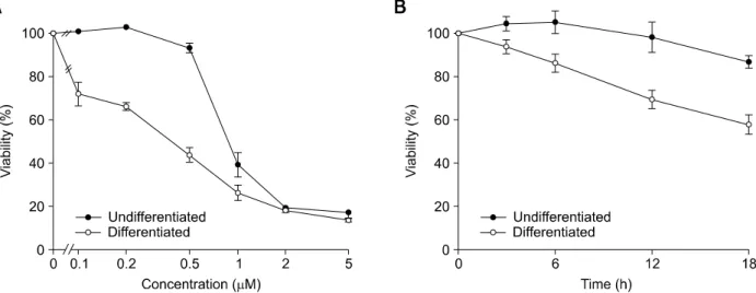
Figure 1. Neuronally differentiated PC12 cells exhibit increased sensitivity to staurosporine-induced cell death. Neuronally differentiated and un-differentiated PC12 cells were treated with indicated concentrations of staurosporine for 12 h (A) or treated with 0.2 μM staurosporine for indicated times (B). The viability of cells was determined by MTT assay. Results are presented as the means ± SD of four independent experiments.
neuronally differentiated PC12 cells exposed to staurosporine. Undifferentiated PC12 cells are more resistant to staurosporine-induced effects.
Results
Staurosporine induces cell death to a great extent in
neuronally differentiated PC12 cells
Previously, neuronally differentiated PC12 cells appeared to be more sensitive to apoptotic stimuli, such as TNFα and ethanol, than undifferentiated cells (Oberdoerster and Rabin, 1999; Zhang et al., 2007). In the present study, we tested if NGF- induced neuronal differentiation in PC12 cells acce-lerates cell death in response to staurosporine, which has been used as a common inducer of cell death in almost all cell types. Staurosporine (at concentrations greater than 0.1 μM) induced the death of neuronally differentiated PC12 cells to a greater extent compared to undifferentiated PC12 cells (Figure 1A). The cell death provoked by 0.2 μM staurosporine progressed more rapidly over time in differentiated PC12 cells than in undi-fferentiated cells (Figure 1B). Therefore, our findings are in agreement with those recently reported by Zhang and colleagues (Oberdoerster and Rabin, 1999; Zhang et al., 2007), but not with those reported by Ekshyyan and Aw (Ekshyyan et al., 2005). Ekshyyan and Aw demonstrated that the transition of undifferentiated PC12 cells into diffe-rentiated cells affords protection against oxidative stress. While the discrepancy between these oppo-site observations has not been resolved, the alte-ration of survival or death signals during neuronal
differentiation might be involved.
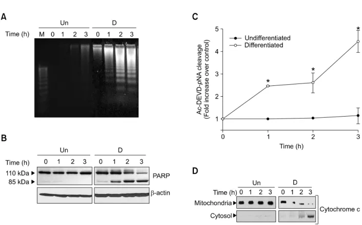
Staurosporine induces DNA fragmentation, caspase
3 activation, and release of mitochondrial
cytochrome c into cytosol in neuronally
differentiated PC12 cells
In an attempt to determine underlying mechanisms responsible for enhanced cell death by stauro-sporine in neuronally differentiated cells, we ana-lyzed DNA fragmentation, a typical characteristic feature of apoptosis. As shown in Figure 2A, 0.2 μM staurosporine induced DNA fragmentation in neuronally differentiated cells, but not in undiffe-rentiated cells. DNA fragmentation was accompa-nied by a sequential 85 kDa cleavage product of PARP, one of the targets associated with caspase activation (Figure 2B) (Lazebnik et al., 1994). Consistent with this, measurement of caspase 3 protease activity using the colorimetric substrate Ac-DEVD-pNA confirmed that staurosporine en-hanced caspase 3 activation in the neuronally differentiated PC12 cells (Figure 2C). Since the mitochondrial cytochrome c released into the cytosol has been identified as an apoptosis initia-tion molecule (Desagher et al., 2000), we examined whether staurosporine accelerated the release of mitochondrial cytochrome c into the cytosol in neuronally differentiated PC12 cells. As shown in Figure 2D, 0.2 μM staurosporine caused a signi-ficant increase in cytosolic cytochrome c, and a decrease in mitochondrial cytochrome c, after treat-ment with staurosporine in neuronally differentiated cells, whereas the content of cytosolic and mito-chondrial cytochrome c remained constant in
undi-Figure 2. Staurosporine induces DNA fragmentation, caspase-3 activation, and cytochrome c release in neuronally differentiated PC12 cells. Neuronally differentiated and undifferentiated PC12 cells were treated with 0.2 μM staurosporine for the indicated times. (A) The fragmented DNA was analyzed by agarose gel electrophoresis. (B) Total cell extracts were immunoblotted with anti-PARP and anti-β-actin antibodies. (C) Caspase-3 activity was measured using a colorimetric substrate Ac-DEVD-pNA. Values are the means ± SD of three independent experiments. *Significantly different from undifferentiated cells (P < 0.05). (D) Cytoplasmic and mitochondrial fractions of extracts were immunoblotted with anti-cytochrome c antibody. D: differentiated PC12 cells, Un: undifferentiated PC12 cells.
fferentiated cells up to 3 h (Figure 2D).
Elevated [Ca
2+]
mand [Ca
2+]
cin differentiated PC12
cells are responsible for staurosporine-induced cell
death
Several lines of evidence indicate that uncontrolled cytosolic or mitochondrial Ca2+ overload mediates
staurosporine-induced cell death in neuronal cells (Prehn et al., 1997; Kruman et al., 1998, 1999). To determine if the alteration of Ca2+ homeostasis is
essential for initiating differentiation-dependent, stau-rosporine-activated cell death signaling, we analyzed [Ca2+]c and [Ca2+]m in undifferentiated and
neu-ronally differentiated PC12 cells.
Exposure of neuronally differentiated PC12 cells to 0.2 μM staurosporine resulted in early and sustained elevation of [Ca2+]
c, whereas exposure of
undifferentiated cells had little effect on [Ca2+] c
(Figure 3A). The [Ca2+]
c increase was completely
prevented by 20 μM of the membrane permeable intracellular Ca2+ chelator EGTA-AM. It is well
esta-blished that sustained overload of cytosolic Ca2+
causes enhanced accumulation of Ca2+ by the
mitochondria, which sensitizes the cytochrome c release pathway (Szabadkai et al., 2004; Dong et al., 2006). Since staurosporine caused early and sustained increase in [Ca2+]
c in the present study,
we speculated that Ca2+ might have accumulated in mitochondria. In this context, we assessed chan-ges in [Ca2+]m microscopically in living cells loaded
with the mitochondrial Ca2+ indicator Rhod 2-AM, which detects free Ca2+ levels in the mitochondrial matrix. Following treatment with 0.2 μM stauro-sporine, we observed a significant increase in [Ca2+]
m in neuronally differentiated PC12 cells, but
not in undifferentiated cells (Figure 3B). While we did not investigate the direct role of mitochondrial Ca2+ overload in cell death in this study, previous reports revealed that it is linked to mitochondrial membrane depolarization and ROS accumulation, which are believed to play a role in cell death (Hajnoczky et al., 2003).
We next analyzed whether chelating of intra-cellular Ca2+ could block the cleavage of PARP in
neuronally differentiated PC12 cells (Figure 3C). Pretreatment of neuronally differentiated PC12 cells with intracellular Ca2+ chelators such as BAPTA-AM
272 Exp. Mol. Med. Vol. 41(4), 269-276, 2009
Figure 3. Staurosporine-induced in-creases in [Ca2+]
m and [Ca2+]c are
in-volved in the death of differentiated PC12 cells. (A) Changes in [Ca2+]
c
were measured in fura-2 loaded neuronally differentiated and un-differentiated PC12 cells using ratio-metric fluorescence recording tech-niques after the application of 0.2 μM staurosporine. In some experi-ments, cells were pretreated with 20 μM EGTA-AM for 10 min. (B) Changes in [Ca2+]
m were monitored
in rhod-2 loaded neuronally differ-entiated (right) and undifferdiffer-entiated PC12 cells (left), by confocal micro-scopy, after treatment with 0.2 μM staurosporine for 1 h. (C) Neuronally differentiated PC12 cells were treat-ed with intracellular Ca2+ chelators
(20 μM BAPTA-AM or 20 μM EGTA-AM) for 30 min followed by 0.2 μM staurosporine for 3 h. Cell lysates were then immunoblotted with PARP and β-actin anti-bodies. (D) Neuronally differentiated PC12 cells were treated with 0.2 μM staurosporine for 24 h either in the presence or absence of 10 μM EGTA-AM, and cell viability was measured by MTT assay. Values are the means ± SD of four in-dependent experiments. *Significan-tly different from cells unexposed to staurosporine (P < 0.05). **Signifi-cantly different from cells exposed to staurosporine alone (P < 0.05).
and EGTA-AM inhibited the cleavage of PARP, suggesting that Ca2+ acts upstream of caspase 3
activation in the staurosporine-induced death pro-cess. Consistent with this, inhibition of [Ca2+]
c
in-crease by EGTA-AM in differentiated cells atte-nuated the staurosporine-induced cell death (Figure 3D). These results indicate that neuronally differentiated PC12 cells are more sensitive to staurosporine-induced cell death than
undifferen-tiated cells due, in part, to the enhanced increases in [Ca2+]
c in the differentiated cells.
Bcl-X
Lprevents staurosporine-induced [Ca
2+]
cincreases and cell death
We next investigated if anti-apoptotic Bcl-XL
anta-gonizes staurosporine-induced cell death in diffe-rentiated PC12 cells as reported previously (Boise
Figure 4. Overexpression of Bcl-XL prevents DNA fragmentation, PARP cleavage, [Ca2+]c increase, and cell death in neuronally differentiated PC12 cells.
(A) Bcl-XL overexpressing (Bcl-XL) and control (pCMV) PC12 cells were differentiated and treated with 0.2 μM staurosporine for 24 h, and cell viability was
measured by MTT assay. Values are the means ± SD of four independent experiments. *Significantly different from cells unexposed to staurosporine (P <0.05). **Significantly different from pCMV-transfected cells exposed to staurosporine (P < 0.05). (B) Bcl-XL overexpressing (Bcl-XL) and control
(pCMV) PC12 cells were differentiated and treated with 0.2 μM staurosporine for indicated times and the fragmented DNA was analyzed by agarose gel electrophoresis. (C) Bcl-XL overexpressing (Bcl-XL) and control (pCMV) PC12 cells were differentiated and treated with 0.2 μM staurosporine for 6 h, and
total cell extracts were immunoblotted with anti-PARP and anti-β-actin antibodies. (D) Changes in [Ca2+]
c were measured in fura-2 loaded, neuronally
dif-ferentiated Bcl-XL overexpressing cells (Bcl-XL) and control (pCMV) using ratiometric fluorescence recording techniques after the application of 0.2 μM
staurosporine.
et al., 1993; Gonzalez-Garcia et al., 1994). Bcl-XL
is an anti-apoptotic member of the Bcl-2 family, which is localized to the membranes of nuclear envelope, ER, and mitochondria (Lithgow et al., 1994). While the mechanism of Bcl-XL is still
de-bated, multiple mechanisms are believed to be involved in the protection of cells from apoptosis. As shown in Figure 4A, 0.2 μM staurosporine- induced neuronal cell death was largely prevented in Bcl-XL overexpressing PC12 cells. The inhibitory
effect of Bcl-XL was also observed on DNA
frag-mentation and the PARP cleavage pattern (Figure 4B and C).
Since our results indicate that an increase in [Ca2+]
c is an early event in the staurosporine-
induced apoptotic process, we next analyzed whe-ther Bcl-XL could inhibit the [Ca2+]c increase in
neuronally differentiated PC12 cells. As shown in Figure 4D, treatment of Bcl-XL overexpressing stable
cells with 0.2 μM staurosporine prevented the increase in [Ca2+]
c, confirming that the
anti-apop-totic action of Bcl-XL is accompanied by the
inhi-bition of [Ca2+]
c increase.
Discussion
PC12 cells differentiate into neuronal cells with neurite extensions in response to NGF (Hatayama et al., 1997). Several controversial reports show that neurotoxic effect was different between undi-fferentiated and diundi-fferentiated phenotypes of PC12 cells (Oberdoerster and Rabin, 1999; Okouchi et al., 2005; Zhang et al., 2007).
274 Exp. Mol. Med. Vol. 41(4), 269-276, 2009
In this study, we analyzed the response of neu-ronally differentiated and undifferentiated PC12 cells to staurosporine to elucidate whether cellular state determines apoptotic sensitization and found that differentiated neuronal cells respond more sensitively to staurosporine than undifferentiated cells. Our results provide evidence that the alte-ration of Ca2+ homeostasis following NGF-induced
differentiation is directly correlated with the accele-ration of staurosporine-induced apoptotic commit-ment in PC12 cells; intracellular Ca2+ overload in response to staurosporine is evident in differen-tiated PC12 cells compared with undifferendifferen-tiated cells. To our knowledge, this is the first report to highlight the role of differentiation-dependent alte-ration of Ca2+ signaling in cell death in response to
staurosporine.
At present, it is unclear why the potency and efficacy of staurosporine to cause increase of [Ca2+]c and [Ca2+]m differs between undifferentiated
and differentiated neuronal cells. We speculate that the expression of proteins involved in the regulation of intracellular Ca2+ homeostasis, inclu-ding Ca2+ channels and ATPase on endoplasmic
reticulum or plasma membrane, is modulated during the neuronal differentiation process. In supports of this, several lines of reports suggested that NGF induces expression of several types of ion chan-nels (Usowicz et al., 1990; Furukawa et al., 1993; Lewis et al., 1993; Jimenez et al., 2001). For example, increased expression of Na+ and Ca2+ channels in PC12 cells, and ryanodine receptor isoform 2 (RyR2) in rat chromaffin cells by the NGF treatment (Furukawa et al., 1993; Jimenez and Hernandez-Cruz, 2001). Thus, alteration of voltage- gated Ca2+ influx and Ca2+ release from
intra-cellular stores might cause the acceleration of staurosporine-induced apoptotic process.
Consistent with actions of staurosporine on alterations of Ca2+ homeostasis following differen-tiation of PC12 cells, we found that Bcl-XL
pre-vented staurosporine-induced neuronal cell death. Bcl-XL, a member of anti-apoptotic Bcl-2 subfamily,
has been shown to interfere with Ca2+-mediated
apoptotic signals by inhibiting Ca2+ release from
ER. In the present study, we showed that the anti- apoptotic effect of Bcl-XL was accompanied by the
inhibition of [Ca2+]
c increases in differentiated PC12
cells exposed to staurosporine. These results are in good agreement with those obtained by Wang and colleagues (Wang et al., 2007). They found that Bcl-XL blocked cytochrome c release and
caspase-3 activation in response to staurosporine in rat hepatocytes. Furthermore, Li and colleagues reported that less Ca2+ was released from the ER
in Bcl-XL expressing cells in response to apoptotic
stimuli due to down-regulation of IP3 receptors (Li
et al., 2002).
In fact, we observed that despite the complete chealation of intracellular Ca2+ and blockage of
caspase 3 activity by EGTA-AM, staurosporine- induced cell death was not completely blocked by EGTA-AM in differentiated cells, raising the po-ssibility that staurosporine could activate death pathway through a mechanism that is independent of Ca2+. Although increase in [Ca2+]c is not the only
death mechanism induced by staurosporine, it is likely that acceleration of death in differentiated PC12 cells is primarily mediated through the Ca2+- dependent pathway.
Collectively, our findings suggest that increased sensitivity to staurosporine-induced death in neu-roanlly differentiated PC12 cells might be caused by the alteration of Ca2+ signaling during the
diffe-rentiation process.
Methods
Cells and reagents
PC 12 cells were obtained from ATCC (Manassas, VA), and PC12 cells overexpressing Bcl-XL were kindly
pro-vided by Dr. Y. J. Oh (Yonsei University, Seoul, Korea). BAPTA-AM, EGTA-AM, anti-poly-(ADP-ribose) polymerase (PARP) antibody, and acetyl-Asp-Glu-Val-Asp-p-nitroanilide (Ac-DEVD-pNA) were purchased from Calbiochem (San Diego, CA). Anti-cytochrome c antibody was purchased from Pharmingen (San Diego, CA). Fura-2 AM and Rhod-2 AM were purchased from Molecular Probes (Eugene, OR). NGF was purchased from Alomone labs (Jerusalem, Israel). Other reagents, including staurosporine, were obtained from Sigma (St. Louis, MO).
Cell culture
Cultures were maintained in DMEM (Life technologies, Inc.) supplemented with 10% heat-inactivated horse serum and 5% FBS (Life technologies, Inc.). To obtain neu-ronally differentiated PC12 cultures, cells were grown on collagen coated plates (10 μg/ml; Upstate Biotechnology, Lake Pacid, NY) supplemented with 2% heat-inactivated horse serum, 1% FBS, and 50 ng/ml NGF for seven days. The medium, including NGF, was replaced every two days. Cultures were maintained at 37oC in a humidified, 5% CO
2
incubator. In differentiated PC12 cells, all experiments were performed in the presence of NGF to exclude the possibility of NGF-deprived cell death signaling pathways.
MTT assay
After each indicated treatment, cells were incubated with MTT at a final concentration of 1 mg/ml for 1 h at 37oC, followed by lysis in solubilizing solution (50% dimethylfor-mamide and 20% SDS, pH 4.8) for 24 h. The absorption value was determined at 570 nm and viability was
determined as percent survival relative to untreated control.
Immunoblot analysis
Cells were lysed in buffer containing 10 mM Tris-Cl (pH 7.5), 150 mM NaCl, 1% Triton X-100, 0.5% Nonidet P-40, 1 mM EDTA, 1 mM EGTA, 1 mM PMSF, 1 μg/ml leupeptin, and 1 μg/ml aprotinin. Equal amounts of proteins were separated on 10% SDS-polyacrylamide gels and subse-quently transferred to nitrocellulose membranes. Specific immunodetection was carried out by incubation with indi-cated antibodies followed by peroxidase-conjugated anti- mouse or anti-rabbit IgG antibody. Blots were evaluated using an ECL detection system.
Measurement of [Ca
2+]
cand [Ca
2+]
m For [Ca2+]c measurements, PC12 cells were loaded with 4
μM fura-2 AM at 37oC in a 5% CO
2 incubator for 20 min in
a HCO3--buffered solution containing [110 mM NaCl, 4.5
mM KCl, 1 mM NaH2PO4, 1 mM MgSO4, 1.5 mM CaCl2, 5
mM HEPES-Na, 5 mM HEPES free acid, 25 mM NaHCO3
and 10 mM D-glucose (pH 7.4)]. Cells were then rinsed twice and incubated in the HCO3--buffered solution for at
least 20 min before use. [Ca2+]
c was measured on the
stage of an inverted microscope (Nikon, Tokyo, Japan) by spectrofluorometry (Photon Technology International, Brun-swick, NJ), while cells were superfused at a constant per-fusion rate of 2 ml/min with the HCO3--buffered solution
equilibrated with 95% O2, 5% CO2 to maintain pH 7.4. All
experiments were performed at 37oC. The excitation wavelength was alternated between 340 and 380 nm and the emission fluorescence was recorded at 510 nm. [Ca2+]
c values were calculated using the equation
descri-bed by Grynkiewicz (Grynkiewicz et al., 1985). Relative [Ca2+]
m was measured with the fluorescent probe rhod
2-AM following methods described previously (Hoth et al., 1997). In brief, cells were loaded with 4 μM rhod-2 AM for 60 min. The residual cytosolic fraction of the dye was eliminated when cells were kept in culture for an additional 6 h after loading, whereas the mitochondrial dye fluores-cence was retained. Cellular fluoresfluores-cence was imaged using a confocal microscope with excitation at 514 nm and emisson at 535 nm.
DNA fragmentation analysis
Cells were lysed in 0.05% Triton X-100, 20 mM EDTA, and 10 mM Tris-Cl (pH. 8.0) for 30 min and the fragmented DNA was precipitated with ethanol. The precipitates were then resuspended in TE buffer and electrophoresed on 1.5% agarose gels.
Caspase 3 assay
Cells were lysed in buffer containing 1 mM KCl, 1.5 mM MgCl, 1 mM DTT, 1 mM PMSF, 5 μg/ml leupeptin, 2 μg/ml aprotinin and 10% glycerol. 20 μg of each protein were added to reaction buffer [25 mM HEPES (pH 7.4), 10 mM DTT, 10% sucrose, 0.1% CHAPS] containing 40 μM Ac-
DEVD-pNA, a colorimetric substrate for caspase-3 pro-tease activity (Stefanis et al., 1996), and incubated at 37oC
for 30 min. DEVD-pNA cleavage was measured at 405 nm.
Subcellular fractionation
Subcellular fractionation was performed according to pre-viously reported methods (Gross et al., 1999). Briefly, PC12 cells were homogenized in five volumes of extraction buffer containing 220 mM mannitol, 68 mM sucrose, 50 mM PIPES-KOH (pH 7.4), 50 mM KCl, 5 mM EGTA, 2 mM MgCl2, 1 mM DTT and 1 mM PMSF. Cells were then spun
at 400 × g for 10 min at 4oC to separate out nuclei and
unbroken cells. Supernatant was centrifuged at 10,000 × g for 10 min at 4oC to collect the mitochondrial-enriched
pellet. The new supernatant was then spun at 100,000 × g for 30 min at 4oC to separate the light membrane ER-
enriched pellet (not used in these experiments) from the supernatant (containing the cytosol).
Statistical analysis
Results are presented as the means ± SD of three or four independent experiments. When comparing two groups, an unpaired Student's t-test was used to address differences. P-values less than 0.05 were considered significant.
Acknowledgements
This work was supported by 2007 Research Grant from Kangwon National University, and by the Brain Korea 21 program (to S.R.S.). This work was also supported by the Korea Research Foundation Grant funded by the Korean Government (MOEHRD) (KRF-2005-041-E00100). This work was originated at Yonsei University College of Den-tistry and was carried out in the facilities of Institute of Bioscience and Biotechnology at Kangwon National Uni-versity.
References
Beal MF. Mitochondrial dysfunction in neurodegenerative diseases. Biochim Biophys Acta 1998;1366:211-23 Boise LH, Gonzalez-Garcia M, Postema CE et al. bcl-x, a bcl-2-related gene that functions as a dominant regulator of apoptotic cell death. Cell 1993;74:597-608
Demaurex N, Distelhorst C. Cell biology. Apoptosis--the calcium connection. Science 2003;300:65-7
Desagher S, Martinou JC. Mitochondria as the central control point of apoptosis. Trends Cell Biol 2000;10:369-77 Dong Z, Saikumar P, Weinberg JM et al. Calcium in cell injury and death. Annu Rev Pathol 2006;1:405-34
Ekshyyan O, Aw TY. Decreased susceptibility of differen-tiated PC12 cells to oxidative challenge: relationship to cellular redox and expression of apoptotic protease activator factor-1. Cell Death Differ 2005;12:066-77
Furukawa K, Onodera H, Kogure K et al. Time-dependent expression of Na and Ca channels in PC12 cells by nerve
276 Exp. Mol. Med. Vol. 41(4), 269-276, 2009
growth factor and cAMP. Neurosci Res 1993;16:43-7 Gonzalez-Garcia M, Perez-Ballestero R, Ding L et al. bcl-XL is the major bcl-x mRNA form expressed during murine development and its product localizes to mitochondria. Development 1994;120:3033-42
Gross A, Yin XM, Wang K et al. Caspase cleaved BID targets mitochondria and is required for cytochrome c release, while BCL-XL prevents this release but not tumor necrosis factor-R1/Fas death. J Biol Chem 1999;274:1156-63 Grynkiewicz G, Poenie M, Tsien RY. A new generation of Ca2+ indicators with greatly improved fluorescence pro-perties. J Biol Chem 1985;260:3440-50
Hajnoczky G, Davies E, Madesh M. Calcium signaling and apoptosis. Biochem Biophys Res Commun 2003;304:445- 54
Hatayama T, Takahashi H, Yamagishi N. Reduced induction of HSP70 in PC12 cells during neuronal differentiation. J Biochem 1997;122:904-10
Hoth M, Fanger CM, Lewis RS. Mitochondrial regulation of store-operated calcium signaling in T lymphocytes. J Cell Biol 1997;137:633-48
Jimenez N, Hernandez-Cruz A. Modifications of intracellular Ca2+ signalling during nerve growth factor-induced neu-ronal differentiation of rat adrenal chromaffin cells. Eur J Neurosci 2001;13:487-500
Johansson AC, Steen H, Ollinger K et al. Cathepsin D mediates cytochrome c release and caspase activation in human fibroblast apoptosis induced by staurosporine. Cell Death Differ 2003;10:1253-9
Kabir J, Lobo M, Zachary I. Staurosporine induces endo-thelial cell apoptosis via focal adhesion kinase depho-sphorylation and focal adhesion disassembly independent of focal adhesion kinase proteolysis. Biochem J 2002;367(Pt 1):145-55
Kruman II, Mattson MP. Pivotal role of mitochondrial calcium uptake in neural cell apoptosis and necrosis. J Neurochem 1999;72:529-40
Kruman I, Guo Q, Mattson MP. Calcium and reactive oxygen species mediate staurosporine-induced mitochondrial dys-function and apoptosis in PC12 cells. J Neurosci Res 1998; 51:293-308
Lazebnik YA, Kaufmann SH, Desnoyers S et al. Cleavage of poly(ADP-ribose) polymerase by a proteinase with pro-perties like ICE. Nature 1994;371:346-7
Lewis DL, De Aizpurua HJ, Rausch DM. Enhanced expression of Ca2+ channels by nerve growth factor and the v-src oncogene in rat phaeochromocytoma cells. J Physiol 1993;465:325-42
Li C, Fox CJ, Master SR et al. Bcl-X(L) affects Ca(2+) homeostasis by altering expression of inositol
1,4,5-trispho-sphate receptors. Proc Natl Acad Sci USA 2002;99:9830-5 Lithgow T, van Driel R, Bertram JF et al. The protein product of the oncogene bcl-2 is a component of the nuclear envelope, the endoplasmic reticulum, and the outer mito-chondrial membrane. Cell Growth Differ 1994;5:411-7 Murray FE, Landsberg JP, Williams RJ et al. Elemental analysis of neurofibrillary tangles in Alzheimer's disease using proton-induced X-ray analysis. Ciba Found Symp 1992;169:201-10; discussion 10-6
Oberdoerster J, Rabin RA. NGF-differentiated and undi-fferentiated PC12 cells vary in induction of apoptosis by ethanol. Life Sci 1999;64:PL 267-72
Okouchi M, Okayama N, Aw TY. Differential susceptibility of naive and differentiated PC-12 cells to methylglyoxal- induced apoptosis: influence of cellular redox. Curr Neuro-vasc Res 2005;2:13-22
Park EK, Kwon KB, Park KI et al. Role of Ca(2+) in diallyl disulfide-induced apoptotic cell death of HCT-15 cells. Exp Mol Med 2002;34:250-7
Prehn JH, Jordan J, Ghadge GD et al. Ca2+ and reactive oxygen species in staurosporine-induced neuronal apop-tosis. J Neurochem 1997;68:1679-85
Rodnitzky RL. Can calcium antagonists provide a neuro-protective effect in Parkinson's disease? Drugs 1999;57: 845-9
Simpson EP, Mosier D, Appel SH. Mechanisms of disease pathogenesis in amyotrophic lateral sclerosis. A central role for calcium. Adv Neurol 2002;88:1-19
Stefanis L, Park DS, Yan CY et al. Induction of CPP32-like activity in PC12 cells by withdrawal of trophic support. Dissociation from apoptosis. J Biol Chem 1996;271:30663- 71
Szabadkai G, Rizzuto R. Participation of endoplasmic reticulum and mitochondrial calcium handling in apoptosis: more than just neighborhood? FEBS Lett 2004;567:111-5 Usowicz MM, Porzig H, Becker C et al. Differential expression by nerve growth factor of two types of Ca2+ channels in rat phaeochromocytoma cell lines. J Physiol 1990;426:95-116
Wang Y, Zhang B, Peng X et al. Bcl-x(L) prevents staurosporine-induced hepatocyte apoptosis by restoring protein kinase B/mitogen-activated protein kinase activity and mitochondria integrity. J Cell Physiol 2007
Witasp E, Gustafsson AC, Cotgreave I et al. Vitamin D fails to prevent serum starvation- or staurosporine-induced apoptosis in human and rat osteosarcoma-derived cell lines. Biochem Biophys Res Commun 2005;330:891-7
Zhang L, Xing D, Liu L et al. TNFalpha induces apoptosis through JNK/Bax-dependent pathway in differentiated, but not naive PC12 cells. Cell Cycle 2007;6:1479-86
![Figure 3. Staurosporine-induced in- in-creases in [Ca 2+ ]](https://thumb-ap.123doks.com/thumbv2/123dokinfo/5070928.71507/4.892.89.641.162.935/figure-staurosporine-induced-creases.webp)
![Figure 4. Overexpression of Bcl-X L prevents DNA fragmentation, PARP cleavage, [Ca 2+ ] c increase, and cell death in neuronally differentiated PC12 cells](https://thumb-ap.123doks.com/thumbv2/123dokinfo/5070928.71507/5.892.181.709.499.996/figure-overexpression-prevents-fragmentation-cleavage-increase-neuronally-differentiated.webp)
