Clinical Evaluation of Rapid Diagnostic Test Kit for Scrub Typhus with Improved Performance
Diagnosis of scrub typhus is challenging due to its more than twenty serotypes and the similar clinical symptoms with other acute febrile illnesses including leptospirosis, murine typhus and hemorrhagic fever with renal syndrome. Accuracy and rapidity of a diagnostic test to Orientia tsutsugamushi is an important step to diagnose this disease. To
discriminate scrub typhus from other diseases, the improved ImmuneMed Scrub Typhus Rapid Diagnostic Test (RDT) was evaluated in Korea and Sri Lanka. The sensitivity at the base of each IgM and IgG indirect immunofluorescent assay (IFA) in Korean patients was 98.6% and 97.1%, and the specificity was 98.2% and 97.7% respectively. The sensitivity and specificity for retrospective diagnosis at the base of IFA in Sri Lanka was 92.1% and 96.1%. ImmuneMed RDT was not reactive to any serum from seventeen diseases including hemorrhagic fever with renal syndrome (n = 48), leptospirosis (n = 23), and murine typhus (n = 48). ImmuneMed RDT shows superior sensitivity (98.6% and 97.1%) compared with SD Bioline RDT (84.4% at IgM and 83.3% at IgG) in Korea. The retrospective diagnosis of ImmuneMed RDT exhibits 94.0% identity with enzyme-linked Immunosorbent assay (ELISA) using South India patient serum samples. These results suggest that this RDT can replace other diagnostic tests and is applicable for global diagnosis of scrub typhus. This rapid and accurate diagnosis will be beneficial for diagnosing and managing scrub typhus.
Keywords: Rapid Diagnostic Test; Scrub Typhus; Orientia tsutsugamushi Young-Jin Kim,1* Sungman Park,1*
Ranjan Premaratna,2 Stephen Selvaraj,3 Sang-Jin Park,1 Sora Kim,1 Donghwan Kim,1 Min Soo Kim,1 Dong Hoon Shin,4 Kyung-Chan Choi,5 Soon-Hwan Kwon,6 Wonjun Seo,7 Nam Taek Lee,8 Seung-Han Kim,9 Heui Keun Kang,9 and Yoon-Won Kim1,10
1Institute of Medical Science, School of Medicine, Hallym University, Chuncheon, Korea; 2Department of Medicine, Faculty of Medicine, University of Kelaniya, Ragama, Sri Lanka; 3Department of Microbiology, Mahatma Gandhi Medical College &
Research Institute, Pondicherry, India; 4Department of Laboratory Medicine, Hallym University College of Medicine, Chuncheon, Korea; 5Department of Pathology, Chuncheon Sacred Heart Hospital, Hallym University College of Medicine, Chuncheon, Korea; 6Department of Infectious Diseases, Research Center for Infectious & Environmental Diseases, Armed Forces Medical Research Institute, Daejeon, Korea; 7Agency for Defense Development, Institute of Civil-Military Technology Cooperation, Daejeon, Korea; 8National Biodefense Research Institute, College of Life Sciences and Biotechnology, Korea University, Seoul, Korea; 9ImmuneMed, Chuncheon, Korea; 10Department of Microbiology, College of Medicine, Hallym University, Chuncheon, Korea
* Young-Jin Kim and Sungman Park contributed equally to this work.
Received: 2 May 2016 Accepted: 29 May 2016 Address for Correspondence:
Yoon-Won Kim, MD
Department of Microbiology, College of Medicine, Hallym University, 39 Hallymdaehak-gil, Chuncheon 24252, Korea E-mail: [email protected]
Funding: This research was supported by the Agency for Defense Development Institute of Civil-Military Technology Cooperation, Dual Use Technology Program, 2014 (14-SF- EB-11).
http://dx.doi.org/10.3346/jkms.2016.31.8.1190 • J Korean Med Sci 2016; 31: 1190-1196
INTRODUCTION
Scrub typhus, also called tsutsugamushi disease, scrub fever, Japanese river fever and trombidiasis, is a disease that causes high fever, headache, and conjunctival hyperemia by proliferation of Orientia tsutsugamushi when larvae of Trombiculidae as a vector in- fected with O. tsutsugamushi bites in the skin of a person and eats the body fluid. It is an acute, febrile, exanthematous illness with a high fatality rate. Scrub typhus is wildly present in the triangle area connecting Japan, India, and Northern Australia (1,2). The vector of scrub typhus in Southeast Asia is Leptotrombidium delicense and, in the case of Japan, the vector contains L. akamushi, L. scutellare, and L. pallidumia (3). Vectors, such as L. pallidum and L. scutellare, are known in Korea (4). During World War II and the Vietnam War, tens of thousands of patients and a large number of deaths were re- ported. The infection is prevalent in Southeast Asian countries such as Taiwan, Malay- sia, the Philippines and Australia (5,6)
The first cases in Korea were confirmed from six soldiers of the United Nations in 1951 (7). An army medical officer of the United States isolated and reported the patho- gen from mites and wild rodents in 1957 (8). In patients, the bacterium was separated after proving serologically in 1986 (9,10). It has been reported that scrub typhus patients of acute febrile diseases during a fall season in Korea hold about 40%-50%. More than 90% of the disease occurrence happens from mid-October to early December (11).
More than twenty serotypes are known in the world. Three serotypes including Gil- liam, Karp, and Kato have been used for serodiagnosis as major serotypes of O. tsutsu- gamushi (12). Until now, Gilliam, Karp, and Boryong serotype have been reported in Korea. However, Kato has not been reported. A number of novel strains have been iso-
lated in Korea (13,14).
An indirect immunofluorescence antibody assay (Immuno- fluorescence Assay, IFA) is a typical assay for tsutsugamushi dis- ease. This assay has an advantage of accurate diagnosis compar- ed with other methods (15). However, it has many disadvantag- es, for example, requiring expensive equipment, a fluorescence microscope, slides with all serotype specimens prevalent in each area, and consuming the time to process many specimens. The assay should be directly confirmed by a professional using an immunofluorescence microscope. There is subjective disagree- ment between the deciphers. Furthermore, it is impossible for accurate diagnosis when a new or unknown serotype is found (16,17). The passive red blood cell agglutination method is de- signed to make it easier to diagnose scrub typhus in the clinic.
This method is simple and widely used due to consuming less time and effort compared with IFA (18). However, it has disad- vantages such as relatively low sensitivity and nondiscrimina- tion of IgG and IgM.
In this study, the developed recombinant antigenic protein can be applied to patients all over the world to determine the presence of specific antibodies against the scrub typhus patho- gen regardless of serotypes. The test kit, using the above anti- gen, can quickly and accurately discriminate IgM and IgG. Both domestic and international clinical institutions have evaluated the clinical performance of the test kit.
MATERIALS AND METHODS
Gene cloning and expression of recombinant proteins Based on the fact that 56 kDa of surface antigenic protein of Orientia tsutsugamushi has antigenicity and diagnostic value, genes encoding the fragment of 56 kDa protein from the major serotypes, including Gilliam (tsg56, GenBank AY335819), Karp (tsa56, GenBank AY956315), and Kato (tst56, GenBank M63382), were amplified by polymerase chain reaction (PCR). The anti- genic region was selected from the 56-kDa outer membrane protein gene which showed more than 30% amino-acid sequen- ce homology with each other at 56-kDa outer membrane pro- teins of O. tsutsugamushi prototype Gilliam, Karp and Kato to make the chimeric 56-kDa protein. The amplified DNAs were connected in series and cloned into protein expression vector (pET-22b+). The cloned DNA was expressed in E. coli as a fusion protein. This fusion antigenic protein (cr56, 103 kDa) which is produced, isolated and purified in a single process at the same time can be used in diagnosing scrub typhus.
In addition to Gilliam, Karp, and Kato, the gene encoding 56 kDa protein (kr56, GenBank AF302990) from O. tsutsugamushi Kangwon strain and the gene encoding 21 kDa protein (r21, Gen- Bank AM494475) from O. tsutsugamushi Boryong, were ampli- fied by PCR and cloned into protein expression vector (pET-30a) to improve the sensitivity of the scrub typhus diagnosis. Each
cloned DNA was expressed in E. coli and purified to use the scrub typhus diagnosis and each protein was added to cr56.
The expressed 3 antigens were purified using His-bind Resin (Novagen, Cat No. 69670-4) and dialyzed with potassium phos- phate buffer. After formulating each antigen as 2 mg/mL, the mixed antigens were applied to the test line.
Collection of serum samples
Control serum specimens (n = 217) from a healthy person were obtained from the Department of Microbiology, College of Med- icine, Hallym University and Korea Bank for Pathogenic Virus- es. The serum specimens from a patient with scrub typhus (n = 141 for IgM, 136 for IgG), leptospirosis (n = 23), murine typhus (n = 48), hemorrhagic fever with renal syndrome (HFRS, n = 48), and rheumatoid factor (n = 3) were provided by the Department of Microbiology, College of Medicine, Hallym University and Korea Bank for Pathogenic Viruses. The serum specimens from patients with mycoplasma (n = 3), tularemia (n = 1), cytomega- lovirus (CMV, n = 3), Epstein-Barr virus (EBV, n = 3), herpes simplex virus (HSV, n = 2), varicella zoster virus (VZV, n = 2), hepatitis A (n = 1), and hepatitis B (n = 3) were kindly provided from Biobank of Gyeongsang National University Hospital. The serum specimens from patients with toxoplasma (n = 2) and human immunodeficiency virus (HIV, n = 1) were obtained from Biobank of Keimyung University Dongsan Medical Cen- ter. The serum specimens from patients with rheumatoid factor (n = 3) and anti-nuclear antibody (ANA, n = 3) were provided by Biobank of Wonkwang University Hospital.
For the serum samples from India, blood samples were collect- ed from clinically suspected scrub typhus cases (n = 87), which were IgM positive at InBios ELISA, during a sixteen months pe- riod from August, 2013 to November, 2014. IFA was performed blindly at Department of Microbiology & Immunology, School of Medicine, Hallym University.
In the case of serum samples (n = 89 for cases, 76 for control) from Sri Lanka, IFA was performed blindly at the Rickettsial Dis- ease Diagnostic and Research Laboratory, Faculty of Medicine, University of Kelaniya for rickettsial antibody testing against O.
tsutsugamushi antigens.
Indirect immunofluorescent antibody assay (IFA)
Serological diagnosis of scrub typhus was performed by the IFA as described by Kim et al. (14). Briefly, O. tsutsugamushi strains Gilliam, Karp, and Kangwon 87-61 were cultured on monolay- ered mouse L929 cells in a humidified 5% CO2 atmosphere at 37°C containing 2% fetal bovine serum (Gibco BRL, Grand Is- land, NY, USA). When the cytopathic effect approached 50%- 70% of the cells, the cells were harvested and then dotted on Teflon-coated spot slides, following fixation with acetone for 10 minutes. The slides can be stored in a freezer at -70°C without exposure to air from outside until used. After applying patient
serum diluted two-fold serially (1:10 to 1:1,280 in phosphate buffered saline [PBS]) to the antigen-coated spot of the slide for 30 minutes, fluorescein isothiocyanate (FITC)-conjugated anti- body, antihuman IgM or IgG (Cappel Laboratories, Cochranville, PA, USA) was applied to determine the positive signal. Endpoint titer against individual serum was represented as the reciprocal of the highest serum dilution at which rickettsiae exhibited clear positive fluorescence at any one of the three strains. From diag- nostic study in Korea, cutoff values of titer at IgM and IgG IFA were ≥ 1:10 and ≥ 1:40 (14).
InBios ELISA for diagnosing scrub typhus
An ELISA plate was coated with O. tsutsugamushi derived re- combinant antigens. ELISA, IgM/IgG targets antibodies to the 56-kDa antigenic protein. The procedure followed with the man- ufacturer’s protocol. The initial serum dilution was 1:100. After incubation and washing of plates, optical density (OD) was read at 450 nm in iMark Microplate Reader (Bio-Rad, Japan). Twenty samples were collected from healthy volunteers and used in the calculation of the cut-off value in both IgM & IgG ELISA tests.
Cut-off values were calculated as follows: Cut-off value = Average of the normal human serum samples (NHS) + three times of standard deviation (SD) from of NHS.
The samples with OD values above the cut-off were consid- ered positive and those below the cut-off were taken as negative.
Borderline samples were retested in triplicate.
Rapid diagnostic Test (RDT)
A mixture of cr56, r21 and kr56 of O. tsutsugamushi as antigen
(0.8 ± 0.08 μg/strip) was applied to RDT membrane (RDT kit manufactured by Immunemed) (19). The test procedure was briefly as follows: 300 μL diluent buffer including 3 μL serum was applied to the sample port of the test kit and then a com- plex of antigen-serum antibody-gold conjugated anti-IgM or IgG was run on each IgM or IgG test strip. The result was read at 15-20 minutes. The red color appearing concurrently on control line (C) and test line (T) was regarded as positive. The test was considered as negative when only the control line appeared and it was invalid if there was no detection of control line.
Data analysis
Variables indicate the number of true positives (TP), true nega- tives (TN), false positives (FP), and false negatives (FN). Accu- racy and reliability were calculated by (TP+TN)/number of all tests and ([TP×TN]-[FP×FN])/([TP+FN][TN+FP]) respectively.
Positive predictive value (PPV) indicates the value of TP/(TP+FP) and negative predictive value (NPV) is TN/(TN+FN).
Ethics statement
The study protocol was approved by the institutional review board of Hallym University (IRB, 2011-83). Informed consent was exempted by the board.
RESULTS
Sensitivity and Specificity of the ImmuneMed RDT of Each IgM and IgG
Positive or negative was determined by subjective visual inter-
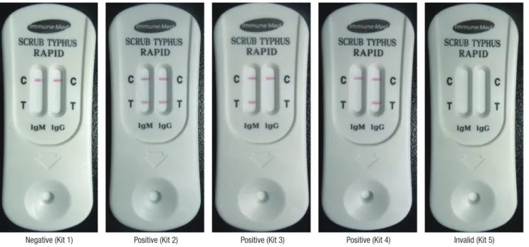
Fig. 1. Representative pictures for RDT indicating negative and positive results. Red colorization of the test line (T) indicates the presence of human antibody against O. tsutsu- gamushi and the red control line (C) represents the valid test result. Demonstration is negative (kit 1), both positive in IgM and IgG (kit 2), IgM positive (kit 3), and IgG positive (kit 4) in RDT results.
Negative (Kit 1) Positive (Kit 2) Positive (Kit 3) Positive (Kit 4) Invalid (Kit 5)
pretation (Fig. 1). Control line (C) represents the validation of test results. Red colorization of test line (T) indicates the pres- ence of human antibody against O. tsutsugamushi.
Table 1 shows the sensitivity and specificity of RDT of each IgM and IgG at the base of IFA performed in Korea. Of the 141 positive serum samples by IFA IgM from Korea, 139 were posi- tive in RDT assay with 98.6% sensitivity for IgM. Out of 136 posi- tive scrub typhus cases evaluated by IFA IgG, 132 cases were identified as positive in RDT assay with 97.1% sensitivity for IgG.
For IgM, four of the 217 controls were found false positive in Ko- rea. Thus, specificity of RDT for IgM was 98.2%. For IgG among 217 control specimens, only five controls were considered as false positive in Korea. Thus, specificity of RDT for IgG was 97.7%.
Examination of cross-reactivity of ImmuneMed RDT to different diseases
To evaluate the cross-reactivity of ImmuneMed RDT to differ- ent diseases, serum samples were collected from Korea Bank for Pathogenic Viruses, and Biobank of several different hospi- tals. Each blood sample was applied to ImmuneMed RDT as serum, plasma, and whole blood to evaluate the performance depending on the state of the sample. Serum samples for mu- rine typhus (n = 48), leptospirosis (n = 23), and hemorrhagic
fever with renal syndrome (HFRS, n = 48) were collectively called as acute febrile illness due to the similarity of clinical symptoms and epidemic patterns.
ImmuneMed RDT did not react with any other disease, in- cluding HFRS (n = 48), leptospirosis (n = 23), murine typhus (n = 48), mycoplasma (n = 3), tularemia (n = 1), CMV (n = 3), EBV (n = 3), HSV (n = 2), VZV (n = 2), hepatitis A (n = 1), hepa- titis b (n = 3), malaria (n = 3), rheumatoid factor (n = 6), ANA (n = 3), toxoplasma (n = 2), and HIV (n = 1).
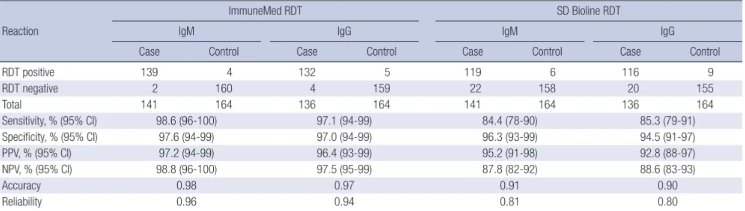
Comparison test of the ImmuneMed RDT and SD Bioline RDT to scrub typhus
To compare with the performance of other scrub typhus RDT, SD Bioline RDT against scrub typhus was used. Table 2 shows the sensitivity and specificity of ImmuneMed RDT and SD Bio- line RDT for each IgM and IgG to O. tsutsugamushi at the base of IFA in Korean patients. One hundred and forty one positive serum samples by IFA IgM were used for evaluating sensitivity for IgM. One hundred and thirty six positive serum samples by IFA IgG were evaluated for examining sensitivity for IgG. Speci- ficity was measured using 164 control specimens for IgM and 164 controls for IgG.
For IgM, the sensitivity and specificity of ImmuneMed RDT was 98.6% and 97.6%, but those of SD bioline RDT was 84.4%
and 96.3% respectively. For IgG, those of ImmuneMed RDT were 97.1% and 97.0%, but those of SD Bioline RDT were 85.3% and 94.5%. This result indicates that the ImmuneMed RDT shows excellent performance of sensitivity and specificity to diagnose scrub typhus compared with SD Biolines.
Sensitivity and specificity of the ImmuneMed RDT in Sri Lanka
Table 3 shows the sensitivity and specificity to scrub typhus at retrospective diagnosis based on IFA in Sri Lanka. Of the 89 (44 with eschars) serum samples from Sri Lanka, 82 were positive in RDT with 92.1% sensitivity. Out of 76 controls evaluated, only 3 cases were identified as false positive in RDT with 96.1% spec- Table 1. Sensitivity and specificity of the ImmuneMed scrub typhus RDT at the base
of each IgM and IgG IFA in Korean patients
Reaction IgM IgG
Case Control Case Control
RDT positive 139 4 132 5
RDT negative 2 213 4 214
Total 141 217 136 217
Sensitivity, % (95% CI) 98.6 (96-100) 97.1 (94-99) Specificity, % (95% CI) 98.2 (96-99) 97.7 (95-99)
PPV, % (95% CI) 97.2 (97-99) 96.4 (93-99)
NPV, % (95% CI) 99.1 (98-100) 98.2 (93-99)
Accuracy 0.98 0.98
Reliability 0.97 0.95
RDT, rapid diagnostic test; PPV, positive predictive value; NPV, negative predictive value.
Table 2. Comparison test of the ImmuneMed RDT and SD Bioline RDT for each IgM and IgG to O. tsutsugamushi at the base of IFA in Korean patients Reaction
ImmuneMed RDT SD Bioline RDT
IgM IgG IgM IgG
Case Control Case Control Case Control Case Control
RDT positive 139 4 132 5 119 6 116 9
RDT negative 2 160 4 159 22 158 20 155
Total 141 164 136 164 141 164 136 164
Sensitivity, % (95% CI) 98.6 (96-100) 97.1 (94-99) 84.4 (78-90) 85.3 (79-91)
Specificity, % (95% CI) 97.6 (94-99) 97.0 (94-99) 96.3 (93-99) 94.5 (91-97)
PPV, % (95% CI) 97.2 (94-99) 96.4 (93-99) 95.2 (91-98) 92.8 (88-97)
NPV, % (95% CI) 98.8 (96-100) 97.5 (95-99) 87.8 (82-92) 88.6 (83-93)
Accuracy 0.98 0.97 0.91 0.90
Reliability 0.96 0.94 0.81 0.80
RDT, rapid diagnostic test; PPV, positive predictive value; NPV, negative predictive value.
ificity. This result indicates that the ImmuneMed RDT can be used in the epidemic region of several serotypes to discriminate O. tsutsugamushi from other diseases.
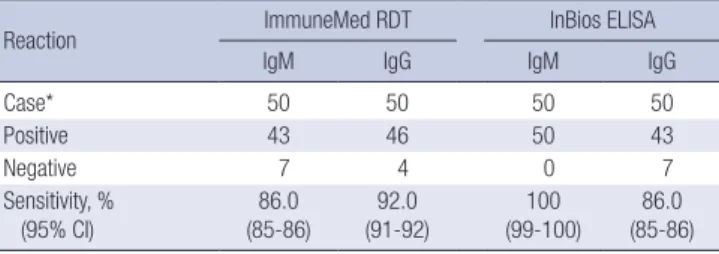
Comparison of ImmuneMed RDT and InBios ELISA for diagnosing scrub typhus in South India
To compare the performance of ImmuneMed RDT with ELISA test, the serum specimens diagnosed with InBios ELISA kit was used. ELISA from InBios was currently used for diagnosing scrub typhus in South India. As shown in Table 4, there is a 94.0% con- cordance rate between ImmuneMed RDT and InBios ELISA to diagnose scrub typhus. This result suggests that ImmuneMed RDT can replace ELISA test for diagnosing scrub typhus with high sensitivity.
DISCUSSION
Scrub typhus is prevalent in rural areas of Asia (20). However, this disease is difficult to diagnose for reasons of the non-spe- cific signs and symptoms, and the difficulty of conventional IFA availability. Although IFA is a gold standard with high sensitivity and specificity, it does not represent perfect sensitivity. Because all epidemic serotypes of O. tsutsugamushi should be provided for IFA diagnosis in each region, accessibility of IFA is difficult in most regions. O. tsutsugamushi is an obligate intracellular pathogen. It is difficult to culture due to a relatively high cost and it requires biosafety level (BSL) 3. For these reasons, clini- cians in underdeveloped countries may choose and use the non- specific and insensitive Well-Felix reaction (18).
In our previous study, we showed the diagnostic sensitivity of scrub typhus using a mixture of recombinant antigens derived from O. tsutusgamushi serotypes (21). In this study, we further improve the sensitivity and specificity of the ImmuneMed RDT and apply it in Korea and Sri Lank to confirm the performance of RDT. Performance of the ImmuneMed RDT was also com- pared with that of SD Bioline RDT. Furthermore, the Immun-
eMed RDT was compared with ELISA test for confirming the performance of the RDT using serum samples in South India.
In this study, the RDT, using recombinant proteins derived from O. tsutsugamushi serotype Gilliam, Karp, Kato, Kangwon, and Boryong, was successfully applied to the different regions including Korea, Sri Lanka and South India. The performance of the ImmuneMed RDT for scrub typhus using Korea serum samples shows 98.6% and 97.1% sensitivity and 98.2% and 97.7%
specificity for IgM and IgG, respectively (Table 1). Two patients for IgM and four patients for IgG from Korea showed false nega- tive in sensitivity. IFA titer for IgM false negative was 1:40 at sam- ples number 98-1221 and 1:10 at 00-253, and IgG false negative was 1:1,280 at sample number 90-110, 1:160 at 99-279 and DK 32, and 1:640 at 00-253. These titer values are low in the IFA pos- itive criterion and do not indicate enough antibody to react with the recombinant proteins in these patients’ serum samples. There- fore, the RDT may not be reacting with patients’ serum.
False positive in specificity was rarely detected in healthy controls and other disease controls. Although this false positive may not be non-specific reaction, we cannot rule out the factor that patients have residual antibody reacting with antigens from a past infection, an unapparent infection, or an infection with another serovar which is not used at IFA, especially in case of Sri Lanka.
Furthermore, a comparison test was performed between Im- muneMed RDT and SD Bioline RDT. The specificity of each RDT was similar at healthy controls and other diseases. However, at sensitivity, the ImmuneMed RDT is superior to that of SD Bio- line RDT. This result indicates that the recombinant proteins (antigens) made from the ImmuneMed RDT have good prop- erties to detect the antibody against O. tsutsugamushi compared with SD Bioline RDT (Table 2). ImmuneMed scrub typhus RDT used antigens from five serotypes of O. tsutsugamushi including Gilliam, Karp, Kato, Kangwon, and Boryong. However, SD Bio- lines tsutsugamushi test utilized antigens from three serotypes of O. tsutsugamushi including Gilliam, Karp, and Kato. Another difference is that ImmuneMed used anti-human IgG as second- ary antibody to enhance the affinity compared with protein A Table 3. Sensitivity and specificity of the ImmuneMed RDT for retrospective diagnosis
at the base of IFA in Sri Lanka Reaction
Sri Lanka IgM/G
Case Control
RDT positive 82 3
RDT negative 7 73
Total 89 76
Sensitivity, % (95% CI) 92.1 (84-97)
Specificity, % (95% CI) 96.1 (88-98)
PPV, % (95% CI) 96.5 (96-97)
NPV, % (95% CI) 91.3 (85-97)
Accuracy 0.94
Reliability 0.88
RDT, rapid diagnostic test; PPV, positive predictive value; NPV, negative predictive value.
Table 4. Comparison test of ImmuneMed RDT and InBios ELISA diagnosing scrub ty- phus in South India patient serum samples
Reaction ImmuneMed RDT InBios ELISA
IgM IgG IgM IgG
Case* 50 50 50 50
Positive 43 46 50 43
Negative 7 4 0 7
Sensitivity, % (95% CI)
86.0 (85-86)
92.0 (91-92)
100 (99-100)
86.0 (85-86) IgM positive and/or IgG positive at IFA were selected and then examined by Immun- eMed RDT and InBios ELISA.
RDT, rapid diagnostic test.
*All patients (n = 87) were IgM positive at InBios ELISA and among them each 50 serum specimens.
for SD biolines. These two factors make differences for sensitiv- ity and specificity for diagnosing scrub typhus.
In Sri Lanka, the performance of the ImmuneMed RDT shows 92.1% of sensitivity and 96.1% of specificity. Although the num- ber of serum samples looks small, the accuracy and reliability of the test is sufficient. Seven false negatives may be due to un- known infection except O. tsutsugamushi in Sri Lanka. Three false positives from healthy controls from Sri Lanka reacted with antigens from a past infection, an inapparent infection, or an infection with an unknown serovar. In addition, as a patient in- fected with a rare or new serovar has not been diagnosed as scrub typhus even though to use IFA, RDT may be a real positive but it might be interpreted as a false positive.
To compare with other detection methods, ELISA test was chosen to evaluate the performance of the ImmuneMed RDT.
Even though only IgM positive at ELISA as sample was selected, there is a 94.0% similarity between ImmuneMed RDT and In- Bios ELISA at retrospective diagnosis from patients of O. tsutsu- gamushi (Table 4). This means that the selected antigens from five different serotypes and other technical improvement rep- resent to similar or better performance with InBios tsutsuga- mushi ELISA. This indicates that the ImmuneMed RDT can re- place ELISA test to diagnose scrub typhus as an easy and time saving diagnostic tool with high sensitivity and specificity.
ImmuneMed Scrub Typhus RDT can detect IgM and IgG to speculate not only a current infection but also the progress of the disease simultaneously. Especially in Sri Lanka and South India, it can be positive interpretation based on an inapparent infection, past infection, or infection of another serovar which is not used in IFA even though IFA titer is not positive.
In this study, the clinical evaluations reveal that ImmuneMed Scrub Typhus RDT is very consistent with IFA in Korea and Sri Lanka. Additionally, the use of ImmuneMed Scrub Typhus RDT as a primary diagnosis for O. tsutsugamushi infection would as- sure easy diagnosis of the disease. Since scrub typhus can be treated with doxycycline, accurate and rapid diagnosis will pro- vide effective management of this disease (3,5). We believe that ImmuneMed Scrub Typhus RDT should be in place not only at hospitals but also other health centers owing to its accuracy, ra- pidity, simplicity, and low necessity for skill. Furthermore, it may improve the rapid detection of scrub typhus and be applicable for global diagnosis after further development.
ACKNOWLEDGMENT
The biospecimens and data used in this study were provided by the KBPV (Korea Bank for Pathogenic Viruses), the Biobank of Chonbuk National University Hospital, Biobank of Gyeongsang National University Hospital, Biobank of Keimyung University Dongsan Medical Center and Biobank of Wonkwang University Hospital. The members of the Korea Biobank Network are sup-
ported by the Ministry of Health, Welfare and Family Affairs. All samples derived from the Korea Biobank Network were obtain- ed with informed consent under institutional review board-ap- proved protocols.
DISCLOSURE
The authors have no potential conflicts of interest to disclose.
AUTHOR CONTRIBUTION
Study design: Kim YJ, Park S, Kim YW. Evaluation of scrub ty- phus rapid kit: Kim YJ, Park S, Premaratna R, Selvaraj S, Seo W, Lee NT. Preparation of scrub typhus antigen: Park SJ, Kim S, Kim D. Preparation of scrub typhus rapid kit: Kim MS, Kim SH, Kang HK. Indirect Immunofluorescent Antibody Assay (IFA):
Choi KC, Kim YW. Collection of samples: Shin DH, Kwon SH.
Writing manuscript: Kim YW. Revision and final approval of manuscript: all authors.
ORCID
Young-Jin Kim http://orcid.org/0000-0003-1996-9367 Sungman Park http://orcid.org/0000-0002-6105-7129 Ranjan Premaratna http://orcid.org/0000-0002-6588-9467 Stephen Selvaraj http://orcid.org/0000-0003-0103-4061 Sang-Jin Park http://orcid.org/0000-0002-4838-2664 Sora Kim http://orcid.org/0000-0002-5314-2569 Donghwan Kim http://orcid.org/0000-0001-5881-9105 Min Soo Kim http://orcid.org/0000-0001-7489-3532 Dong Hoon Shin http://orcid.org/0000-0002-3513-6333 Kyung-Chan Choi http://orcid.org/0000-0001-6134-8631 Soon-Hwan Kwon http://orcid.org/0000-0002-1582-303X Wonjun Seo http://orcid.org/0000-0002-7109-3799 Nam Taek Lee http://orcid.org/0000-0002-8352-4760 Seung-Han Kim http://orcid.org/0000-0003-1310-8136 Heui Keun Kang http://orcid.org/0000-0002-3216-4961 Yoon-Won Kim http://orcid.org/0000-0002-2242-2664 REFERENCES
1. Brown GW, Robinson DM, Huxsoll DL, Ng TS, Lim KJ, Sannasey G. Scrub typhus: a common cause of illness in indigenous populations. Trans R Soc Trop Med Hyg 1976; 70: 444-8.
2. Chang WH, Kang JS, Lee WK, Choi MS, Lee JH. Serological classification by monoclonal antibodies of Rickettsia tsutsugamushi isolated in Korea.
J Clin Microbiol 1990; 28: 685-8.
3. Ebisawa I. Current epidemiology and treatment of tsutsugamushi disease in Japan. J Travel Med 1995; 2: 218-20.
4. Ree HI, Lee IY, Jeon SH, Yoshida Y. Geographical distribution of vectors and sero-strains of tsutsugamushi disease at mid-south inland of Korea.
Korean J Parasitol 1997; 35: 171-9.
5. Watt G, Walker DH. Scrub typhus. In: Guerrant RL, Walker DH, Weller PF, editors. Tropical Infectious Diseases: Principles, Pathogens, and Practice:
Vol 1. Philadelphia, PA: Churchill Livingstone, 1999, p592-7.
6. Brown GW. Scrub typhus: pathogenesis and clinical syndrome. In: Walk- er DH, editor. Biology of Rickettsial Diseases: Vol I. Boca Raton, FL: CRS Press, 1988, p93-100.
7. Munro-Faure AD, Andrew R, Missen GA, Mackay-Dick J. Scrub typhus in Korea. J R Army Med Corps 1951; 97: 227-9.
8. Groves MG. Scrub typhus. In: Beran GW, editor. Handbook of Zoonoses:
Section A: Bacterial, Rickettsial, Chlamydial and Mycotic. 2nd ed. Boca Raton, FL: CRC Press, 1994, p663-8.
9. Lee JS, Ahn C, Kim YK, Lee MH. Thirteen cases of rickettsial infection in- cluding nine cases of tsutsugamushi disease, first confirmed in Korea. J Korean Med Assoc 1986; 29: 430-8.
10. Chang WH, Kang JS. Isolation of Rickettsia tsutsugamushi from Korean patients. J Korean Med Assoc 1987; 30: 999-1008.
11. Ree HI, Cho MK, Lee IY, Jeon SH. Comparative epidemiological studies on vector/reservoir animals of tsutsugamushi disease between high and low endemic areas in Korea. Korean J Parasitol 1995; 33: 27-36.
12. Kitaoka M, Tanaka Y. Rickettsial toxin and its specificity in 3 prototype strains, Karp, Gilliam and Kato, of Rickettsia orientalis. Acta Virol 1973;
17: 426-34.
13. Lee YM, Kim DM, Lee SH, Jang MS, Neupane GP. Phylogenetic analysis of the 56 kDa protein genes of Orientia tsutsugamushi in Southwest Area of Korea. Am J Trop Med Hyg 2011; 84: 250-4.
14. Min CH, Chang WH, Kang JS, Cho SI, Choi MK, Cho MK, Yoon CS, Kim YW. Murine typhus and scrub typhus in Kangwon-do Korea. Korean J Infect Dis 1988; 20: 105-16.
15. Bozeman FM, Elisberg BL. Serological diagnosis of scrub typhus by indi- rect immunofluorescence. Proc Soc Exp Biol Med 1963; 112: 568-73.
16. Elisberg BL, Campbell JM, Bozeman FM. Antigenic diversity of Rickettsia tsutsugamushi: epidemiologic and ecologic significance. J Hyg Epidemi- ol Microbiol Immunol 1968; 12: 18-25.
17. Jiang J, Marienau KJ, May LA, Beecham HJ 3rd, Wilkinson R, Ching WM, Richards AL. Laboratory diagnosis of two scrub typhus outbreaks at Camp Fuji, Japan in 2000 and 2001 by enzyme-linked immunosorbent assay, rapid flow assay, and Western blot assay using outer membrane 56-kD recombinant proteins. Am J Trop Med Hyg 2003; 69: 60-6.
18. Brown GW, Shirai A, Rogers C, Groves MG. Diagnostic criteria for scrub typhus: probability values for immunofluorescent antibody and Proteus OXK agglutinin titers. Am J Trop Med Hyg 1983; 32: 1101-7.
19. Kim YW, Kim IS, Chang IA, Woo SD, Kim YJ, Chun JM, Kim WC, Byun YH, Cho MK, Inventors; IMMUNEMED Inc., assignee. Diagnostic formu- lation for tsutsugamushi disease. Korea patent KR 10-2006-0084490. 2008 Jan 15.
20. Rapmund G. Rickettsial diseases of the Far East: new perspectives. J Infect Dis 1984; 149: 330-8.
21. Kim YJ, Yeo SJ, Park SJ, Woo YJ, Kim MW, Kim SH, Chang IA, Jeon SH, Park BJ, Song GJ, et al. Improvement of the diagnostic sensitivity of scrub ty- phus using a mixture of recombinant antigens derived from Orientia tsu- tsugamushi serotypes. J Korean Med Sci 2013; 28: 672-9.