381
Copyrights © 2015 The Korean Society of Radiology
INTRODUCTION
Carcinoma of the ampulla of Vater (AoV) is a relatively rare neoplasm, comprising 15–37% of surgically resected pancreato- duodenal tumors and 0.2% of routine autopsy cases; and the most common tumors affecting the AoV are adenocarcinomas (1, 2). Signet ring cell carcinoma (SRCC) of the AoV is rare, and only 21 cases have previously been described in the English lan- guage literature (3). Most of the previously reported articles have been published in the clinical or pathology literatures, and little attention has been paid to radiological features of SRCC of the AoV. Here, we present a case of SRCC in the AoV, with the emphasis on imaging features.
CASE REPORT
A 41-year-old man was hospitalized for 20 days with jaundice.
Upon physical examination, he presented with icteric sclera and visible jaundice. Liver function tests showed total bilirubin 7.5
mg/dL, aspartate aminotransferase 142 IU/L, alanine aminotrans- ferase 159 IU/L, alkaline phosphatase 420 IU/L, and γ-glutamyl transferase 1048 IU/L. Tumor marker, carbohydrate antigen 19-9 was elevated (53.6 U/mL). The urine color was amber and con- tained 3.0 mg/dL of bilirubin.
Dynamic contrast-enhanced 64-channel multidetector com- puted tomography of the liver showed a 1.2-cm sized lobulating contoured mass with enhancement in the AoV. The upstream intrahepatic and extrahepatic bile ducts were proportionally di- lated (Fig. 1A, B). These image findings suggested a carcinoma of the AoV with direct invasion of the distal common bile duct (CBD). There was no evidence of distant metastases. Endoscop- ic retrograde cholangiopancreatography revealed an ulcerofun- gating mass in the AoV. Diffuse dilatation of the bile duct was noted, and there was a focal nodular contrast filling defect in the far-distal CBD (Fig. 1C, D). Dilated bile duct was decompressed with endoscopic nasobiliary drainage (ENBD). Endoscopic ret- rograde cholangioscope-guided biopsy samples from the mass of the AoV showed moderately differentiated adenocarcinoma.
Case Report
pISSN 1738-2637 / eISSN 2288-2928 J Korean Soc Radiol 2015;72(6):381-384 http://dx.doi.org/10.3348/jksr.2015.72.6.381
Received October 27, 2014 Accepted January 12, 2015
Corresponding author: Yoon Young Jung, MD Department of Radiology, Eulji Hospital, Eulji University School of Medicine, 68 Hangeulbiseok-ro, Nowon-gu, Seoul 139-872, Korea.
Tel. 82-2-970-8290 Fax. 82-2-970-8346 E-mail: [email protected]
This is an Open Access article distributed under the terms of the Creative Commons Attribution Non-Commercial License (http://creativecommons.org/licenses/by-nc/3.0) which permits unrestricted non-commercial use, distri- bution, and reproduction in any medium, provided the original work is properly cited.
Most malignant tumors affecting the ampulla of Vater (AoV) are adenocarcinomas, and signet ring cell carcinoma (SRCC) of the AoV is rare. We report a case of SRCC of the AoV in a 41-year-old man who presented with a small tumor in the AoV, which was discovered by multiple imaging modalities. Since most of the previously reported articles have been published in the clinical pathology literature, we mainly described the imaging features.
Index terms
Signet Ring Cell Carcinoma Ampulla of Vater
CT
Positron Emission Tomography/CT
Imaging Findings of Signet Ring Cell Carcinoma of the Ampulla of Vater: A Case Report
1바터 팽대부에 발생한 반지세포암의 영상 소견: 증례 보고1
Minchul Kim, MD
1, Yoon Young Jung, MD
1, Myung-Won You, MD
1, Dong Hee Kim, MD
2, Won Mee Lee, MD
3, Yun Sun Choi, MD
1Departments of 1Radiology, 2Surgery, 3Pathology, Eulji Hospital, Eulji University School of Medicine, Seoul, Korea
Imaging Findings of Signet Ring Cell Carcinoma of the Ampulla of Vater
382
J Korean Soc Radiol 2015;72(6):381-384 jksronline.orgthese findings, the final histopathologic diagnosis was SRCC.
The tumor cells were found in two regional lymph nodes. Ac- cording to the TNM classification, tumor of the AoV was T2N1M0 (Stage IIB).
After the surgery, the patient underwent five cycles of postop- erative chemotherapy with combination of 5-fluorouracil and cisplatin chemotherapy regimen. On the follow-up CT scans, local recurrence or metastatic disease were not detected. The pa- tient was alive and disease-free at the 13 months post-operative follow-up.
DISCUSSION
Carcinoma of the AoV is a relatively rare neoplasm, and the most common tumors affecting the AoV are adenocarcinomas (1, 2). Ampullary carcinomas typically manifest as small tumors at the time of diagnosis, because of the relatively early onset of symptoms; therefore, the mass itself is often not apparent on im- Gadoxetic acid (Gd-EOB-DTPA, Primovist; Bayer HealthCare,
Berlin, Germany) enhanced magnetic resonance cholangiogra- phy revealed a distinct nodular enhancing lesion in the AoV similar to CT, and bilary system was well decompressed by ENBD. The 18-fluoro-2-deoxy-D-glucose positron emission to- mography/computed tomography (18F-FDG PET/CT) showed increased FDG uptake (maximum standardized uptake value 4.1) in the distal CBD (Fig. 1E).
The patient underwent a pylorus preserving pancreaticoduo- denectomy. On gross evaluation of the resected specimen, mu- cosal surface of the duodenum showed a polypoid mass with central ulceration, measuring 2.0 × 1.6 cm in diameter in the AoV. The cut surface of the ampullary tumor revealed a whitish tumor with pushing margins, measuring 1.1 × 1 cm in diameter.
The tumor invaded through the duodenal wall but the pancreas was grossly unremarkable (Fig. 1F). The histology of tumor was composed of usual signet ring cells, and the lower half was con- sisted of eosinophilic signet ring cells (Fig. 1G). As a result of
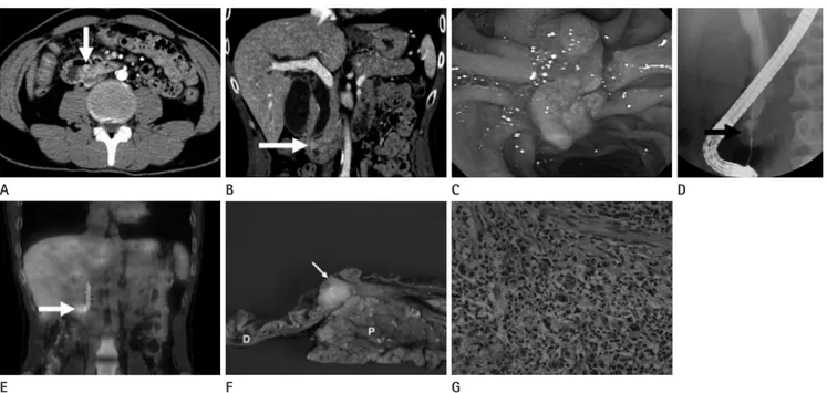
Fig. 1. 41-year-old man with signet ring cell carcinoma of the ampulla of Vater.
A, B. Axial (A) and coronal (B) reformatted contrast-enhanced CT images in the portal venous phase show a lobulating mass with enhancement (arrows) in the ampulla of Vater. Upstream intrahepatic and extrahepatic bile ducts are dilated.
C. Duodenoscopy shows an ulcerofungating mass in the ampulla of Vater, macroscopically.
D. Endoscopic retrograde cholangiography shows nodular filling defect (arrow) in the far-distal common bile duct, corresponding to the intralu- minal nodular enhancing lesion found on CT. Diffuse dilatation of common bile duct is seen.
E. Coronal 18F-FDG PET/CT image shows increased FDG uptake (arrow) in the distal common bile duct without evidence of distant metastases.
F. The gross cut surface of surgical specimen shows a whitish periampullary tumor (arrow). The tumor invaded through the duodenal wall but the pancreas is grossly unremarkable.
G. Microscopic photomicrograph shows diffusely infiltrative adenocarcinoma. Upper half is composed of usual signet ring cells and lower half is consisted of eosinophilic signet ring cells (hematoxylin & eosin stain, × 400).
D = duodenum, P = pancreas, 18F-FDG PET/CT = 18-fluoro-2-deoxy-D-glucose positron emission tomography/computed tomography A
E
B
F
C
G
D
Minchul Kim, et al
383
jksronline.org J Korean Soc Radiol 2015;72(6):381-384
on PET/CT.
In conclusion, little is known about the imaging features of SRCC in the AoV; but considering the poor prognosis of SRCCs of other sites, radiologists and clinicians should be aware of the possibility of a SRCC when hyper-enhancing tumor in the AoV is encountered.
REFERENCES
1. Kamisawa T, Fukayama M, Koike M, Tabata I, Egawa N, Isa- wa T, et al. Carcinoma of the ampulla of Vater: expression of cancer-associated antigens inversely correlated with prognosis. Am J Gastroenterol 1988;83:1118-1123
2. Albores-Saavedra J, Schwartz AM, Batich K, Henson DE.
Cancers of the ampulla of vater: demographics, morphol- ogy, and survival based on 5,625 cases from the SEER pro- gram. J Surg Oncol 2009;100:598-605
3. Talebi A, Mohammadizadeh F, Hani M, Bagheri M, Bagheri A. Signet ring carcinoma of ampulla of vater. Adv Biomed Res 2014;3:30
4. Kim S, Lee NK, Lee JW, Kim CW, Lee SH, Kim GH, et al. CT evaluation of the bulging papilla with endoscopic correla- tion. Radiographics 2007;27:1023-1038
5. Mosconi S, Beretta GD, Labianca R, Zampino MG, Gatta G, Heinemann V. Cholangiocarcinoma. Crit Rev Oncol Hema- tol 2009;69:259-270
6. Lee JH, Park MS, Kim KW, Yu JS, Kim MJ, Yang SW, et al. Ad- vanced gastric carcinoma with signet ring cell carcinoma versus non-signet ring cell carcinoma: differentiation with multidetector CT. J Comput Assist Tomogr 2006;30:880-884 7. Taghavi S, Jayarajan SN, Davey A, Willis AI. Prognostic sig- nificance of signet ring gastric cancer. J Clin Oncol 2012;
30:3493-3498
8. Lee EY, Kim C, Kim MJ, Park JY, Park SW, Song SY, et al.
Signet ring cell carcinoma of the extrahepatic bile duct.
Gut Liver 2010;4:402-406
9. Gao JM, Tang SS, Fu W, Fan R. Signet-ring cell carcinoma of ampulla of Vater: contrast-enhanced ultrasound find- ings. World J Gastroenterol 2009;15:888-891
10. Chang S, Lim JH, Choi D, Kim SK, Lee WJ. Differentiation of ampullary tumor from benign papillary stricture by thin- section multidetector CT. Abdom Imaging 2008;33:457-462 aging. However, secondary findings such as marked bile duct
dilatation, in association with mild to moderate dilatation of the pancreatic duct, can usually be seen on CT. Large ampullary tu- mors usually manifest as an infiltrative or polypoid mass (4).
SRCC can arise in many organs, but it usually occurs in the gastrointestinal tract, especially in the stomach. It has been re- ported that 90% of SRCC occurs in the stomach, with the rest arising in several other organs, including the breast, gallbladder, pancreas, urinary bladder, and colon (5). When it arises from the stomach, SRCC shows stronger enhancement than non-sig- net ring cell carcinoma (6) and is generally known for its ad- vanced stage and poor prognosis (7). SRCC of the biliary system is rare and most SRCCs of the biliary system are originated from gallbladder (8). Because of its rareness, little is known about the origin of SRCCs in the periampullary area, and two possible ex- planations for this histologic variation were suggested. One ex- planation is that the tumors may arise from ectopic gastric mu- cosa. Indeed, there are some reported cases of SRCCs with ectopic gastric mucosa in the ampullary tumors. Another theory holds that these carcinomas arise from the areas of gastric-type meta- plastic epithelium, which are considered to be a protective re- sponse to elevated acidity and are observable in the duodenal bulb of patients with peptic ulcer patients (7, 8).
Several modalities such as CT, MRI, and US (including con- trast-enhanced US) were used to diagnose the SRCC of the AoV in the previously reported cases. All accessible literatures only showed dilated bile ducts without an apparent mass lesion in the AoV, which are unable to reveal malignant etiology, except for Gao et al. (9) who reported with contrast-enhanced US. Unlike these previously reported cases, we were able to detect the tumor.
According to Chang et al. (10), mean CT attenuation values of ampullary tumor was 107 ± 22 Hounsfield unit (HU) (in the ar- terial phase) and 103 ± 17 HU (in the portal venous phase). In our case, the tumor showed 120 HU and 148 HU, respectively.
The attenuation numbers were higher than average ampullary tumor. This enhancement pattern is similar to the gastric SRCC pattern (6). But more cases are needed to confirm hyperenhance- ment as a feature of SRCC of the AoV, since this is the first case to describe it. In previously reported 21 cases, only two cases had lymph node metastasis at the diagnosis, and this presenting case also had two metastatic lymph nodes. The nodes were neither significantly enlarged on CT nor showed increased FDG uptake
Imaging Findings of Signet Ring Cell Carcinoma of the Ampulla of Vater
384
J Korean Soc Radiol 2015;72(6):381-384 jksronline.org바터 팽대부에 발생한 반지세포암의 영상 소견: 증례 보고1
김민철
1· 정윤영
1· 유명원
1· 김동희
2· 이원미
3· 최윤선
1바터 팽대부에 발생하는 악성 종양은 대부분 선암이며, 반지세포암의 발생은 드물다. 저자들은 여러 영상 기법에서 바터 팽대부에 종양으로 보이며, 반지세포암으로 확진된 41세 남자 환자의 증례를 보고하고자 한다. 이전에 출판된 연구물들은 대부분 임상 및 병리학적 소견에 대한 문헌들이기 때문에, 저자들은 주로 영상 소견을 기술하고자 한다.
을지대학교 의과대학 을지병원 1영상의학과, 2외과, 3병리과