Sarcoid-Like Reaction after Complete Remission of
Malignancy: CT and
18 F-FDG PET/CT Features for the Differential Diagnosis from Lymph Node
Metastasis
악성종양의 완전관해 후 발생한 사르코이드증 유사 반응:
림프절 전이와의 감별진단에 유용한 CT와
18F-FDG PET/CT 소견
Hyun Ji Kang, MD1 , Yookyung Kim, MD1* , June Young Bae, MD1 , Jung Hyun Chang MD2 , Soo-Hyun Lee, MD3
Department of 1Radiology, 2Division of Pulmonary and Critical Care Medicine, Department of Internal Medicine, College of Medicine, Ewha Womans University, Seoul, Korea
3Department of Radiology, National Cancer Center, Goyang, Korea
Purpose To identify the imaging features indicative of sarcoid-like reactions in patients with in- trathoracic lymphadenopathy after complete remission of malignancies.
Materials and Methods This study enrolled five patients with histopathologically confirmed sarcoid-like reactions that developed after cancer remission. The clinical features and findings of CT and 18F-fluorodeoxyglucose (FDG) PET/CT were assessed.
Results The underlying malignancies included breast, nasopharyngeal, colon, and endometri- al cancer and lymphoma. The time intervals between complete remission of malignancy and the diagnosis of sarcoid-like reaction ranged from 6 to 78 months. CT findings of sarcoid-like re- action included bilateral hilar and mediastinal lymphadenopathies (n = 5), pulmonary nodules (1–15 mm) with peribronchovascular, fissural, or subpleural distribution, and interlobular inter- stitial thickening in the lungs (n = 4). 18F-FDG PET/CT revealed hypermetabolic uptake in the me- diastinal and hilar lymph nodes and both lungs in the absence of extrathoracic uptake (n = 3).
The sarcoid-like reactions resolved in all patients after corticosteroid treatment.
Conclusion In patients with complete remission of malignancies, newly developed bilateral hi- lar and mediastinal lymphadenopathies with or without pulmonary nodules of perilymphatic
Received May 5, 2020 Revised August 15, 2020 Accepted September 15, 2020
*Corresponding author Yookyung Kim, MD Department of Radiology, Ewha Womans University Mokdong Hospital, 1071 Anyangcheon-ro, Yangcheon-gu, Seoul 07985, Korea.
Tel 82-2-2650-5380 Fax 82-2-2650-5302 E-mail yookkim@ewha.ac.kr This is an Open Access article distributed under the terms of the Creative Commons Attribu- tion Non-Commercial License (https://creativecommons.org/
licenses/by-nc/4.0) which permits unrestricted non-commercial use, distribution, and reproduc- tion in any medium, provided the original work is properly cited.
ORCID iDs Hyun Ji Kang https://
orcid.org/0000-0001-6162-4552 Yookyung Kim
https://
orcid.org/0000-0002-7247-7815 June Young Bae
https://
orcid.org/0000-0001-6400-4105 Jung Hyun Chang
https://
orcid.org/0000-0003-1000-2491 Soo-Hyun Lee
https://
orcid.org/0000-0003-4647-2137
distribution, in the absence of recurrence at the primary tumor site and extrathoracic metastasis, may suggest a sarcoid-like reaction. Such cases warrant histologic evaluation of the lymph nodes to pre- vent unnecessary systemic chemotherapy.
Index terms Sarcoidosis; Malignancy; Lymphoma
INTRODUCTION
Sarcoidosis is a systemic granulomatous disorder of unknown origin, which is character- ized by chronic noncaseating granulomatous inflammation. It commonly affects young- and middle-aged adults and predominantly involves the lung, eye, and skin (1).
The presence of noncaseating granulomas in local lymph nodes or organs is rarely observed in patients with hematological malignancies or solid cancers. This local reaction is known as
“sarcoid-like reaction” and presents with radiological and histological features similar to clas- sic thoracic sarcoidosis, without clinical manifestations. Different from the idiopathic sarcoid- osis, sarcoid-like reaction is considered as an immunologic abnormality related to a malignant condition, and can occur in 4.4% of carcinoma, 7.3% of non-Hodgkin lymphoma, and 13.8%
of Hodgkin disease (2).
In patients with a history of malignancy, radiologic aspects of sarcoid-like reaction might mimic metastatic lymphadenopathy and therefore, the diagnostic suggestion of possibility of sarcoid-like reaction is essential to prevent unnecessary systemic chemotherapy. The objec- tive of this study was to identify CT and 18F-fluorodeoxyglucose (FDG) PET/CT features of sar- coid-like reaction which may be helpful for the differential diagnosis from lymph node me- tastasis in patients with a history of malignancy.
MATERIALS AND METHODS PATIENTS
The present study was approved by the Institutional Review Board of our hospital and in- formed consent was waived due to the retrospective nature of this study (IRB No. EUMC 2020-04-002). Five patients were enrolled who were diagnosed to have histopathologically confirmed sarcoid-like reaction during the surveillance for disease progression after the achievements of complete remission of malignancies from May 2012 to August 2018. All the patients did not have prior history or evidence of sarcoidosis before the diagnosis of malig- nancy and were not treated with immunotherapy. The Clinical and CT follow-up for the ma- lignancy and sarcoid-like reaction were performed in all patients until March 2020.
Chest CT scan (n = 5) and 18F-FDG PET/CT scans (n = 3) were obtained at the time of diagno- sis of sarcoid-like reaction and all the patients underwent follow-up chest CT scans. Initial chest CT scan and 18F-FDG PET/CT scan had been also obtained at the time of diagnosis of ma- lignancy, before the onset of sarcoid-like reaction. We retrospectively reviewed the clinical features using the electronic medical record system and analyzed CT and 18F-FDG PET/CT find- ings on picture archiving and communication system (PACS).
CT EXAMINATION AND IMAGING ASSESSMENT
Chest CT scans were performed with a 16-channel, multi-detector, row CT scanner (SO- MATOM Sensation 16; Siemens Medical Solutions, Forchheim, Germany) or a 64-channel, multi-detector, row CT scanner (SOMATOM Sensation 64; Siemens Medical Solutions) or 2 × 128-channel, dual-source CT scanner (SOMATOM Definition Flash, Siemens Medical Solu- tions). The CT parameters were as follows: tube voltage: 120 kVp; tube current: 40 to 100 mAs (effective); collimation: 1 × 16 × 1.5 mm (16-channel) or 1 × 64 × 1.2 mm (64-channel) or 2 × 128 × 0.6 mm (dual source); pitch: 1.0 or 1.4; and gantry rotation time: 0.5 s. CT scans were obtained with or without contrast-enhancement from the thoracic inlet to the upper abdomen, including both adrenal glands. Contrast-enhanced CT scans were obtained after injection of 100 mL of the iodinated contrast agent (Ultravist 300; iopromide, Schering, Berlin, Germany).
Chest CT scans were reviewed by two radiologists (Kim Y, with 23 years of experience; and Kang HJ, with 3 years of experience) by consensus on monitors using a PACS. They recorded the presence of lymph node enlargements with their location (mediastinum, hilum, other sites of the thorax) and size (short diameter of the largest lymph node), pulmonary nodules with their size, location (pulmonary lobes), and distribution (peribronchial, subpleural, fissural), pulmonary interstitial thickening, and other abnormalities in the thorax.
18
F-FDG PET/CT AND IMAGING ASSESSMENT
Whole-body PET/CT imaging was performed from the skull base to the mid-thigh. Patients fasted for at least 6 h prior to PET/CT examination. After ensuring a normal blood glucose lev- el in peripheral blood, the patients received an intravenous injection of 370 MBq (10 mCi) of
18F-FDG and were then rested for approximately 1 hour prior to imaging. Image acquisition was conducted using a PET/CT device (Discovery LS, GE Medical Systems, Milwaukee, WI, USA) consisting of an Advance NXi PET scanner and an eight-slice Light Speed Plus CT scan- ner. CT was conducted using a standardized protocol involving the following: 140 kV, 80 mA, a tube-rotation time of 0.5 s per rotation, a pitch of 6, and a section thickness of 5 mm, which matched the PET image section thickness. No contrast material was administered. Immediate- ly after CT, PET was conducted in the identical axial field of view. CT data were resized from a 512 × 512 matrix to a 128 × 128 matrix in order to match the PET data to allow the images to be fused and the CT transmission maps to be generated. The co-registered images were displayed using eNTEGRA software (GE Medical Systems).
One nuclear medicine physician evaluated the integrated PET/CT datasets. Lymph nodes and nodules with increased glucose uptake [to a level greater than that of mediastinal back- ground as determined by qualitative analysis, to have a maximum standardized uptake value (SUVmax) adjusted to the patient’s body weight of more than 3.5 as determined by quantita- tive analysis] with distinct margins were considered positive.
RESULTS
CLINICAL FEATURES
Patient characteristics and clinical courses are summarized in Table 1. There were four fe- male and one male and their age ranged from 42 to 56 years (mean, 47.8 years) at the time of
diagnosis of sarcoid-like reaction. Their underlying malignancies included breast cancer, en- dometrical cancer, nasopharyngeal cancer, colon cancer, and lymphoma of the nasal cavity and they were treated by various methods.
The time interval between the diagnosis of the malignancy and sarcoid-like reaction was 9–84 months [mean ± standard deviation (SD), 30.2 ± 31.1 months]. All patients were diag- nosed to have sarcoid-like reaction after the complete remission of malignancies and the time interval between the diagnosis of complete remission and sarcoid-like reaction was 7–78 months (25.6 ± 30.0 months).
All of them presented without symptoms and new bilateral symmetric mediastinal and hi- lar lymphadenopathies were incidentally detected on chest CT during the surveillance for previous malignancy. At the time of new development of mediastinal and hilar lymphade- nopathies on CT scan, possibility of sarcoid-like reaction was suggested by radiologists in two patients whose underlying malignancies were breast and nasopharyngeal cancer, based on that they had neither recurrent tumor at the previous primary tumor site nor other distant metastasis. However, in other two patients, metastasis was the first radiologic impression.
Lymph node biopsy was performed in all patients (endobronchial ultrasound-guided biopsy in two, mediastinoscopic biopsy in two, video-assisted thoracoscopic biopsy in one) and his- topathologic examination revealed noncaseating granulomatous inflammation with negative real-time polymerase chain reaction for tuberculous or nontuberculous mycobacterium, which is consistent with sarcoid-like reaction in those patients who did not have any systemic mani- festation of sarcoidosis.
After corticosteroid treatment, mediastinal and hilar lymphadenopathies were resolved in all patients and there was no recurrence of lymphadenopathies until the last follow-up CT scan (range of follow-up period, 11–94 months; mean ± SD, 38.2 ± 33.7 months).
Table 1. Demographics and Clinical Features of Five Patients with Histopathologically Confirmed SRs
Patient No.
Age (years)/
Sex
Primary
Malignancy Treatment
Dx of SR:
Biopsy of Mediastinal or
Hilar Lymph Nodes Via
Time Interval (months) Complete Resolution of
SR after Corticosteroid
Treatment
F/U Period after Dx of SR (months)/
Recurrence of SR Dx of
Malignancy – Dx of SR
Remission of Malignancy –
Dx of SR
1 43/F Breast cancer
BCS cyclophosphamide, methotrexate, 5-FU
EBUS 84 78 Yes 11/No
2 56/M Nasopharyngeal cancer
CCRT
5-FU, cisplatin Mediastinoscopy 9 7 Yes 35/No
3 42/F Endometrial cancer
TAH with BSO
Paclitaxel, carboplatin EBUS 22 16 Yes 39/No
4 49/F Colon cancer Capecitabine Mediastinoscopy 9 6 Yes 12/No
5 49/F Nasal cavity lymphoma
Cyclophosphamide,
vincristine, prednisone VATS 27 21 Yes 94/No
BCS = breast conserving surgery, CCRT = concurrent chemoradiation therapy, Dx = diagnosis, EBUS = endobronchial ultrasound, F/U = follow- up, SR = sarcoid-like reaction, TAH with BSO = total abdominal hysterectomy with bilateral salpingo-oophorectomy, VATS = video-assisted thoracoscopic surgery, 5-FU = fluorouracil
CT FEATURES
Image findings of sarcoid-like reaction on chest CT and PET/CT are summarized in Table 2.
All five patients showed symmetric bilateral hilar and mediastinal lymphadenopathy on CT scans. In one patient, enlarged lymph nodes are also noted in both supraclavicular and lower cervical areas. Lymph nodes are homogeneous without necrosis or calcification and short diameter of the largest lymph node in each patient ranged from 13?23 mm (mean, 17.0 ± 5.9 mm) (Figs. 1, 2).
Pulmonary lesions are noted in four of the five patients. All the four patients showed pul- monary nodules of peribronchovascular, fissural or subpleural distribution, but the predom- inant sizes and locations of pulmonary nodules were different from each other. Patient 2 and 4 demonstrated poorly-defined tiny (usually less than 3 mm) nodules of predominantly cen- trilobular location, suggestive of peribronchovascular distribution (Fig. 1D). In patient 3, pul- monary nodules showed peribronchovascular distribution but they were larger (4–15 mm) and well defined (Fig. 2C, D). Pulmonary nodules in patient 5 had predominantly subpleural or fissural location, and they had intermediate sizes (4–10 mm) and well defined margin (Fig. 3).
Interlobular septal thickenings were noted in all the three patients, but they were observed only in small areas of the lung (Fig. 2D).
Lymphadenopathy and pulmonary nodules disappeared on follow-up CT scans (time inter- val from the diagnosis of sarcoid-like reaction: range, 2–11 months; mean ± SD 6.8 ± 3.7 months) obtained after corticosteroid treatment in all patients.
18
F-FDG PET/CT FEATURES
In three of the five patients, 18F-FDG PET/CT scan was performed at the time of diagnosis of underlying malignancy and two or more scans were additionally obtained during the follow- up periods. Sarcoid-like reaction was diagnosed on follow-up PET/CT scans in all patients.
On initial PET/CT scans, abnormal FDG uptakes were noted at the primary tumor sites in
Table 2. Chest CT and 18F-FDG PET/CT Findings of Five Patients with Histopathologically Confirmed Sarcoid-Like Reactions
Patient No.
CT 18F-FDG PET/CT
Lymphadenopathies Pulmonary Lesions SUVmax of
Lymph Nodes
Pulmonary FDG Uptake
Extrathoracic FDG Uptake
1 Bilateral hilar and mediastinal (–) Not performed
2 Bilateral hilar and mediastinal
Poorly-defined micronodules (< 3 mm) with peribronchovascular, fissural or subpleural
distribution, interlobular interstitial thickening
Up to 17.5 Mild uptakes in
both lungs (–)
3 Bilateral hilar and mediastinal
Well-defined nodules (4–15 mm) with peribronchovascular, fissural or subpleural
distribution, interlobular interstitial thickening
Up to 6.2
Uptakes in multiple nodules in both
lungs
(–)
4 Bilateral hilar and mediastinal
Poorly-defined micronodules (< 3 mm) with peribronchovascular or fissural distribution,
interlobular interstitial thickening
Not performed
5
Bilateral hilar and mediastinal, bilateral supraclavicular and
lower cervical
Well-defined nodules (4–10 mm) with peribronchovascular, fissural or subpleural
distribuion, interlobular interstitial thickening
Up to 7.2
Uptakes in multiple nodules in both
lungs
(–)
FDG = fluorodeoxyglucose, SUVmax = maximum standardized uptake value
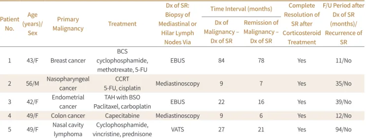
Fig. 1. A 56-year-old male with nasopharyngeal cancer and a sarcoid-like reaction (patient 2).
A. Initial 18F-FDG PET/CT scan obtained at the time of diagnosis of nasopharyngeal cancer shows a hypermetabolic nasopharyngeal mass and bilateral cervical lymph nodes. The patient was treated with concurrent chemoradiation therapy and complete remission was achieved.
B. Follow-up 18F-FDG PET/CT scan performed 9 months after (A) demonstrates hypermetabolic activity in the bilateral hilar and mediastinal lymph nodes (maximum standardized uptake value, 17.5) without FDG uptake in the extrathoracic sites.
C. Contrast-enhanced CT scan shows homogeneous bilateral hilar and mediastinal lymphadenopathy.
D. Lung window image reveals poorly-defined micronodules with peribronchovascular (arrows) and subpleural (arrowheads) distributions in the right upper lobe, especially in the posterior segment.
FDG = fluorodeoxyglucose A
C
B
D
two patients (patient 2 and 3) (Figs. 1, 2) and absent in one who underwent PET/CT scan after chemotherapy (patient 5). Hypermetabolic activity was not noted in the hilum and mediasti- num in all patients.
PET/CT scan performed at the diagnosis of sarcoid-like reaction revealed symmetric hyper-
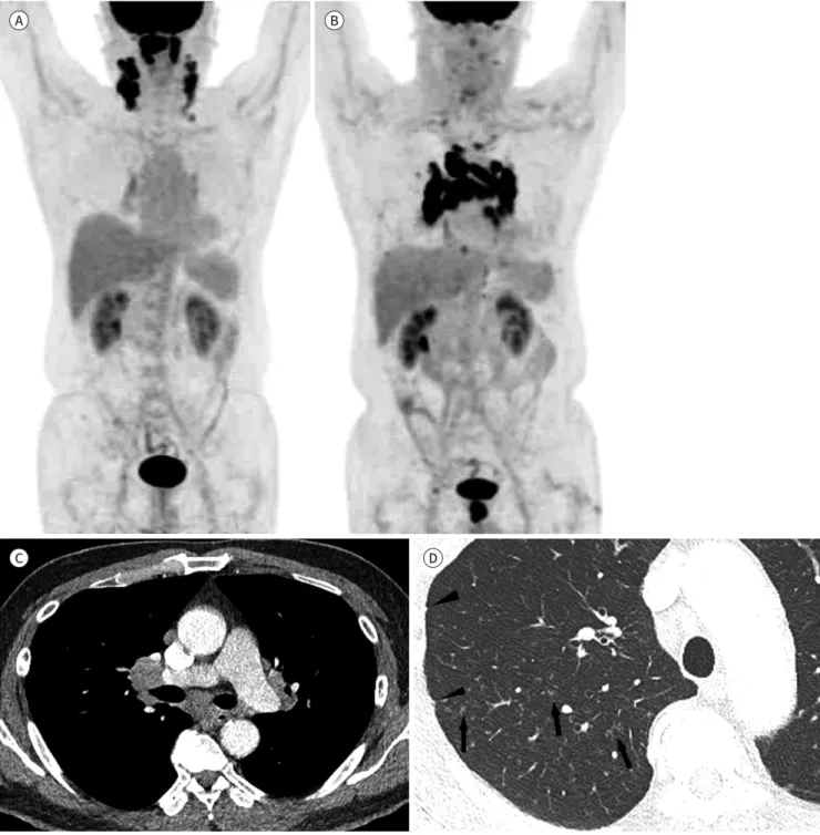
Fig. 2. A 42-year-old female with endometrial cancer and a sarcoid-like reaction (patient 3).
A. Initial 18F-FDG PET/CT scan performed at the time of diagnosis of endometrial cancer demonstrates hypermetabolic activity in the uterus and a metastatic left ovarian mass. The patient achieved complete remission after surgery and chemotherapy.
B. 18F-FDG PET/CT scan obtained 22 months after (A) shows FDG uptake in the bilateral hilar and mediastinal lymph nodes (maximum stan- dardized uptake value, 6.2). FDG uptake is not observed elsewhere.
C, D. CT scans show well-defined nodules with peribronchovascular distribution (arrows) and interlobular septal thickening (small arrows) in both the lungs. In addition, bilateral hilar lymphadenopathy is noted.
FDG = fluorodeoxyglucose A
C
B
D
metabolic activity in the hilum and mediastinum (SUVmax: range, 6.2–17.5; mean ± SD, 10.3
± 6.3) in all patients. In patient 2 who had tiny peribronchovascular nodules in the lung, mild FDG uptakes were noted in both lungs. In patient 3 and 4 who had larger nodules, ab- normal FDG uptakes were observed in the pulmonary nodules. In all patients, hypermetabol- ic activity was not noted neither at the previous primary tumor sites nor extrathoracic sites.
Only one of them (patient 5) underwent follow-up PET/CT scan after corticosteroid treat- ment for sarcoid-like reaction and abnormal FDG uptake in the hilum and mediastinum dis- appeared).
DISCUSSION
The term sarcoid-like reaction is used for cases of sarcoidosis that develop in the setting of malignancy, inflammatory bowel disease, interferon therapy for hepatitis C infection or im- munotherapy for inflammatory bowel disease or malignancy (2, 3).
Sarcoid-like reaction may occur in patients with variable types of malignancies including lymphoma, breast, prostate, uterine cervix, colon, stomach, bladder, and ovary cancer (3-8).
Sarcoid-like reaction is caused by antigenic factors derived from the tumor cells, eliciting an immunological hypersensitivity reaction leading to epithelioid-cell granuloma formation. It may be a marker of an immunologically mediated antitumor response of macrophages acti- vated by T lymphocytes, and there is evidence that patients with Hodgkin’s disease and sar- coid-like reaction had better prognosis (2). Recently, sarcoid-like reaction has been increasing- ly reported as immune-related adverse events during anti-PD-1/PD-L1 antibody treatment in patients with melanoma, lung cancer, or bladder cancer (9-11).
The onset time of sarcoid-like reaction is variable. It may occur during the chemotherapy or a long-time after the completion of treatment (3, 12). In the study by Kaneko et al. (12), it ap- pears 9–86 months after the initiation of therapy, and 44% of patients had persistent or recur- rent tumor at the time of diagnosis of sarcoid reaction. In our study, sarcoid-like reaction devel-
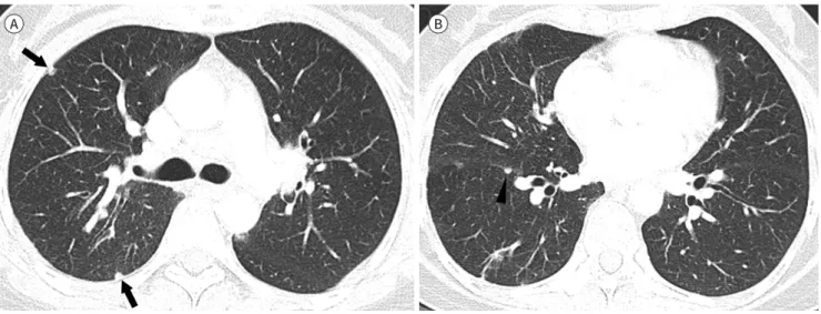
Fig. 3. A 49-year-old female with marginal zone B-cell lymphoma in the nasal cavity and a sarcoid-like reaction (patient 5).
A, B. Lung window CT scan images show well-defined subpleural (arrows) (A) and fissural (arrowhead) (B) nodules in the right lung.
A B
oped from 6 months to 70 months after the diagnosis of complete remission of malignancies.
Imaging features of sarcoid-like reaction associated with malignancies are similar to idio- pathic sarcoidosis, which are hardly distinguishable from each other (3, 13). In a study compar- ing lymph nodes of sarcoid-like reaction and metastasis by Koo et al. (14), sarcoid-like reaction had more total number of enlarged lymph nodes and tended to be bilateral and homogeneous, and their total volume was smaller than metastatic lymph nodes.
Lung parenchymal lesions are associated in 64–75% of the patients with sarcoid-like reac- tion (3, 14) and their CT findings are usually the typical features of idiopathic sarcoidosis. The dominant finding is multiple lung nodules which are usually small (less than 5 mm) and have perilymphatic distribution. Sometimes lung nodules are very small and confluent, and thus mimic ground glass opacity. Other imaging findings include ground-glass opacity or intersti- tial thickening but they are not the dominant pattern in most patients (3). These associated lung parenchymal lesions of typical features of sarcoidosis might be helpful distinguishing sarcoid-like reaction from disease progression of underlying malignancy.
In two patients of our study, the lung nodules were unique. They were large and well-defined than those usually described in sarcoid-like reaction or sarcoidosis with maximum diameter of 10–15 mm, but showed definite perilymphatic distribution with peribronchovascular, sub- pleural or fissural location (Figs. 2, 3). They seemed to be enlarged intrapulmonary lymph nodes more likely rather than lung parenchymal lesions.
Although the lung is the most commonly involved organ in sarcoid-like reaction, any organ can be involved in more extensive cases. Recently, extensive sarcoid-like reactions have been reported in melanoma patients receiving anti-PD-1 antibody therapy, involving skin, deeper soft tissues, visceral organs including the spleen, liver, pancreas and bony structures at differ- ent time points during immunotherapy, which were detected as extrathoracic 18F-FDG PET/CT abnormalities (15-17). For the clinician, it is of utmost importance to consider the possibility of the appearance of sarcoid-like during anti-PD-1 antibody therapy, to be able to distinguish it from progressive disease.
On 18F-FDG PET/CT, lymph nodes of sarcoid-like reaction show hypermetabolic uptake mim- icking malignant lesions and even the SUVmax of lymph nodes cannot differentiate those from metastatic lymph nodes (12, 14). However, as in our cases, PET/CT findings of new hypermet- abolic bilateral hilar and mediastinal lymph nodes in the absence of uptakes at the primary tumor sites and extrathoraic areas may be highly suggestive of sarcoid-like reaction.
Biopsy is essential for differential diagnosis between sarcoid-like reaction and disease pro- gression of malignancies. Endobronchial ultrasound-guided transbronchial needle aspiration is safe and minimally invasive and provides a histologic diagnosis with high sensitivity (83–
89%) and specificity (100%) (18, 19). However, the diagnosis of sarcoid-like reaction via biop- sy does not definitively rule out the possibility of coexisting malignancy and especially when there is an accompanying persistent or progressive disease, or suspicious findings such as asymmetric thoracic lymphadenopathy, coexisting extrathoracic lymphadenopathy, or a new- ly developed lung nodule, pathological reevaluation should be strongly recommended (14).
Sarcoid-like reaction is completely resolved in most cases treated with corticosteroids and spontaneous complete or partial resolution can occur in patients who are not treated (3, 12).
In our study, all patients had complete resolution after corticosteroid treatment and there was
no recurrence on follow-up studies.
Limitations of our study include its small sample size, retrospective design and selection bias. The sample size was small because of the low incidence of sarcoid-like reaction and we enrolled only the patients who were diagnosed to have sarcoid-like reaction during the surveil- lance for disease progression after the achievements of complete remission of malignancies.
In conclusion, we report relatively rare cases of sarcoid-like reaction developed after com- plete remission of malignancy in patients with several types of malignancy and variable types of antineoplastic therapy. Homogeneous bilateral hilar and mediastinal lymphadenopathy with or without small pulmonary nodules of perilymphatic distribution which are hypermet- abolic on FDG-PET/CT in the absence of tumor recurrence or other metastasis may suggest possibility of sarcoid-like reaction, and histologic confirmation of lymph nodes is needed to prevent inappropriate therapy.
Author Contributions
Conceptualization, K.H.J., K.Y.; data curation, K.H.J., B.J.Y.; formal analysis, K.H.J., K.Y., B.J.Y.; inves- tigation, K.H.J., K.Y., B.J.Y.; methodology, K.H.J., K.Y.; project administration, K.H.J., K.Y.; resources, all authors; supervision, K.Y.; visualization, K.H.J., K.Y.; writing—original draft, K.H.J., K.Y.; and writ- ing—review & editing, all authors.
Conflicts of Interest
The authors have no potential conflicts of interest to disclose.
Funding None
REFERENCES
1. Jain R, Yadav D, Puranik N, Guleria R, Jin JO. Sarcoidosis: causes, diagnosis, clinical features, and treatments.
J Clin Med 2020;9:1081
2. Brincker H. Sarcoid reactions in malignant tumours. Cancer Treat Rev 1986;13:147-156
3. Lau RK, Takasugi JE, David Godwin J, Pipavath SN. Sarcoid-like reaction-computed tomography features in 12 patients. J Comput Assist Tomogr 2015;39:143-148
4. London J, Grados A, Fermé C, Charmillon A, Maurier F, Deau B, et al. Sarcoidosis occurring after lymphoma:
report of 14 patients and review of the literature. Medicine (Baltimore) 2014;93:e121
5. Tolaney SM, Colson YL, Gill RR, Schulte S, Duggan MM, Shulman LN, et al. Sarcoidosis mimicking metastat- ic breast cancer. Clin Breast Cancer 2007;7:804-810
6. Kim MH, Lee K, Kim KU, Park HK, Lee MK, Suh DS. Sarcoidosis mimicking cancer metastasis following che- motherapy for ovarian cancer. Cancer Res Treat 2013;45:354-358
7. Powell JL, Cunill ES, Gajewski WH, Novotny DB. Sarcoidosis mimicking recurrent endometrial cancer. Gy- necol Oncol 2005;99:770-773
8. Beutler BD, Cohen PR. Sarcoma-associated sarcoid reaction: report of cutaneous sarcoid reaction in a pa- tient with liposarcoma. World J Clin Cases 2015;3:988-992
9. Cheshire SC, Board RE, Lewis AR, Gudur LD, Dobson MJ. Pembrolizumab-induced sarcoid-like reactions during treatment of metastatic melanoma. Radiology 2018;289:564-567
10. Lainez S, Tissot C, Cottier M, Vergnon JM. EBUS-TBNA can distinguish sarcoid-like side effect of nivolumab treatment from tumor progression in non-small cell lung cancer. Respiration 2017;94:518-521
11. Mitchell MA, Hogan K, Amjadi K. Atezolizumab-induced sarcoid-like granulomatous reaction in a patient with urothelial cell carcinoma. Immunotherapy 2018;10:1189-1192
12. Kaneko Y, Kato H, Matsuo M. Hilar and mediastinal sarcoid-like reaction after the treatment of malignant tumors: imaging features and natural course on 18F-FDG-PET/CT. Jpn J Radiol 2019;37:88-94
13. Hunsaker AR, Munden RF, Pugatch RD, Mentzer SJ. Sarcoidlike reaction in patients with malignancy. Radi- ology 1996;200:255-261
14. Koo HJ, Kim MY, Shin SY, Shin S, Kim SS, Lee SW, et al. Evaluation of mediastinal lymph nodes in sarcoid- osis, sarcoid reaction, and malignant lymph nodes using CT and FDG-PET/CT. Medicine (Baltimore) 2015;
94:e1095
15. Sanan P, Lu Y. Multiorgan involvement of chemotherapy-induced sarcoidosis mimicking progression of lymphoma on FDG PET/CT. Clin Nucl Med 2017;42:702-703
16. Van Willigen WW, Gerritsen WR, Aarntzen EHJG. 18F-FDG PET/CT of multiorgan sarcoid-like reaction during anti-PD-1 treatment for melanoma. Clin Nucl Med 2019;44:905-906
17. Lu Y. FDG PET/CT course of pembrolizumab-associated multiorgan sarcoidosis. Clin Nucl Med 2019;44:167- 168
18. ParmaksIz ET, Caglayan B, Salepci B, Comert SS, Kiral N, Fidan A, et al. The utility of endobronchial ultra- sound-guided transbronchial needle aspiration in mediastinal or hilar lymph node evaluation in extratho- racic malignancy: benign or malignant? Ann Thorac Med 2012;7:210-214
19. Tremblay A, Stather DR, MacEachern P, Khalil M, Field SK. A randomized controlled trial of standard vs en- dobronchial ultrasonography-guided transbronchial needle aspiration in patients with suspected sarcoid- osis. Chest 2009;136:340-346
악성종양의 완전관해 후 발생한 사르코이드증 유사 반응:
림프절 전이와의 감별진단에 유용한 CT와
18F-FDG PET/CT 소견
강현지1 · 김유경1* · 배준영1 · 장중현2 · 이수현3
목적 악성 종양의 완전 관해 후 발생한 흉강 내 림프절병증 환자에서 사르코이드증 유사 반 응을 시사할 수 있는 영상의학적 특징을 알아보고자 한다.
대상과 방법 이 연구는 악성 종양의 완전 관해 상태에서 발생한 조직병리학적으로 확인된 사 르코이드증 유사 반응을 보인 5명의 환자의 임상적 특징과 CT 및 18F-fluorodeoxyglucose (이하 FDG) PET/CT 소견을 분석하였다.
결과 기저 악성 종양으로는 유방암, 비인두암, 자궁내막암 및 림프종이 포함되며. 악성 종양 의 완전 관해와 사르코이드증 유사 반응 진단 사이의 시간 간격은 6~78개월이었다. CT 소견 으로는 양측 폐문 및 종격동 림프절병증(n = 5), 기관지혈관주위, 엽간열주위 또는 흉막하 폐 결절(1~15 mm) 및 소엽내 간질비후가 포함되었다(n = 4). 18F-FDG PET/CT는 흉강 외 FDG 흡수 없이 양측 폐문 및 종격동 림프절의 대사항진을 나타냈다(n = 3). 모든 환자에서 코르티 코스테로이드 치료 후 사르코이드증 유사 반응이 호전되었다.
결론 악성 종양의 완전 관해를 달성한 환자에서, 원발성 종양의 재발과 흉강 외 원격 전이 없 는 상태에서 새로 발견된 양측 폐문 및 종격동 림프절병증은 림프관주위 폐결절의 유무에 관 계없이 사르코이드증 유사 반응의 가능성을 시사할 수 있으며 불필요한 전신 화학요법을 예 방하기 위해 림프절의 조직병리학적 확인이 필요하다.
이화여자대학교 의과대학 1영상의학과, 2호흡기내과,
3국립암센터 영상의학과