Radiologic Evaluation for Resectability of
Pancreatic Adenocarcinoma
췌장 선암의 절제 가능성 평가
Shin Hye Hwang, MD
1,2, Mi-Suk Park, MD
3*
1Department of Radiology, National Health Insurance Service Ilsan Hospital, Goyang, Korea
2Department of Radiology, Yongin Severance Hospital, Yongin, Korea
3Department of Radiology, Severance Hospital, Yonsei University College of Medicine, Seoul, Korea
Imaging studies play an important role in the detection, diagnosis, assessment of resectability, staging, and determination of patient-tailored treatment options for pancreatic adenocarcino- ma. Recently, for patients diagnosed with borderline resectable or locally advanced pancreatic cancers, it is recommended to consider curative-intent surgery following neoadjuvant or pallia- tive therapy, if possible. This review covers how to interpret imaging tests and what to consider when assessing resectability, diagnosing distant metastasis, and re-assessing the resectability of pancreatic cancer after neoadjuvant or palliative therapy.
Index terms Carcinoma, Pancreatic Ductal; Neoadjuvant Therapy; Surgery
서론
췌장암은 전체 5년 생존율이 6.8~12.6%으로 낮은 악성 종양으로(1, 2), 대한민국 암 발생 자 수 8위이며 5번째로 흔한 암 관련 사망 원인이다(3, 4). 외과적 절제술이 완치를 기대할 수 있는 유일한 치료법이나 대부분 환자는 국소 진행 혹은 전이 상태에서 진단되기 때문에, 새 로 진단된 환자의 15~20%만이 절제술의 대상이 된다(5). 그러나 절제술 이후에도 5년 생존 율이 20% 미만이며(5), 수술에 따른 주요 합병증을 동반하는 비율이 높다. 따라서 수술 대상 환자를 선별하고 적절한 치료 방법을 결정하는 것이 중요하다. 전산화단층촬영(computed tomography; 이하 CT)은 췌장암의 진단, 절제 가능성 및 원격 전이 여부를 평가하는데 일 차적인 검사이며, 선행 보조치료(neoadjuvant therapy) 후 절제 가능성을 재평가하는 데에 도 널리 사용된다. 자기공명영상(magnetic resonance imaging; 이하 MRI)은 CT에 보조적 혹은 대안적으로 시행된다. 본 종설에서는 췌장암의 국소 절제 가능성 평가, 원격 전이의 진 단 및 선행 보조 치료 후 절제 가능성의 재평가에 대한 최근까지의 영상의학 분야의 연구 결
Received January 29, 2021 Revised March 15, 2021 Accepted March 17, 2021
*Corresponding author Mi-Suk Park, MD Department of Radiology, Severance Hospital, Yonsei University College of Medicine,
50-1 Yonsei-ro, Seodaemun-gu, Seoul 03722, Korea.
Tel 82-2-2228-7400 Fax 82-2-393-3035 E-mail [email protected] This is an Open Access article distributed under the terms of the Creative Commons Attribu- tion Non-Commercial License (https://creativecommons.org/
licenses/by-nc/4.0) which permits unrestricted non-commercial use, distribution, and reproduc- tion in any medium, provided the original work is properly cited.
ORCID iDs Shin Hye Hwang https://
orcid.org/0000-0003-2367-0413 Mi-Suk Park
https://
orcid.org/0000-0001-5817-2444
과를 소개하고자 한다.
영상 검사 전산화단층촬영
CT는 뛰어난 시간 및 공간 해상도로 복강 전체를 시각화하고 3차원 재구성을 하여 다각도에서 병변과 주변 구조의 관계를 평가할 수 있기 때문에, 췌장암의 진단, 절제 가능성 평가, 병기 설정에 일차적으로 권장되는 검사이다(5, 6). 췌장암은 빠른 진행을 보일 수 있으므로, 수술을 시행하기 4 주 이내에 검사를 시행할 것이 권장된다(6).
권장되는 췌장 프로토콜 CT는 연속되는 가능하면 얇은(< 3 mm, 0.5~1 mm이 선호됨) 절편으 로 64채널 이상의 다중 검출기를 사용하는 나선형 스캔을 시행하며(helical CT), 2~5 mm 두께의 축상(axial) 절편 및 3 mm 이하의 두께로 관상면(coronal) 재건을 할 것이 권장된다. 기관에 따라 시상면(sagittal) 재건, 혈관의 평가를 위한 최대 강도 투영(maximal intensity projection) 또는 3차원 체적 두꺼운 단면 영상(three-dimensional volumetric thick section)을 추가할 수 있다.
정맥을 통해 고농도의 요오드를 함유한 조영제(> 300 mgI/L 권장)를 초당 3~5 mL 속도로 투여하 고, 췌장 실질 단계(조영제 주입 후 40~50초) 및 정맥 단계(조영제 주입 후 65~70초) 영상을 필수적 으로 촬영한다. 췌장 실질 단계에서는 췌장의 원발 종괴가 주변의 실질과 최대 대조를 보이며 인 접한 혈관 구조와의 관계를 평가하기 용이하다. 정맥 단계에서는 간을 포함한 원격 전이를 평가한 다. 대부분의 기관에서 정맥 조영제의 주입 전 검사도 함께 시행한다(Table 1).
자기공명영상
MRI는 기본적으로 CT에서 정확히 판단할 수 없는 소견이 있는 경우 시행하는 검사(problem-
Table 1. Recommended CT Protocol for Evaluation of Pancreatic Adenocarcinoma
Parameter Details
Scan type Helical (preferably at least 64 multidetector row scanner)
Acquisition section thickness Thinnest possible (< 3 mm), preferably submillimeter (0.5–1.5 mm) if available and saving source images for problem solving
Oral contrast Neutral contrast (positive contrast may compromised the 3D and MIP reformatted images)
Intravenous contrast Iodine-containing contrast agent, preferably high concentration (> 300 mgI/mL). Lower concentration contrast can be used if low kVp setting is applied
Injection rate 3–5 mL/s
Acquisition timing Pancreatic parenchymal phase (40–50 s) and portal venous phase (65–70 s) after intravenous contrast injection
Image reconstruction
Axial and multiplanar reformats (in the coronal, and per institutional preference, sagittal plane) at 2–3 mm thickness
MIP or 3D volumetric thick section images may be helpful for vascular evaluation
Adapted from National Comprehensive Cancer Network. NCCN Clinical Practive Guideline in Oncology (NCCN GuidelinesⓇ): Pancreatic adeno- carcinoma, version 1.2021. Available at. https://www.nccn.org/store/login/login.aspx?ReturnURL=https://www.nccn.org/professionals/physi- cian_gls/pdf/pancreatic.pdf (6).
solving tool)이다. 췌장암이 의심되나 CT에서 직접적으로 종괴가 관찰되지 않고 2차 징후만을 보 일 때(7), 췌장 선암 이외의 감별 진단이 필요한 비전형적인 소견이 있는 종괴일 때, 혹은 임상적인 의미가 불분명한 간 병변이 있을 때 시행된다(8). 그러나 요오드화 조영제에 심한 과민반응이 있는 등 CT를 시행하기 어려운 환자에서는 일차 검사로서 시행될 수 있다. 중증의 신부전이 있는 경우 비조영 MRI 검사를 시행할 수 있다. 권장되는 MRI 프로토콜은 Table 2와 같다.
초음파 내시경
초음파 내시경(endoscopic ultrasound; 이하 EUS)은 공간 분해능이 높고 세침흡입검사를 시 행하여 병리학적인 진단이 가능하므로, CT에서 췌장 종괴의 존재가 불분명하나 의심되는 경우, 혹은 췌장 선암 이외의 감별 진단이 필요한 경우 진단에 도움이 된다(9). 모호한 혈관 침범 혹은 림 프절 비대가 있을 때에도 절제 가능성을 평가하기 위한 추가 정보를 얻을 수 있으나(10), 병변의 위치 및 시행자에 따라 진단 성능이 다른 한계가 있으며 CT에 비교하여 우위에 있다는 확실한 근 거가 없다. 대부분의 가이드라인은 EUS를 종양 평가 과정의 일상적인(routine) 단계로 권장하지 는 않으며, 추가 정보 및 조직을 얻기 위해 시행하기를 권고한다(5, 6, 11).
췌장암의 병기
췌장암의 병기는 일반적으로 American Joint Committee on Cancer에서 개발한 TNM 시스템 을 이용한다(12). 원발 종양의 병기(T 병기)는 종양의 크기 및 주요 동맥[상장간막동맥(superior mesenteric artery; 이하 SMA), 복강 동맥(celiac artery; 이하 CA) 및 총간동맥(common hepat- ic artery; 이하 CHA)]의 침범 여부를 반영한다. 2017년에 개정된 8판에서는 췌장 주변 침범 여부 가 기준에서 제외되었고, N 병기는 전이된 림프절의 수에 따라 N1과 N2로 세분화되었다. 주요 동 맥에 침범이 없으며(T 0~3) 전이 림프절이 3개 이하인 경우(N 0~1)가 0~2기에 해당하며, 수술 후 보 조항암치료(adjuvant chemotherapy)가 일차 치료이다. 주요 동맥 침범이 있는 경우(T4) 혹은 4개
Table 2. Recommended MRI Protocol for Evaluation of Pancreatic AdenocarcinomaSequence Plane Slice Thickness
T2-weighted single shot fast SE (SSFSE/HASTE) Coronal with or without axial < 6 mm
T1-weighted In- and opposed- phase GRE Axial < 6 mm
T2-weighted fat-suppressed fast SE Axial < 6 mm
DWI Axial < 6 mm
Pre and dynamic post intravenous contrast administration 3D T1-weighted
fat-suppressed GRE (in pancreatic, portal venous, and equilibrium phasese) Axial Thinnest possible 2–3 mm (4–6 mm if overlapping)
T2-weighted MRCP (prefereably 3D, FRFSE) Coronal < 3 mm
Adapted from National Comprehensive Cancer Network. NCCN Clinical Practive Guideline in Oncology (NCCN GuidelinesⓇ): Pancreatic adeno- carcinoma, version 1.2021. Available at. https://www.nccn.org/store/login/login.aspx?ReturnURL=https://www.nccn.org/professionals/physi- cian_gls/pdf/pancreatic.pdf (6).
DWI = diffusion-weighted imaging, FRFSE = fast relaxation fast SE, GRE = gradient echo, HASTE = half-Fourier acquisition single-shot turbo SE, MRCP = magnetic resonance cholangiopancreatography, SE = spin echo, SSFSE = single-shot fast SE, 3D = three-demensional
이상의 림프절 전이가 있는 경우(N2) 3기, 전이가 있는 경우 4기(M1)이다. 3기 환자는 영상 검사에 서 평가된 절제 가능성 및 환자의 전신 상태에 따라 다양한 치료 옵션을 선택할 수 있다.
국소 절제 가능성의 평가
췌장암 수술의 일차적 목표는 잔존 종양이 없는 완전한 종양의 절제이며, 절제연 음성(margin- negative, R0)은 현미경으로 관찰하여 절제연의 1-mm 이내에 종양 세포가 없는 것을 의미한다 (13). R0 절제술 이후에도 5년 생존율은 18~24%로 불량하나 절제연 양성(R1 또는 R2) 절제술을 시 행한 경우보다는 예후가 좋다(14). 2006년에 MD Anderson Cancer Center에서 처음으로 국소 절제 가능성을 평가하는 기준을 발표하였고(15), 현재 가장 널리 사용되는 기준은 미국 국립 종합 암센터 네트워크(National Comprehensive Cancer Network; 이하 NCCN)에서 제안하였다(6).
시스템 간에 다소 정의의 차이는 있으나, 기본적으로 원격 전이가 없는 췌장암은 1) 표준 술식으로 R0 절제술을 달성할 수 있을 것으로 기대되는 경우(절제 가능, resectable), 2) 기술적으로 절제는 가능하나 주변의 중요 구조를 병합 절제하지 않으면 절제연 양성(R1)일 가능성이 높다고 생각되 는 경우(경계절제성, borderline resectable), 그리고 3) 국소 절제가 어려운 경우(국소 진행, lo- cally advanced)로 분류된다. 절제연 양성일 경우, 조기 재발이나 혈관 병합 절제로 인한 수술 후 합병증의 가능성이 높아진다(15). 최근 경계절제성 혹은 국소 진행 상태의 췌장암 환자군을 대상 으로 선행 보조 치료 또는 완화 치료가 발전함에 따라 선행 보조 치료 후 R0 절제를 시도하는 경우 가 많아지고 있으며, 주요 가이드라인들도 경계절제성일 경우 선행 보조 치료를 우선 고려할 것을 추천한다(5, 6, 16, 17).
혈관 절제술
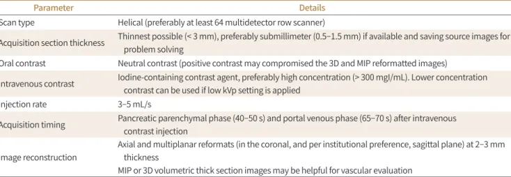
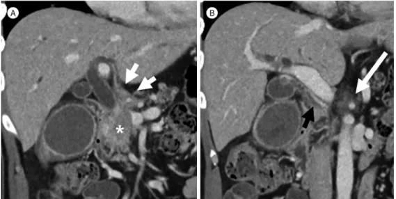
CT에서 혈관과 종양으로 의심되는 연조직 사이에 지방 경계면이 소실되었을 때, 혈관 직경의
Fig. 1. The abutment of the SMA.A, B. Axial (A) and coronal (B) images of CT show a hypoattenuating mass (asterisks) in the pancreatic head and perivascular fat stranding (arrows) abutting posterior aspect of SMA less than 180°. The CBD is dilated (arrowhead) due to tumor invasion of intrapancreatic CBD. Margin-negative resection was achieved.
CBD = common bile duct, SMA = superior mesenteric artery
A B
180°를 기준으로 “abutment (≤ 180°, Fig. 1)”와 “encasement (> 180°)”로 분류한다(Fig. 2) (15).
1) abutment가 있으면서 혈관 모양의 변형이 있는 경우, 혹은 2) encasement가 있는 경우는 혈 관을 침범하였을 가능성이 높아서 혈관을 절제하지 않으면 R0 절제가 어려울 것으로 예상한다. 혈 관 모양의 변형 없이 abutment만 있는 경우는 혈관 침범의 가능성이 불확실하여(equivocal) 절 제 후 R0를 달성할 수도 있는 상태이다.
SMA의 절제는 일부 기관에서 자체적인 기준에 따라 시도하기도 하지만, 일반적으로는 시행되 지 않는다. CT에서 encasement가 있을 경우 절제 불가이며, abutment가 있을 경우 경계절제성 이다. SMA는 많은 림프절 및 신경총에 둘러싸여 있어 침범이 있을 경우 근치적 절제가 어렵다. 또 한 절제 후 문합부에서 혈전이 발생하여 장 허혈이 생기거나 혈관 주위 신경을 광범위하게 절제하 여 조절되지 않는 설사 등의 합병증이 발생하면 수술 후 환자의 삶의 질이 낮아지고 보조 항암치 료를 시행하기가 어렵다. 현재까지의 연구 결과에서 절제에 따른 생존율의 이득은 밝혀지지 않았
Fig. 2. Vascular encasement and distant metastasis.A. Pancreatic head cancer (asterisk) encases the superior mesenteric vein (long arrow) and invades the right gastroepiploic vessels. The possibility of carcinomatosis is suspected due to subtle nodular lesions of the omentum (short arrow).
B. There is no evidence of liver metastasis in CT.
C. A tiny hyperintense hepatic lesion is detected in the MRI (arrow).
D. On follow-up CT image after 2 months, the hepatic lesion is proven to be metastatic (arrow).
C
A B
D
으며, 수술 후 합병증 및 사망률은 증가한다(18-20).
CA의 경우, 두부암 환자에서는 abutment가 있을 경우 경계절제성, encasement가 있을 경우 절제 불가능하다. 두부암 수술 시에는 위십이지장동맥(gastroduodenal artery; 이하 GDA)을 반 드시 결찰해야 하기 때문에, SMA에서 위와 간에 도달하는 혈류 우회로가 차단되기 때문이다. 체 부 및 미부암 환자에서는 CA encasement가 있을 때에도 뒤에서 설명할 복강동맥 절제를 포함하 는 원위췌장절제술(distal pancreatectomy with en-bloc celiac axis resection; 이하 DP-CAR, Appleby procedure)을 시행할 조건을 만족하는 경우, NCCN 가이드라인은 경계절제성으로 분류 한다. 그러나 기관과 전문가에 따라 절제 불가능으로 간주할 수 있다고 제시한다. 메타 분석상 DP- CAR를 시행한 경우 R0 절제율은 72.8%이며(21), CA 침범이 없이 표준적인 절제술을 시행한 환자 에 비하여 3년 생존율에 유의미한 차이가 없었고(22, 23), 절제술을 시행하지 못한 환자에 비하여 서는 1년 생존율이 높았다(22, 23). 그러나 수술에 관련된 합병증(간, 담낭 및 위 허혈, 신경총의 박 리로 인한 지연 위배출 등)의 발생 위험이 높다(21, 22, 24). 또한 관련된 연구들은 종양이 혈관을 침범한 정도나 수술 술기가 다양하였고, 일부 연구는 DP-CAR 후에 재발률이 높다고 보고하였다 (25). 어떤 환자군에서 선행 보조 치료 후 절제술보다 일차적인 DP-CAR가 도움이 될지는 아직 정 해진 기준이 없다.
간문맥(portal vein; 이하 PV) 및 상장간막정맥(superior mesenteric vein; 이하 SMV)의 병합 절제(Fig. 3)는 수술 의사의 재량에 따라 재건이 가능한 범위에서 시행된다. 약 4~5 cm까지는 단 단문합(end-to-end anastomosis)을 시행할 수 있고, 그 이상의 경우 이식편(graft)을 사용한다 (26). 정맥절제술을 받은 환자는 표준적인 절제술을 받은 환자에 비해 생존율이 낮고(27) 수술 관 련 합병증이 많은 경향이 있다(27, 28). 그러나 정맥절제술을 받는 환자군의 수술 전 종양이 좀 더 크고 국소적으로 진행된 상태인 경우가 많아, 예후에 영향을 주는 것으로 해석된다(27). 정맥병합 절제를 시행한 환자만을 대상으로 살펴보면 R0 절제를 달성할 경우 R1 절제보다 5년 생존율이 좋 다(29). 따라서 R0 절제가 가능할 것으로 예측되는 환자군에서는 예후 향상을 위해 정맥병합절제
Fig. 3. Deformity and segmental resection of the SMV.
A-C. Axial (A, B) and coronal (C) images of CT show deformity with segmental luminal narrowing of SMV (arrows) due to the invasion of the pancreatic head cancer (asterisk). Pylorus preserving pancreaticoduodenectomy with segmental resection of SMV achieved margin-negative.
SMV = superior mesenteric vein
A B C
를 고려할 수 있다.
절제 가능성을 분류하는 시스템 간의 주요 차이
가장 널리 사용되는 NCCN 가이드라인은 미국 복부영상의학회(Society of Abdominal Radiol- ogy)와 미국 췌장협회(American Pancreatic Association)의 합의문, 그리고 유럽 종양학회(Eu- ropean Society for Medical Oncology) 가이드라인에서 채택되었다(5, 30). 이외에 MD Ander- son Cancer Center 기준(15), American Hepato-Pancreato-Biliary Association (이하 AHPBA)/
Society of Surgical Oncology (이하 SSO)/Society for Surgery of the Alimentary Tract (이하 SSAT)가 공동으로 제안한 분류(31), ALLIANCE trial의 분류(32), Japan Pancreas Society 분류 (11) 등이 있다. 2016년에는 International Association of Pancreatology (이하 IAP)에서 해부 학적인 기준은 일본 가이드라인과 동일하나, 이외에 혈청 carbohydrate antigen (이하 CA 19-9)
> 500 U/mL이나 환자의 상태(Eastern Cooperative Oncology Group performance status) 등
Table 3. Criteria Defining Resectability Status According to National Comprehensive Cancer Network Guideline (Version 1.2021)
Resectability Status Arterial Venous
Resectable No arterial tumor contact (CA, SMA, or CHA) No tumor contact with the SMV or PV or ≤ 180°
contact without vein contour irregularity
Borderline resectable
Pancreatic head/uncinated process:
Solid tumor contact with CHA without extension to CA or hepatic artery bifurcation allowing for safe and complete resesection and reconstruction
Solid tumor contact with SMA of ≤180°
Solid tumor contact with variant arterial anatomy (ex: accessory right hepatic artery, replaced right hepatic artery, replaced CHA, and the origin of replaced or accessory artery) and the presence and degree of tumor should be noted if present, as it may affect surgical planning Pancreatic body/tail:
Solid tumor contact with the CA of ≤ 180°
Solid tumor contact with the CA of > 180° without involvement of the aorta and with intact and uninvolved GDA thereby permitting a modified Appleby procedure (some panel members prefer these criteria to be in the locally advanced category)
Solid tumor contact with the SMV or PV of > 180°, contact of ≤ 180° with contour irregularity of the vein or thrombosis of the vein but with suitable vessel proximal and distal to the site of involvement allowing for safe and complete resection and vein reconstruction
Solid tumor contact with the IVC
Locally advanced
Head/on cine weight process:
Solid tumor contact with SMA > 180°
Solid tumor contact with the CA > 180°
Pancreatic body/tail:
Solid tumor contact of > 180° with the SMA or CA Solid tumor contact with the CA and aortic involvement
Unreconstructible SMV/PV due to tumor involvement or occlusion
(can be due to tumor or bland thrombus)
Adapted from National Comprehensive Cancer Network. NCCN Clinical Practive Guideline in Oncology (NCCN GuidelinesⓇ): Pancreatic adeno- carcinoma, version 1.2021. Available at. https://www.nccn.org/store/login/login.aspx?ReturnURL=https://www.nccn.org/professionals/physi- cian_gls/pdf/pancreatic.pdf (6).
CA = celiac axis, CHA = common hepatic artery, GDA = gastroduodenal artery, IVC = inferior vena cava, PV = portal vein, SMA = superior mesen- teric artery, SMV = superior mesenteric vein
임상적인 기준을 추가한 기준을 제시하였다(33).
가이드라인 간에 기본적인 절제 가능성 판단 기준은 거의 비슷하다. 가장 큰 차이는 NCCN 가이 드라인에 따르면 CA의 encasement가 있는 환자의 일부가 경계절제성으로 분류(DP-CAR 적응 증) 될 수 있다는 점이다(Table 3). MD Anderson, AHPBA/SSAT/SSO 및 IPA의 기준은 이를 절제 불가능으로 분류하며, AHPBA/SSAT/SSO 기준은 CA의 abutment도 절제 불가능으로 분류한다.
NCCN이 비교적 최근에 개정이 되었고 이 항목은 2017년 개정에서 변경된 부분이기 때문에, 그동 안의 수술 기술 및 연구 성과를 반영하여 발생한 차이로 보인다. 그리고 NCCN 가이드라인 역시 이러한 경우 전문가에 따라 절제 불가능으로 보기도 한다고 기술하고 있다. SMA 및 CHA의 침범 은 가이드라인마다 기술 방식에 차이가 있으나 CHA의 분절 절제를 고려할 수 있다는 내용상에는 큰 차이가 없다. 정맥 침범 역시 외과적으로 재건 가능한지가 공통적인 판단의 기준이다. 다만 NCCN 가이드라인은 모양 변화가 없는 abutment를 절제 가능으로 분류하였고 AHPBA/SSAT/
SSO 가이드라인은 이를 경계절제성으로 분류한다. IAP 및 일본 가이드라인은 정맥 침범이 십이 지장의 하연보다 아래로 확장되면 재건이 어렵다고 기술하고 있다.
실제로는 경계절제성의 정의는 기관의 경험 및 수술 기술에 따라 다르게 적용된다.
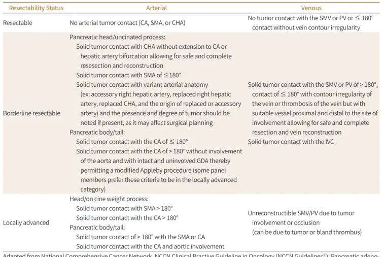
혈관 침범의 평가
기본적으로 CHA, CA, SMA 및 대동맥에 관하여 각각 평가가 이루어져야 한다(Fig. 4A). 췌장 체 부나 미부암이 다른 주요 동맥은 침범하지 않고 비장동맥(splenic artery)만 침범한 경우는 비장 의 병합 절제가 가능하므로 이는 절제 가능성 평가에 영향을 미치지 않는다. GDA의 기시부 침범 은 절제연 음성을 얻으며 CHA와 분리해내기 어렵기 때문에 CHA 침범과 같은 등급으로 간주한 다. 마찬가지로 CA의 대동맥 기시부가 침범된 경우도 대동맥 침범에 준한다. NCCN 가이드라인 에서 제안하는 경계절제성과 국소 진행암의 판정 기준을 요약하면 Table 3과 같다.
동맥의 병합 절제는 합병증을 증가시키나 환자의 예후를 개선하지는 못한다(18, 19). 수술 전 영
Fig. 4. Usual arterial anatomy and variations in CT angiography.
A-C. Usual anatomy of CA (A), replaced right hepatic artery (arrow) from the SMA (B), and (C) common celiaco-mesenteric (CA-SMA) trunk (ar- rowhead).
CA = celiac artery, SMA = superior mesenteric artery
A B C
Fig. 5. Hepatic artery invasion in CT.
A. Pancreatic head cancer (asterisk) encases the common and left hepatic artery (arrows).
B. The celiac artery is also encased by cancer (white arrow). Replaced right hepatic artery from the superior mesenteric artery is intact (black arrow).
상 검사에서 주요 혈관에 분명한 침범이 의심될 경우에는 NCCN 가이드라인(6)을 기준으로 다음 의 제한적인 경우에만 동맥 절제를 포함한 수술을 고려할 수 있다(경계절제성). 췌장 두부암이 CHA를 2 cm 미만으로 침범하고 있을 경우에는 CHA의 분절 절제(segmental resection) 및 재건 을 시도할 수 있다. 이 경우에는 간동맥 분기부(bifurcation) 혹은 CA의 침범은 없어야 한다(Fig. 5).
또한 체부 및 미부암 환자의 일부에서 선택적으로 DP-CAR (Appleby procedure)를 일차 치료로 고려할 수 있다. 이 수술을 시행한 후 간과 위로 가는 혈류는 SMA 및 췌십이지장동맥궁(pancre- atoduodenal arcade)의 우회로를 통해 전달되기 때문에, GDA, 고유간동맥(proper hepatic ar- tery) 및 좌·우간동맥에 침범이 없는지와 SMA 기시부에 심한 협착이 없는지를 확인하여야 한다.
CA의 기시부까지 침범하여 대동맥에서 분리하여 절제하기 어려운 경우는 DP-CAR의 적응증이 되지 않는다.
주요 혈관의 변이 혹은 CA나 SMA의 기시부에 협착이 있는 경우 수술 계획에 영향을 주므로 반 드시 평가되어야 한다. 이 부위에서 가장 흔한 해부학적 변이는 replaced right hepatic artery이 며, 이외에도 replaced or accessory left hepatic artery, common celiaco-mesenteric (CA-SMA) trunk, common hepatico-mesenteric (CHA-SMA) trunk의 여부 등을 확인한다(Fig. 4B, C).
절제에 영향을 미치는 주요 정맥은 PV과 SMV이다. 이들은 기본적으로 침범이 의심되더라도 병합 절제 및 재건을 시도할 수 있다(절제 가능 혹은 경계절제성). 그러나 종양의 침범 혹은 내부 의 혈전이 매우 긴 분절을 차지하는 경우 재건이 어렵기 때문에 절제 불가능으로 분류된다. 하대 정맥(inferior vena cava)과 종양의 abutment가 있는 경우 NCCN 가이드라인은 경계절제성으로 분류한다.
A B
국소 절제 가능성을 평가하는 영상 검사의 성능
Bipat 등(34)이 보고한 메타 분석(1990~2003)에서는 절제 가능성에 대한 나선형 CT와 MRI의 민 감도[81%(95% 신뢰구간 76~85) vs. 82%(95% 신뢰구간 69~91)] 및 특이도[82%(95% 신뢰구간 77~87) vs. 78%(95% 신뢰구간 63~87)]는 유의한 차이가 없었다. 다른 메타 분석(1990~2013)에서 도 혈관 침범을 진단하는 CT와 MRI의 민감도[68%(95% 신뢰구간 55~79) vs. 62%(95% 신뢰구간 48~74)] 및 특이도[97%(95% 신뢰구간 94~98) vs. 96%(95% 신뢰구간 93~98)]는 유의한 차이가 없 었다(35). 하지만 한 전향적 연구에서 CT는 혈관 침범과 절제 가능성을 평가하는 음성 예측도(neg- ative predictive value) 및 정확도가 MRI나 EUS에 비하여 우수했다(36).
수술 전 CT의 절제 가능성을 NCCN (version 1.2017) 가이드라인에 따라 평가한 최근 연구에 따 르면 절제 가능, 경계절제성 및 국소 진행성 카테고리에서 R0 절제율이 각각 73%, 55%, 및 16%였 다(37). 주요 정맥의 abutment가 있는 경우(NCCN 에서 절제 가능암으로 분류), 정맥과 abut- ment가 없는 환자에 비해 R0 절제율이 낮았다(59% vs. 80%). 혈관 침범을 기준으로 한 절제 가능 성 평가 이외에도, 종양의 크기(> 4 cm)가 R1 절제와 유의미한 상관이 있었다.
절제 가능성을 분류하는 관찰자 간의 일치(interobserver agreements)에 대하여서는 서로 상 반된 연구 결과가 보고되어 있다. 한 연구는 NCCN 가이드라인을 이용하였을 때 관찰자의 경험에 상관없이 높은(very strong agreement, intraclass correlation coefficient = 0.82) 수준의 관찰자 간 일치를 보인다고 보고했으나(38), 다른 연구에서는 중간(moderate, κ = 0.48) 수준의 관찰자 간 일치를 보였고, 경험이 많은 영상의학과 의사 간의 일치도가 더 높았다[κ = 0.55 (95% 신뢰구간 0.50~0.60) vs. 0.43 (95% 신뢰구간 0.38~0.49)] (39).
림프절 및 원격 전이의 평가
림프절 전이는 주로 단경을 측정하여 평가한다. 원격 전이는 간과 복막에 호발하며, 복강 외에 서는 폐 전이가 가장 흔하다(40).
림프절 전이
표준적인 췌십이지장절제술 시에 십이지장 및 췌장 주위, 전·후 췌십이지장 림프절, 간십이지장 인대(hepatoduodenal ligament) 및 SMA의 우측의 림프절이 함께 절제된다. 수술 후 재발률을 낮추기 위하여 CA 및 하장간막동맥 기시부 상방까지의 림프절을 포함하는 확대림프절절제술(ex- tended lymphadenectomy)이 시도되었다. 그러나 생존율에 이득이 없으며 합병증과 사망률은 증가하기 때문에(41), 현재 일상적인 확대절제술은 권장되지 않는다(6).
영상의 발달에도 불구하고, 수술 전 영상에서 림프절 전이를 평가하는 것은 여전히 한계가 있
다. 고전적으로 CT에서 단축 직경이 10-mm 이상인 림프절을 양성으로 보고하기 때문에 작은 림
프절 전이를 진단하기 어려우며(Fig. 6), 이 경우 진단의 민감도는 14~22%, 특이도는 57~85%이다
(42, 43). Positron emission tomography/computed tomography (이하 PET/CT)는 림프절 전이
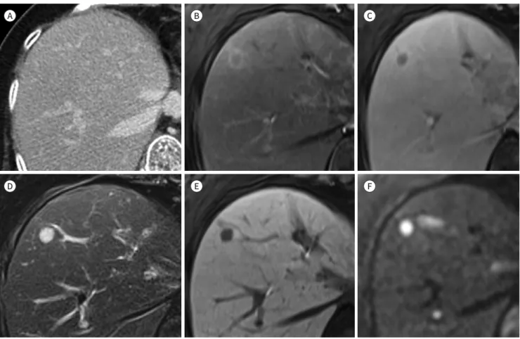
Fig. 7. Added-value of MRI for the detection of liver metastasis.
A-F. There is no evidence of hepatic lesion in CT (A). In MRI, arterial (B) and delayed phase (C) images show a rim-enhancing hepatic lesion at segment 8 of the liver. It shows hyperintensity in T2-weighted images (D), hypointensity in the hepatobiliary phase (E), and diffusion restric- tion (F).
A B C
CT를 보조적으로 사용할 경우 작은 림프절 전이 평가에 도움이 될 수 있다.
간 전이
간 전이는 주로 저혈관성 종괴로 관찰되며, 췌장 두부암의 경우에는 담도 폐쇄에 동반된 미세
Fig. 6. Margin-negative resection of borderline resectable pancreatic head cancer with aortocaval lymph node metastasis.
A, B. Axial (A) and coronal (B) images of CT show encasement of the gastroduodenal artery (arrows) by the pancreatic head cancer sparing the common hepatic artery.
C. An oval lymph node (long arrow) with a diameter of 9-mm is seen in aortocaval space.
A
D
B
E
C
F
간농양과의 감별이 필요하다. 간 전이의 평가는 MRI가 CT보다 우수한 성적을 보인다(Figs. 2, 7).
Diffusion-weighted imaging를 포함하는 간 MRI (세포 외 조영제)를 이용하면 CT에서 간 병변 이 보이지 않는 환자의 약 1.5~10.2%에서 간 전이가 발견되었다(8, 46). 또한 CT에서 전이 여부가 불분명한 간 병변이 있는 환자가 MRI를 시행할 경우 약 10.5~13.6%(세포 외 조영제)에서 32%
(간세포 특이 조영제, gadolinium ethoxybenzyl diethylene-triamine pentaacetic acid; Gd- EOB-DTPA)가 전이로 진단되었다(46, 47). MRI는 특히 작은 병변(≤ 1 cm)에서 도움이 된다(46).
메타 분석에 따르면, 통계적인 유의성을 보이지는 않았으나 MRI의 간 전이 진단의 민감도 [85%(95% 신뢰구간 74~92)]는 CT [75 (95% 신뢰구간 55~94)]보다 높고, 두 검사의 특이도는 유의 한 차이가 없었다[98%(95% 신뢰구간 78~100)] vs. 94%(95% 신뢰구간 84~100)] (48).
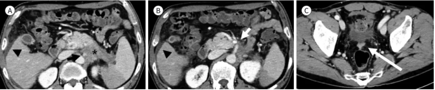
복막 전이
복막 전이는 국소 종괴, 복막 지방의 불규칙적인 결절성 침윤, 혹은 복수를 동반하거나 동반하 지 않는 복막의 결절성 비후 등으로 관찰된다(Fig. 8). 환자의 호흡, 장의 운동 인공물(motion arti- fact), 검사의 해상도 및 스캔 범위의 차이로 인하여 CT가 복막 전이의 진단에 MRI보다 우수한 성 능을 보인다(49). PET/CT가 진단에 도움이 될 수 있다(50, 51).
폐 전이
췌장암이 새로 진단된 환자에서 폐 전이를 배제하기 위하여 흉부 X선 혹은 흉부 CT를 촬영할 수 있다. 병기 설정에 흉부 검사를 필수적으로 시행해야 하는가에 대해서는 가이드라인마다 권고 사항에 차이가 있다. 미국 NCCN 가이드라인과 영국 National Institute for Health and Care Ex- cellence 가이드라인, 프랑스 가이드라인은 흉부 CT를 기본 병기 설정 검사로 추천한다(6, 52, 53).
American Society of Clinical Oncology 가이드라인은 잠재적으로 완치 가능한(potentially cur- able) 환자의 경우 흉부 X선을 시행할 수 있다고 제시하였으며, 국소 절제가 불가능하거나 전이가 있는 경우에는 흉부 CT를 반드시 시행하여야 한다고 권고한다(16, 54). 이 주제에 관련하여서는 소수의 연구들이 시행되었다. 췌장암의 진단 당시 다른 곳에 전이 없이 분명한 폐 전이가 관찰되 는 경우는 3.3%였다(55). 국소적으로 절제 가능 혹은 경계절제성인 경우 흉부 CT에서 폐 전이 등
Fig. 8. Pancreatic tail cancer with peritoneal metastasis in CT.
A, B. Pancreatic tail cancer (asterisk) encases the celiac axis (black arrow) and superior mesenteric artery (white arrow). A small amount of perihepatic ascites is noted (arrowheads).
C. The peritoneal metastatic nodule is seen in the rectovesical pouch (long arrow).
A B C
치료 방침에 영향을 주는 소견이 발견되는 경우는 1% 미만이었다(56). 초진단 당시 불확정(inde- terminate) 폐결절은 약 10%의 환자에서 발견되며, 그중 약 16%가 수술 후 추적 검사상 크기가 증가하여 폐 전이로 확인되었다(57). 그러나 진단 당시 불확정 결절을 가진 환자군과 대조군 간에 추적관찰 기간 중 폐 전이 발생 비율(4~16% vs. 1~13%) (55, 58) 및 전체 생존율에는(55, 57) 의미 있는 차이가 발견되지 않았다. 위 연구들은 모두 단일 기관 후향적 연구이며 비균질한 환자군을 대상으로 한다. 원발 종양의 T 병기나 종양표지자 수치 등에 따라 원격 전이 위험도를 평가하여, 흉부 CT를 수행하여 이득이 될 만한 환자들을 선별할 수 있는 잘 설계된 후속 연구가 필요하다.
수술 전 선행 보조 치료
진단 당시 불완전 절제의 위험이 높은 경우에는 수술 전 선행 보조 치료가 권장된다(5, 6). 선행 항암방사선치료(neoadjuvant chemoradiotherapy) 혹은 선행 항암치료(neoadjuvant chemo- therapy)는 경계절제성암에서 절제연 음성(R0)의 근치적 절제 가능성을 높이고, 수술 후 조기 전 이 혹은 빠르게 재발할 위험이 높은 환자들에게 불필요한 수술의 위험을 감소시키려는 목적으로 시행된다(59). 또한, 췌장암은 R1 절제율 및 수술 후 재발률이 높아(60) 보조 항암치료가 필수적임 에도 불구하고 전신 상태의 악화 등으로 인하여 전체 수술 환자의 1/3~1/2 가량이 보조 항암치료 를 받지 못하기 때문에(61, 62), 수술 전에 항암치료를 시행하여 생존율을 높이려는 목적으로 시행 되기도 한다(63, 64).
110명의 경계절제성 췌장암 환자를 대상으로 한 국내의 전향적 무작위 배정 2/3상 다기관 연구 에서 선행 항암방사선치료는 R0 절제율 및 2년 생존율을 증가시켰다(65). 246명의 환자를 대상으 로 한 유럽의 전향적 무작위 배정 3상 연구(PROPENAC)는 통계적으로 유의한 전체 생존율 향상 을 입증하지는 못했으나( p = 0.096), R0 절제율 및 무병 생존율(disease-free survival)이 향상됨을 보고하였다(66). 선행 항암치료도 역시 긍정적인 성적을 보고하고 있으나(67-69), 사용된 항암치료 의 종류 및 대상 환자군이 불균질하여 추가 연구가 필요하다.
진단 당시 영상의학적으로 절제 가능한 경우에서도 CA 19-9 수치가 매우 높거나 종양의 크기가 큰 경우, 큰 국소 림프절 전이 등으로 재발의 위험이 높을 것으로 예상되는 경우 선행 보조 치료를 시도할 수 있다(6). 또한 영상 소견에서 원격 전이가 의심되나 명확하지는 않은 경우, 혹은 내과적 동반 질환으로 인하여 전신 상태가 악화되어 즉각적인 수술이 어려운 경우에 호전을 기대하며 선 행 보조 치료를 시행할 수 있다. 절제 가능한 췌장암 환자들을 대상으로 한 선행 보조 치료와 우선 적 수술을 비교하는 연구는 아직 후향적 혹은 2상 연구 결과만이 보고되어 있으며, 3상 연구는 진 행 중이다(70).
선행 보조 치료 후 절제 가능성의 평가
췌장암은 일반적으로 종양 자체가 현저한 결합조직 형성반응(desmoplastic stromal re-
sponse) 보이며 선행 보조 치료 후에 종양 내 섬유화가 발생하여 잔존암이 있을 경우 곳곳에 산재
하게 된다. 또한 인접한 비종양성 췌장에도 역시 위축 및 섬유화가 발생하기 때문에, 선행 보조 치
료 후 발생하는 염증 반응 및 섬유화와 잔존암을 구별하는 것은 병리학적으로도 쉽지 않다(71, 72).
따라서 선행 보조 치료 후 영상에서 절제 가능성을 평가하는 것은 도전적인 과제이다. 재평가에는 주로 CT가 사용된다. 선행 보조 치료 후 영상 검사에서 병기가 개선되는(downstaging) 경우는 드 물며(73-75), CT 소견은 절제 불가능성을 과대평가하는 경향이 있다(76-78). Cassinotto 등(77)은 총 80명의 환자를 포함한 환자-대조군 연구에서, 국소 진행암으로 선행 보조 치료를 받은 후 R0 절 제 가능성을 예측하는 CT의 정확도는 58%이고 절제 가능하여 우선적 수술 치료를 시행한 경우 (83%) 보다 정확도가 낮으며, 주로 혈관 침범을 과대평가하게 되고 치료에 반응이 있더라도 영상 에서 종양의 크기가 크게 감소하지 않기 때문이라고 보고하였다. 진단 시 경계절제성으로 선행 보 조 치료 후 영상에서 호전이 없으나 수술을 시행했던 환자의 77.7%에서(73), 그리고 국소 진행암 으로 fluorouracil, leucovorin, irinotecan, and oxaliplatin (FOLFIRINOX) 선행 보조 치료 후에 도 영상에서는 절제 불가능해 보이나 수술을 시행 받은 환자의 92%에서(78) R0 절제가 가능했다 고 보고되었다. Dhir 등(79)은 96개 연구의 메타 분석에서, 선행 보조 치료 후 전체의 64%의 환자 들이 탐색적 수술(surgical exploration)을 시행 받아 그중 대부분(91%, 전체의 55%)에서 절제술 이 시행되었으며, 절제술을 시행한 환자의 85%(전체 환자의 45%)에서 R0 절제가 가능하였다고 보고하였다. R0 절제율은 초 진단 시 절제 가능성 평가 기준에 따라 차이를 보였다.
NCCN 가이드라인은 선행 보조 치료 후 원격 전이가 없을 경우 수술을 고려할 수 있다고 권고 한다(6). 진단 당시 절제 가능 혹은 경계절제성 췌장암의 경우, 선행 보조 치료 후 영상에서 저명한 암의 진행이 없이 혈청 CA 19-9 수준이 비슷하거나 감소한 경우, 수술을 고려할 수 있다(6, 80). 진 단 당시 국소 진행암이었던 경우, 혈중 CA 19-9 수준이 초기의 절반 이하로 감소하고 임상적인 호 전이 있는 경우 수술을 고려할 수 있다.
일부 연구자들은 선행 보조 치료 후 절제 가능성을 평가하는 새로운 영상 기준을 제시하였다.
종양과 주요 혈관 (SMA, SMV 및 PV)의 접촉이 변화 없거나 일부 감소하는 경우(81, 82), 선행 보 조 치료 후 NCCN 가이드라인의 경계절제성의 진단기준을 만족시키는 경우(83), 혈관과 접하는 각도, 길이 및 혈관의 변형을 함께 고려하는 경우(84), 혹은 개별 혈관을 평가하는 것이 아니라 전 반적인 판독자의 판단에 따라 결정하는 경우(85), NCCN 가이드라인 등 기존의 기준을 이용하는 것보다 절제 가능성을 더 정확히 평가할 수 있다고 보고되었다. 메타 분석에서, 선행 보조 치료 후 에 기존 가이드라인의 기준을 사용하는 경우 CT의 절제 가능성을 진단하는 민감도와 특이도는 각 각 45%(95% 신뢰구간 19~73) 및 85%(95% 신뢰구간 65~94)로, 민감도가 낮다(86). 반면에 여러 연구자들이 제시한 변경된 기준을 사용하는 경우 민감도 81%(95% 신뢰구간 71~98) 및 특이도 42%(95% 신뢰구간 28~57)로 절제 가능한 환자를 선별할 수 있다.
치료 후 종양-혈관면 경계를 평가하는 새로운 영상 기준을 제시하는 현재까지의 연구들은 평가
기준이 각자 다양하고, 연구 대상 환자군 및 사용한 선행 보조 치료가 일관되지 않아 후속 연구가
필요하다.
결론
영상 검사는 췌장암의 절제 가능성 평가 및 병기 설정에서 핵심적인 역할을 담당하고 있다. 췌 장암은 발견 당시 절제연 음성 수술이 가능한 경우가 적으며 수술 후에도 재발률이 높고 전체 5년 생존율이 낮아서, 수술로 예후를 향상시킬 수 있는 환자를 선별해 내는 것이 중요하다. 췌장암의 치료 방침은 병변 주변의 해부학적 구조, 환자의 나이 및 전신 상태 등을 고려하여 소화기내과, 외 과, 종양내과, 방사선종양학과, 영상의학과, 핵의학과, 병리과, 노인 의학 및 완화 치료에 숙련된 전문가 간의 종합적인 토론과 상담을 통해 결정되어야 한다. 다학제 간의 원활한 의사소통을 위하 여 구조화된 양식을 사용하여 영상 검사 결과를 보고할 것이 권장된다. 특히 경계절제성을 보이는 환자의 경우, 선행 보조 치료가 예후 개선에 긍정적인 가능성을 보여주고 있다. 다양한 치료 전략 을 이용하여 근치적 절제 가능성을 높이고 불필요한 수술은 줄일 수 있다. 그러나 췌장암은 선행 보조 치료 이후에 임상적 호전이 있는 경우에도 영상 소견상 호전을 보이는 경우가 적어, 선행 보 조 치료 이후 절제 가능성의 재평가하는 영상 기준에 관한 후속 연구가 필요하다. 또한 첫 진단 시 폐 전이를 배제하기 위한 흉부 검사의 일상적 사용에 대한 가이드라인 간의 합의가 없는 실정으 로, 이에 대한 연구가 필요하다.
Author Contributions
Conceptualization, P.M.; data curation, H.S.H.; methodology, all authors; supervision, P.M.; visual- ization, all authors; writing—original draft, H.S.H.; and writing—review & editing, P.M.
Conflicts of Interest
The authors have no potential conflicts of interest to disclose.
Funding None
REFERENCES
1. Papavasiliou P, Arrangoiz R, Zhu F, Chun YS, Edwards K, Hoffman JP. The anatomic course of the first jejunal branch of the superior mesenteric vein in relation to the superior mesenteric artery. Int J Surg Oncol 2012;
2012:538769
2. De Angelis R, Sant M, Coleman MP, Francisci S, Baili P, Pierannunzio D, et al. Cancer survival in Europe 1999- 2007 by country and age: results of EUROCARE--5-a population-based study. Lancet Oncol 2014;15:23-34 3. National Cancer Center. Korea Cancer Registration Statistics, 2018. Korea Central Cancer Registry, Ministry
of Health and Welfare. Available at. https://ncc.re.kr/cancerStatsList.ncc?searchKey=total&searchValue=&pa geNum=1. Published 2020. Accessed Jan 10, 2021
4. Jung KW, Won YJ, Kong HJ, Lee ES. Cancer statistics in Korea: incidence, mortality, survival, and prevalence in 2016. Cancer Res Treat 2019;51:417-430
5. Ducreux M, Cuhna AS, Caramella C, Hollebecque A, Burtin P, Goéré D, et al. Cancer of the pancreas: ESMO Clinical Practice Guidelines for diagnosis, treatment and follow-up. Ann Oncol 2015;26 Suppl 5:v56-v68 6. National Comprehensive Cancer Network. NCCN Clinical Practive Guideline in Oncology (NCCN Guide-
lines®): pancreatic adenocarcinoma, version 1.2021. Available at. https://www.nccn.org/store/login/login.
aspx?ReturnURL=https://www.nccn.org/professionals/physician_gls/pdf/pancreatic.pdf. Published 2020.
Accessed Jan 10, 2021
7. Kim JH, Park SH, Yu ES, Kim MH, Kim J, Byun JH, et al. Visually isoattenuating pancreatic adenocarcinoma at dynamic-enhanced CT: frequency, clinical and pathologic characteristics, and diagnosis at imaging ex-
aminations. Radiology 2010;257:87-96
8. Marion-Audibert AM, Vullierme MP, Ronot M, Mabrut JY, Sauvanet A, Zins M, et al. Routine MRI with DWI se- quences to detect liver metastases in patients with potentially resectable pancreatic ductal carcinoma and normal liver CT: a prospective multicenter study. AJR Am J Roentgenol 2018;211:W217-W225
9. Gonzalo-Marin J, Vila JJ, Perez-Miranda M. Role of endoscopic ultrasound in the diagnosis of pancreatic can- cer. World J Gastrointest Oncol 2014;6:360-368
10. Nawaz H, Fan CY, Kloke J, Khalid A, McGrath K, Landsittel D, et al. Performance characteristics of endoscopic ultrasound in the staging of pancreatic cancer: a meta-analysis. JOP 2013;14:484-497
11. Yamaguchi K, Okusaka T, Shimizu K, Furuse J, Ito Y, Hanada K, et al. Clinical practice guidelines for pancreat- ic cancer 2016 from the Japan Pancreas Society: a synopsis. Pancreas 2017;46:595-604
12. Amin MB, Edge S, Greene F, Byrd DR, Brookland RK, Washington MK, et al. AJCC cancer staging manual. 8th ed. Chicago: Springer International Publishing 2017
13. Konstantinidis IT, Warshaw AL, Allen JN, Blaszkowsky LS, Castillo CF, Deshpande V, et al. Pancreatic ductal adenocarcinoma: is there a survival difference for R1 resections versus locally advanced unresectable tu- mors? What is a “true” R0 resection? Ann Surg 2013;257:731-736
14. Gilbert JW, Wolpin B, Clancy T, Wang J, Mamon H, Shinagare AB, et al. Borderline resectable pancreatic can- cer: conceptual evolution and current approach to image-based classification. Ann Oncol 2017;28:2067-2076 15. Varadhachary GR, Tamm EP, Abbruzzese JL, Xiong HQ, Crane CH, Wang H, et al. Borderline resectable pancre-
atic cancer: definitions, management, and role of preoperative therapy. Ann Surg Oncol 2006;13:1035-1046 16. Khorana AA, Mangu PB, Berlin J, Engebretson A, Hong TS, Maitra A, et al. Potentially curable pancreatic can-
cer: American Society of Clinical Oncology clinical practice guideline. J Clin Oncol 2016;34:2541-2556 17. Delpero JR, Sauvanet A. Vascular resection for pancreatic cancer: 2019 French recommendations based on
a literature review from 2008 to 6-2019. Front Oncol 2020;10:40
18. Malˊczak P, Sierz.e˛ga M, Stefura T, Kacprzyk A, Dros´ J, Skomarovska O, et al. Arterial resections in pancreatic cancer - Systematic review and meta-analysis. HPB (Oxford) 2020;22:961-968
19. Mollberg N, Rahbari NN, Koch M, Hartwig W, Hoeger Y, Büchler MW, et al. Arterial resection during pancre- atectomy for pancreatic cancer: a systematic review and meta-analysis. Ann Surg 2011;254:882-893 20. Jegatheeswaran S, Baltatzis M, Jamdar S, Siriwardena AK. Superior mesenteric artery (SMA) resection dur-
ing pancreatectomy for malignant disease of the pancreas: a systematic review. HPB (Oxford) 2017;19:483- 490
21. Gong H, Ma R, Gong J, Cai C, Song Z, Xu B. Distal pancreatectomy with en bloc celiac axis resection for locally advanced pancreatic cancer: a systematic review and meta-analysis. Medicine (Baltimore) 2016;95:e3061 22. Yamamoto Y, Sakamoto Y, Ban D, Shimada K, Esaki M, Nara S, et al. Is celiac axis resection justified for T4
pancreatic body cancer? Surgery 2012;151:61-69
23. Wu X, Tao R, Lei R, Han B, Cheng D, Shen B, et al. Distal pancreatectomy combined with celiac axis resection in treatment of carcinoma of the body/tail of the pancreas: a single-center experience. Ann Surg Oncol 2010;17:1359-1366
24. Yamamoto T, Satoi S, Kawai M, Motoi F, Sho M, Uemura KI, et al. Is distal pancreatectomy with en-bloc celiac axis resection effective for patients with locally advanced pancreatic ductal adenocarcinoma?-Multicenter surgical group study. Pancreatology 2018;18:106-113
25. Miura T, Hirano S, Nakamura T, Tanaka E, Shichinohe T, Tsuchikawa T, et al. A new preoperative prognostic scoring system to predict prognosis in patients with locally advanced pancreatic body cancer who undergo distal pancreatectomy with en bloc celiac axis resection: a retrospective cohort study. Surgery 2014;155:457- 467
26. Zaky AM, Wolfgang CL, Weiss MJ, Javed AA, Fishman EK, Zaheer A. Tumor-vessel relationships in pancreatic ductal adenocarcinoma at multidetector CT: different classification systems and their influence on treat- ment planning. Radiographics 2017;37:93-112
27. Giovinazzo F, Turri G, Katz MH, Heaton N, Ahmed I. Meta-analysis of benefits of portal-superior mesenteric vein resection in pancreatic resection for ductal adenocarcinoma. Br J Surg 2016;103:179-191
28. Peng C, Zhou D, Meng L, Cao Y, Zhang H, Pan Z, et al. The value of combined vein resection in pancreatico- duodenectomy for pancreatic head carcinoma: a meta-analysis. BMC Surg 2019;19:84
29. Yu XZ, Li J, Fu DL, Di Y, Yang F, Hao SJ, et al. Benefit from synchronous portal-superior mesenteric vein resec- tion during pancreaticoduodenectomy for cancer: a meta-analysis. Eur J Surg Oncol 2014;40:371-378
30. Al-Hawary MM, Francis IR, Chari ST, Fishman EK, Hough DM, Lu DS, et al. Pancreatic ductal adenocarcinoma radiology reporting template: consensus statement of the Society of Abdominal Radiology and the Ameri- can Pancreatic Association. Radiology 2014;270:248-260
31. Callery MP, Chang KJ, Fishman EK, Talamonti MS, William Traverso L, Linehan DC. Pretreatment assessment of resectable and borderline resectable pancreatic cancer: expert consensus statement. Ann Surg Oncol 2009;16:1727-1733
32. Katz MH, Marsh R, Herman JM, Shi Q, Collison E, Venook AP, et al. Borderline resectable pancreatic cancer:
need for standardization and methods for optimal clinical trial design. Ann Surg Oncol 2013;20:2787-2795 33. Isaji S, Mizuno S, Windsor JA, Bassi C, Fernández-Del Castillo C, Hackert T, et al. International consensus on
definition and criteria of borderline resectable pancreatic ductal adenocarcinoma 2017. Pancreatology 2018;18:2-11
34. Bipat S, Phoa SS, van Delden OM, Bossuyt PM, Gouma DJ, Laméris JS, et al. Ultrasonography, computed to- mography and magnetic resonance imaging for diagnosis and determining resectability of pancreatic ade- nocarcinoma: a meta-analysis. J Comput Assist Tomogr 2005;29:438-445
35. Treadwell JR, Zafar HM, Mitchell MD, Tipton K, Teitelbaum U, Jue J. Imaging tests for the diagnosis and stag- ing of pancreatic adenocarcinoma: a meta-analysis. Pancreas 2016;45:789-795
36. Soriano A, Castells A, Ayuso C, Ayuso JR, de Caralt MT, Ginès MA, et al. Preoperative staging and tumor re- sectability assessment of pancreatic cancer: prospective study comparing endoscopic ultrasonography, he- lical computed tomography, magnetic resonance imaging, and angiography. Am J Gastroenterol 2004;99:
492-501
37. Hong SB, Lee SS, Kim JH, Kim HJ, Byun JH, Hong SM, et al. Pancreatic cancer CT: prediction of resectability according to NCCN criteria. Radiology 2018;289:710-718
38. Loizou L, Duran CV, Axelsson E, Andersson M, Keussen I, Strinnholm J, et al. Radiological assessment of lo- cal resectability status in patients with pancreatic cancer: interreader agreement and reader performance in two different classification systems. Eur J Radiol 2018;106:69-76
39. Joo I, Lee JM, Lee ES, Son JY, Lee DH, Ahn SJ, et al. Preoperative CT classification of the resectability of pan- creatic cancer: interobserver agreement. Radiology 2019;293:343-349
40. Kamisawa T, Isawa T, Koike M, Tsuruta K, Okamoto A. Hematogenous metastases of pancreatic ductal carci- noma. Pancreas 1995;11:345-349
41. Wang W, He Y, Wu L, Ye L, Yao L, Tang Z. Efficacy of extended versus standard lymphadenectomy in pancre- atoduodenectomy for pancreatic head adenocarcinoma. An update meta-analysis. Pancreatology 2019;19:
1074-1080
42. Roche CJ, Hughes ML, Garvey CJ, Campbell F, White DA, Jones L, et al. CT and pathologic assessment of prospective nodal staging in patients with ductal adenocarcinoma of the head of the pancreas. AJR Am J Roentgenol 2003;180:475-480
43. Müller MF, Meyenberger C, Bertschinger P, Schaer R, Marincek B. Pancreatic tumors: evaluation with endo- scopic US, CT, and MR imaging. Radiology 1994;190:745-751
44. Kim HR, Seo M, Nah YW, Park HW, Park SH. Clinical impact of fluorine-18-fluorodeoxyglucose positron emis- sion tomography/computed tomography in patients with resectable pancreatic cancer: diagnosing lymph node metastasis and predicting survival. Nucl Med Commun 2018;39:691-698
45. Santhosh S, Mittal BR, Bhasin DK, Rana SS, Gupta R, Das A, et al. Fluorodeoxyglucose-positron emission to- mography/computed tomography performs better than contrast-enhanced computed tomography for me- tastasis evaluation in the initial staging of pancreatic adenocarcinoma. Ann Nucl Med 2017;31:575-581 46. Jeon SK, Lee JM, Joo I, Lee DH, Ahn SJ, Woo H, et al. Magnetic resonance with diffusion-weighted imaging
improves assessment of focal liver lesions in patients with potentially resectable pancreatic cancer on CT. Eur Radiol 2018;28:3484-3493
47. Kim HW, Lee JC, Paik KH, Kang J, Kim YH, Yoon YS, et al. Adjunctive role of preoperative liver magnetic reso- nance imaging for potentially resectable pancreatic cancer. Surgery 2017;161:1579-1587
48. Hong SB, Choi SH, Kim KW, Kim SY, Kim JH, Kim S, et al. Meta-analysis of MRI for the diagnosis of liver me- tastasis in patients with pancreatic adenocarcinoma. J Magn Reson Imaging 2020;51:1737-1744
49. Laghi A, Bellini D, Rengo M, Accarpio F, Caruso D, Biacchi D, et al. Diagnostic performance of computed to- mography and magnetic resonance imaging for detecting peritoneal metastases: systematic review and meta-analysis. Radiol Med 2017;122:1-15
50. Dirisamer A, Schima W, Heinisch M, Weber M, Lehner HP, Haller J, et al. Detection of histologically proven peritoneal carcinomatosis with fused 18F-FDG-PET/MDCT. Eur J Radiol 2009;69:536-541
51. Kim SJ, Lee SW. Diagnostic accuracy of 18F-FDG PET/CT for detection of peritoneal carcinomatosis; a sys- tematic review and meta-analysis. Br J Radiol 2018;91:20170519
52. O’Reilly D, Fou L, Hasler E, Hawkins J, O’Connell S, Pelone F, et al. Diagnosis and management of pancreatic cancer in adults: a summary of guidelines from the UK National Institute for Health and Care Excellence.
Pancreatology 2018;18:962-970
53. Neuzillet C, Gaujoux S, Williet N, Bachet JB, Bauguion L, Colson Durand L, et al. Pancreatic cancer: French clinical practice guidelines for diagnosis, treatment and follow-up (SNFGE, FFCD, GERCOR, UNICANCER, SFCD, SFED, SFRO, ACHBT, AFC). Dig Liver Dis 2018;50:1257-1271
54. Balaban EP, Mangu PB, Khorana AA, Shah MA, Mukherjee S, Crane CH, et al. Locally advanced, unresectable pancreatic cancer: American Society of Clinical Oncology clinical practice guideline. J Clin Oncol 2016;34:
2654-2668
55. Mehtsun WT, Chipidza FE, Fernández-Del Castillo C, Hemingway K, Fong ZV, Chang DC, et al. Are staging computed tomography (CT) scans of the chest necessary in pancreatic adenocarcinoma? Ann Surg Oncol 2018;25:3936-3942
56. Pappas SG, Christians KK, Tolat PP, Mautz AP, Lal A, McElroy L, et al. Staging chest computed tomography and positron emission tomography in patients with pancreatic adenocarcinoma: utility or futility? HPB (Ox- ford) 2014;16:70-74
57. Chang ST, Nguyen DC, Raptis C, Menias CO, Zhou G, Wang-Gillam A, et al. Natural history of preoperative subcentimeter pulmonary nodules in patients with resectable pancreatic adenocarcinoma: a retrospective cohort study. Ann Surg 2015;261:970-975
58. Poruk KE, Kim Y, Cameron JL, He J, Eckhauser FE, Rezaee N, et al. What is the significance of indeterminate pulmonary nodules in patients undergoing resection for pancreatic adenocarcinoma? J Gastrointest Surg 2015;19:841-847
59. Tzeng CW, Tran Cao HS, Lee JE, Pisters PW, Varadhachary GR, Wolff RA, et al. Treatment sequencing for re- sectable pancreatic cancer: influence of early metastases and surgical complications on multimodality therapy completion and survival. J Gastrointest Surg 2014;18:16-24; discussion 24-25
60. Esposito I, Kleeff J, Bergmann F, Reiser C, Herpel E, Friess H, et al. Most pancreatic cancer resections are R1 resections. Ann Surg Oncol 2008;15:1651-1660
61. Rau BM, Bochow J, Kutsch AL, Weinrich M, Alsfasser G, Klar E. Low completion rate of adjuvant chemothera- py in resected pancreatic cancer in clinical routine care. Pancreatology 2015;15:S107
62. DePeralta DK, Ogami T, Zhou JM, Schell MJ, Powers BD, Hodul PJ, et al. Completion of adjuvant therapy in patients with resected pancreatic cancer. HPB (Oxford) 2020;22:241-248
63. Chawla A, Molina G, Pak LM, Rosenthal M, Mancias JD, Clancy TE, et al. Neoadjuvant therapy is associated with improved survival in borderline-resectable pancreatic cancer. Ann Surg Oncol 2020;27:1191-1200 64. Sutton JM, Abbott DE. Neoadjuvant therapy for pancreas cancer: past lessons and future therapies. World J
Gastroenterol 2014;20:15564-15579
65. Jang JY, Han Y, Lee H, Kim SW, Kwon W, Lee KH, et al. Oncological benefits of neoadjuvant chemoradiation with gemcitabine versus upfront surgery in patients with borderline resectable pancreatic cancer: a prospec- tive, randomized, open-label, multicenter phase 2/3 trial. Ann Surg 2018;268:215-222
66. Versteijne E, Suker M, Groothuis K, Akkermans-Vogelaar JM, Besselink MG, Bonsing BA, et al. Preoperative chemoradiotherapy versus immediate surgery for resectable and borderline resectable pancreatic cancer:
results of the Dutch randomized phase III PREOPANC trial. J Clin Oncol 2020;38:1763-1773
67. Michelakos T, Pergolini I, Castillo CF, Honselmann KC, Cai L, Deshpande V, et al. Predictors of resectability and survival in patients with borderline and locally advanced pancreatic cancer who underwent neoadju- vant treatment with FOLFIRINOX. Ann Surg 2019;269:733-740
68. Miyasaka Y, Ohtsuka T, Kimura R, Matsuda R, Mori Y, Nakata K, et al. Neoadjuvant chemotherapy with gem- citabine plus nab-paclitaxel for borderline resectable pancreatic cancer potentially improves survival and facilitates surgery. Ann Surg Oncol 2019;26:1528-1534
69. Masui T, Doi R, Kawaguchi Y, Sato A, Nakano K, Ito T, et al. Concurrent gemcitabine+S-1 neoadjuvant chemo- therapy contributes to the improved survival of patients with small borderline-resectable pancreatic cancer tumors. Surg Today 2016;46:1282-1289
70. Motoi F, Unno M. Neoadjuvant treatment for resectable pancreatic adenocarcinoma: what is the best proto- col? Ann Gastroenterol Surg 2020;4:100-108
71. Pai RK, Pai RK. Pathologic assessment of gastrointestinal tract and pancreatic carcinoma after neoadjuvant therapy. Mod Pathol 2018;31:4-23
72. Kalimuthu SN, Serra S, Dhani N, Chetty R. The spectrum of histopathological changes encountered in pancre- atectomy specimens after neoadjuvant chemoradiation, including subtle and less-well-recognised changes. J Clin Pathol 2016;69:463-471
73. Xia BT, Fu B, Wang J, Kim Y, Ahmad SA, Dhar VK, et al. Does radiologic response correlate to pathologic re- sponse in patients undergoing neoadjuvant therapy for borderline resectable pancreatic malignancy? J Surg Oncol 2017;115:376-383
74. Dudeja V, Greeno EW, Walker SP, Jensen EH. Neoadjuvant chemoradiotherapy for locally advanced pancre- as cancer rarely leads to radiological evidence of tumour regression. HPB (Oxford) 2013;15:661-667 75. Katz MH, Fleming JB, Bhosale P, Varadhachary G, Lee JE, Wolff R, et al. Response of borderline resectable
pancreatic cancer to neoadjuvant therapy is not reflected by radiographic indicators. Cancer 2012;118:5749- 5756
76. White RR, Paulson EK, Freed KS, Keogan MT, Hurwitz HI, Lee C, et al. Staging of pancreatic cancer before and after neoadjuvant chemoradiation. J Gastrointest Surg 2001;5:626-633
77. Cassinotto C, Cortade J, Belleannée G, Lapuyade B, Terrebonne E, Vendrely V, et al. An evaluation of the ac- curacy of CT when determining resectability of pancreatic head adenocarcinoma after neoadjuvant treat- ment. Eur J Radiol 2013;82:589-593
78. Ferrone CR, Marchegiani G, Hong TS, Ryan DP, Deshpande V, McDonnell EI, et al. Radiological and surgical implications of neoadjuvant treatment with FOLFIRINOX for locally advanced and borderline resectable pancreatic cancer. Ann Surg 2015;261:12-17
79. Dhir M, Malhotra GK, Sohal DPS, Hein NA, Smith LM, O’Reilly EM, et al. Neoadjuvant treatment of pancreatic adenocarcinoma: a systematic review and meta-analysis of 5520 patients. World J Surg Oncol 2017;15:183 80. Naumann P, Ottensmeier F, Farnia B, Ben-Josef E, Liermann J, Maier-Hein KH, et al. Radiographic response
of vessel involvement and resectability after neoadjuvant chemoradiation in patients with locally advanced pancreatic cancer. Am J Clin Oncol 2020;43:776-783
81. Cassinotto C, Mouries A, Lafourcade JP, Terrebonne E, Belleannée G, Blanc JF, et al. Locally advanced pan- creatic adenocarcinoma: reassessment of response with CT after neoadjuvant chemotherapy and radiation therapy. Radiology 2014;273:108-116
82. Joo I, Lee JM, Lee ES, Ahn SJ, Lee DH, Kim SW, et al. Preoperative MDCT assessment of resectability in bor- derline resectable pancreatic cancer: effect of neoadjuvant chemoradiation therapy. AJR Am J Roentgenol 2018;210:1059-1065
83. Kim BR, Kim JH, Ahn SJ, Joo I, Choi SY, Park SJ, et al. CT prediction of resectability and prognosis in patients with pancreatic ductal adenocarcinoma after neoadjuvant treatment using image findings and texture analysis. Eur Radiol 2019;29:362-372
84. Ahmed SA, Mourad AF, Hassan RA, Ibrahim MAE, Soliman A, Aboeleuon E, et al. Preoperative CT staging of borderline pancreatic cancer patients after neoadjuvant treatment: accuracy in the prediction of vascular invasion and resectability. Abdom Radiol (NY) 2021;46:280-289
85. Morgan DE, Waggoner CN, Canon CL, Lockhart ME, Fineberg NS, Posey JA 3rd, et al. Resectability of pancre- atic adenocarcinoma in patients with locally advanced disease downstaged by preoperative therapy: a challenge for MDCT. AJR Am J Roentgenol 2010;194:615-622
86. Park S, Jang JK, Byun JH, Kim JH, Lee SS, Kim HJ, et al. CT in the prediction of margin-negative resection in pancreatic cancer following neoadjuvant treatment: a systematic review and meta-analysis. Eur Radiol 2020 Oct [Epub]. https://doi.org/10.1007/s00330-020-07433-0
췌장 선암의 절제 가능성 평가
황신혜
1,2· 박미숙
3*
췌장 선암의 발견, 진단, 절제 가능성 평가, 병기 설정 및 치료 방침 결정에 영상 검사는 중요 한 역할을 담당하고 있다. 최근에는 진단 당시 경계절제성 혹은 국소 진행성 췌장암을 진단 받은 경우 선행 보조 치료 또는 완화 치료 후 가능하면 근치적 절제술을 고려할 것이 권고된 다. 이 종설에서는 췌장 선암의 절제 가능성 평가, 원격 전이의 진단 및 선행 보조 치료 또는 완화 치료 후 절제 가능성을 재평가할 때 영상 검사의 해석, 판독 기준 및 고려할 사항에 대하 여 설명하고자 한다.
1국민건강보험 일산병원 영상의학과,
2용인세브란스병원 영상의학과,
3연세대학교 의과대학 세브란스병원 영상의학과