Copyright © 2015. Anatomy & Cell Biology
ligament. There are few structures blended to the stylos pro- cess, which are in relation to the nerves and vessels. The styl- opharyngeus, stylohyoid and styloglossus are the muscles which attach to the base, middle part and tip of the styloid process respectively. These muscles get the innervations from the 9th, 7th, and 12th cranial nerves [2]. Spinal accessory and vagus nerves run medial to the styloid process. The facial nerve runs anteromedial to this process before piercing the substance of the parotid gland. Glossopharyngeal nerve curves in close proximity to the stylos process. The styloid process and the hyoid bone are connected by the stylohyoid ligament, which forms the anatomical basis for the glossopharyngeal neurological symptoms which are seen in styloid process syn- drome [3]. The clinical features and the patient complaints associated with the long styloid process are referred as Eagle’s syndrome. The objective of the present investigation was to study the prevalence of elongated styloid process in the Indian population and to study its morphology. The morphometric
Introduction
The word styloid process has been originated from the word ‘stylos,’ which means, the pillar, in Greek language [1].
This process belongs to the temporal bone of the skull and it lies anterior to the stylomastoid foramen. Being cylindrical in shape, the styloid process gradually tapers towards the apex just like a pinnacle. Its apex is located next to the tonsillar area in the lateral wall of pharynx, between external and internal carotid arteries. Its tip provides attachment to the stylohyoid
Corresponding author:
B. V. Murlimanju
Department of Anatomy, Kasturba Medical College, Manipal Univer
sity, Mangalore 575004, Karnataka, India
Tel: +918242211746, Fax: +918242421283, Email: flutesnowmm@
gmail.com
Morphological study of styloid process of the temporal bone and its clinical implications
Rajanigandha Vadgaonkar, B. V. Murlimanju, Latha V. Prabhu, Rajalakshmi Rai, Mangala M. Pai, Mamatha Tonse, P. J. Jiji
Department of Anatomy, Kasturba Medical College, Manipal University, Mangalore, Karnataka, India
Abstract: The objective of this study was to study the morphometry of the styloid process of temporal bone and prevalence of elongated styloid process. The morphology of elongated styloid process along with its embryological and clinical importance are discussed. The present study included 110 human dry skulls which were procured from the bone collections of the department of anatomy. The styloid process was observed macroscopically on both sides of all the skulls, the elongations if any were noted.
All the styloids were measured for their length, thickness at different levels and interstyloid distance at various levels. Out of 110 specimens, only 5 skulls (4.5%) exhibited the elongated styloid process. Among them, 3 skulls (2.7%) had unilateral elongation and 2 skulls (1.8%) had bilateral elongation of the styloid process. The mean length of the styloid process was 17.8±9.3 mm and 18.2±5.6 mm for the right and left sides, respectively. The prevalence of elongated styloid process in the present study was 4.5%. The clinical anatomy of this congenital variant is important to the neurosurgeon and radiologist, while interpreting the computed tomogram and magnetic resonance image scans. The morphological knowledge of elongated styloid process is clinically important since the course of the vertebral artery may be distorted in such situations.
Key words: Eagle’s syndrome, Styloid process, Temporal bone
Received August 20, 2014; Revised December 24, 2014; Accepted August 19, 2015
data of the styloid process were collected at various points and variations of the dimensions of styloid process are studied.
The embryological and clinical implications of the elongated styloid process are discussed.
Materials and Methods
The present study included 110 human dried skulls of In- dian origin, which were available in the department of ana- tomy. The dried skulls which had damaged styloid process were excluded from the present study. The styloid processes were measured for their length, thickness at the base, mid- point and the tip. Interstyloid distance of the skull was also measured at their base, mid-point and the tip. The skulls were macroscopically observed on both the sides for the elongation of the temporal bone, styloid process. Lengths of the styloid processes were measured by using the digital vernier caliper.
The measurements were taken from the point of emergence of the process (base) until to the tip. The data were recorded
and tabulated. The thickness of the styloid process was measured at its base, midpoint and the tip by using the digital Vernier caliper. The interstyloid distance of skull between the right and left styloid process was also measured at the same 3 points. The values were statistically analyzed among the right and left sides by using the student t test (paired samples t test). The significance is given as the P-value less than 0.05.
The SPSS version 15 (SPSS Inc., Chicago, IL, USA) was uti- lized for performing the student t test. The data were given as mean±standard deviation. The ossified stylohyoid ligament was also considered as the continuation of the styloid process.
The process was considered elongated if its length is more than 30 mm.
Results
The morphometric data of the styloid process obtained in the present study is represented in Table 1. The data was statistically compared among the right and left sides (Table 1). The analysis was statistically not significant (P>0.05). The mean interstyloid distance of the skull at the base, mid-point and tip of the styloid processes is given in Table 2.
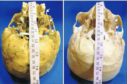
Fig. 1. The human skull bones exhibi
ting the unilateral elongated styloid process (arrows).
Table 1. The morphometric data of the styloid process of temporal bone (n=220)
Styloid process Right side Left side
Length 17.8±9.3 18.2±5.6
Thickness at base 4.4±1.2 4.4±0.9
Thickness at midpoint 3.2±0.4 3.8±0.7
Thickness at tip 1.5±0.6 1.4±0.5
The mean measurements are given in mean±SD (mm), statistical comparison was not significant (P>0.05); paired t test.
Table 2. The data on interstyloid distance of the skull (n=110) at various levels
Interstyloid distance Mean±SD (mm)
At the base of styloid processes 68.9±4.3 At the mid-point of styloid processes 62.7±1.1 At the tip of styloid processes 60.7±2.4
Among our specimens, only 5 skulls (4.5%) exhibited the elongated styloid process. Three skulls (2.7%) had unila- teral elongation (Fig. 1) and 2 skulls (1.8%) showed bilateral elongation (Fig. 2). All the 3 unilateral elongations were observed on the left side. The variant styloid processes were
unusually long (more than 30 mm), stout and strong. The elongated styloid processes were rounded up to half of their length and gradually tapered with a pointed tip (Figs. 1, 2).
Table 3 show the morphometric data of the unilateral elon- gated styloids which were observed in the present study. The data of the bilaterally elongated styloid process is given in Table 4.
Discussion
The elongation of styloid process with or without ossified stylohyoid ligament is considered as Eagle’s syndrome. This syndrome is also called as styloid neuralgia, elongated styloid syndrome, styloid-carotid syndrome and styloid-stylohyoid syndrome [4]. The stylohyoid chain extends between the temporal and hyoid bones and is generally divided into four sections as tympanohyal, stylohyal, ceratohyal, and hypohyal [1]. Eagle, who described this syndrome complex, divided it into two categories. The classical type is presented as foreign body sensation in the throat, pain in the throat and the ear ache. The other type is the styloid process compressing the carotid arterial system and presenting as dizziness and head- ache [5]. A variety of head and neck signs and symptoms are related to the elongated styloid process and its stylohyoid chain component. The dimension of styloid process usually varies, ranging up to 25 mm. The elongated styloid process can be clinically detected by palpating the tonsillar fossa and is diagnosed by taking the X-ray lateral view of the neck, orthopantomogram (OPG) or a computerized axial Fig. 2. The human skull bone exhibiting the bilateral elongated styloid
process (arrows).
Table 3. The morphometric data of the unilaterally elongated styloid process (n=3)
Specimen Right side (mm) Left side (mm) Specimen 1
Length 22 50
Thickness at base 6 5
Thickness at mid-point 4 4
Thickness at the tip 2 1
Interstyloid distance at base 68 -
Interstyloid distance at mid-point 62 -
Interstyloid distance at the tip 59 -
Specimen 2
Length 26 42
Thickness at base 3 6
Thickness at mid-point 4 5
Thickness at the tip 1 1
Interstyloid distance at base 71 -
Interstyloid distance at mid-point 66 -
Interstyloid distance at the tip 62 -
Specimen 3
Length 22 41
Thickness at base 4 6
Thickness at mid-point 3 4
Thickness at the tip 1 1
Interstyloid distance at base 74 -
Interstyloid distance at mid-point 68 -
Interstyloid distance at the tip 59 -
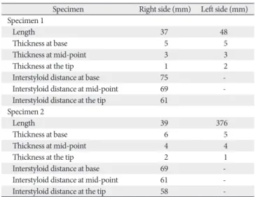
Table 4. The morphometric data of the bilaterally elongated styloid process (n=2)
Specimen Right side (mm) Left side (mm) Specimen 1
Length 37 48
Thickness at base 5 5
Thickness at mid-point 3 3
Thickness at the tip 1 2
Interstyloid distance at base 75 -
Interstyloid distance at mid-point 69 -
Interstyloid distance at the tip 61 Specimen 2
Length 39 376
Thickness at base 6 5
Thickness at mid-point 4 4
Thickness at the tip 2 1
Interstyloid distance at base 69 -
Interstyloid distance at mid-point 61 -
Interstyloid distance at the tip 58 -
tomogram (CT). Although there is no gender predilection for the elongated styloid process, the symptoms tend to be more common in the middle aged females [6, 7]. Eagle’s syndrome is a congenital anomaly which is sometimes misdiagnosed and is found incidentally during the radiological imaging.
The clinical symptoms in patients with Eagle’s syndrome are because of compression of the surrounding trigeminal, fa- cial and glossopharyngeal nerves. During the development, the hyoid smaller horn, styloid process, and the stylohyoid ligament are developed from the cartilage of second pharyn- geal arch (Riechert’s cartilage). On some occasions, the stylo hyoid ligament goes for ossification and will lead to the for mation of elongated styloid process [2]. Elongated styloid pro cess results in a variety of symptoms which range from chronic facial and neck pain, dysphagia, tinnitus, referred pain in the ear, glossopharyngeal neuralgia, orbital pain and radiating pain into the maxillary regions [8-10]. It may also cause stroke when it compresses the carotid arterial system [11, 12]. The inflammation and degeneration changes of the stylohyoid ligament which occurs in its tendon and the rheu- matic styloiditis may also contribute to this syndrome [13].
The length of styloid process of temporal bone varies from population to population. Eagle [3] reported that a normal styloid process measures between 25 mm to 30 mm and any length more than the above mentioned values, is considered as the pathogenic factor for Eagle syndrome.
Keur et al. [2] suggested that the styloid process length and its mineralized stylohyoid ligament, if appears more than 30 mm in a radiograph film, is considered as a significant pre- disposing factor. However, Jung et al. [14] suggested that, a styloid process of only more than 45 mm length should be considered to be elongated. In the present study, the length of styloid processes ranged from 18 mm until 50 mm. The present study observed that the mean length of the styloid process was 17.8±9.3 mm and 18.2±5.6 mm for the right and left sides, respectively. This data is smaller in comparison to the data from North Indian population reported by Rathva et al. [15]. Rathva et al. [15] reported that the length of styloid process was 43.8±11.1 mm and 43.5±10.4 mm for the right and left sides in their specimens. This variation in the data from Indian samples may be because the difference in the method which was used to measure the parameter as they performed the measurements by using digital image analysis using the adobe photoshop. In the present study, digital Ver- nier caliper was used to perform the measurements. However, in an another study by Patil et al. [16], which used the Adobe
photoshop for the measurements, the data were 13.9±8.1 mm and 12.9±8.7 mm for the right and left sides, respectively. The findings of the present study are similar to Patil et al.’s data [16].
Among all the skulls, seven of the styloid processes measured more than 30 mm and were considered as elongated. This was observed in 5 among 110 skulls with a prevalence rate of 4.5%.
The prevalence of elongated styloid process in the earlier studies were 1% [4], 4% [17], 8.2% [18], and 28% [19]. The pre valence rate of the present study (4.5%) is almost similar to the rate observed by Eagle (4%) [19]. Other Indian studies by Rathva et al. [15] reported the prevalence of elongated styloid process as up to 2%. The present study observed that the sty- loid process elongation was observed more commonly on the left (5:2).
The present study performed the thickness of the styloid process at the base, mid-point, and the tip. This much detailed measurements on the thickness was not reported earlier in the literature. The thickness data was compared statistically and it was found that the data was not significant statistically with respect to the right and left sides. The mean thickness at the styloid process base was 4.4±1.2 mm and 4.4±0.9 mm on the right and left sides. The same parameters were 3.2±0.4 mm and 3.8±0.7 mm; 1.5±0.6 mm and 1.4±0.5 mm at the mid-point and tip of the styloid processes, respectively. There were few reports [20] which observed 6-12 mm thickness at the base of styloid process. The present study observed 4.5 mms as the maximum thickness. In the present study, the interstyloid distance of the skull at the base, mid-point, and the tip of styloid process were 68.9±4.3 mm, 62.7±1.1 mm, and 60.7±2.4 mm, respectively. These data are different in comparison to the data by Rathva et al. [15]. Rathva et al. [15]
reported the same parameters as 75 mm, 55 mm, and 30 mm, respectively. This difference is may be because of the methods used as the present study was performed by using the digital Vernier caliper and Rathva et al. [15] used the scion image analyzer. Our findings are almost similar to the data from Patil et al. [16]. According to Patil et al. [16], the mean of distance between the bases of two styloid process was 69±5.2 mm and the same parameter between the tips of two styloid processes was 64±6.1 mm.
The impression of the styloid process elongation can be made by analyzing the clinical history given by the patient and by digitally palpating the tonsillar fossa. Touching the tonsillar area exacerbates the symptoms and is relieved by the local infusion of a local anesthetic agent into the tonsillar area, which is indicative of Eagle’s syndrome [6, 7, 21]. The
diagnosis is confirmed taking the X-ray, OPG and the skull base CT scan. The treatment of elongated styloid process is performed by the surgery, the elongation will be made short by cutting it with the trans-tonsillar and external approaches, considering avoiding injury to the surrounding neurovascular structures [22]. It has been reported that, inspite of the pre- cautions taken during the surgery, the structures attached to this process get injured at their attachments, which may compromise the stability of hyoid bone.
The etiology and pathogenesis of Eagle’s syndrome is poorly understood and is same with the calcification of the stylohyoid ligament. However, there are many theories which are described. The well accepted one is the ossification of the stylohyoid ligament or mineralization of the cartilage which is present embryologically during the development of the styloid process. The solidification of these structures may result in a variety of symptoms referred as Eagle’s syndrome. Steinmann [23] proposed various theories to explain the ossification of stylohyoid ligament which include the theory of reactive- hyperplasia-metaplasia-anatomical variation. The most accepted theory is the congenital theory. According to the congenital theory, mechanical stresses during the intrauterine life may sometimes lead to stretching of the second branchial arch and the styloid process elongation.
The symptoms of elongated styloid process include dys- phagia, pain in the throat, foreign body sensation and pain in the face. It may lead to transient ischemic attacks and cere- brovascular accidents due to the compression of the internal carotid artery and lack of arterial supply to the brain. It is also responsible for the difficult endotracheal intubations which are experienced by the anesthesiologists [24]. The cli- nical implication also includes the course of the vertebral artery which may be distorted in such situations. The Eagle’s syndrome should not be misdiagnosed as trigeminal neu- ralgia [24]. This can be easily differentiated by taking the pain history as the Eagle’s syndrome pain is persistent and the trigeminal neuralgia pain is intermittent in nature. Eagle’s syndrome may mimick symptoms of phantom limb (globus hystericus) and may cause psychosomatic stress to the patient [25]. Differential diagnoses of Eagle’s syndrome include unerupted molar tooth, dental prosthesis implantation, di- seases of the temporo mandibular joint, tumors in the oro- pharynx and laryngopharynx [26]. It has been reported that there is a minimal quantity of elongation in the temporal bone styloid process as the age advances. This is especially noticed in the postmenopausal ladies and is due to the hormonal
changes which occur after the menopause [27].
The morphology and the variations of the craniofacial bones have been studied since the beginning of the century [28]. The morphometric data of the styloid process is im- portant to the neurologists, neurosurgeons, radiologists and otorhinolaryngologists. The data on the interstyloid dis- tance is clinically important as this space accommodates im portant structures of neck like cranial nerves, larynx, eso- phagus, arteries, and veins. The surgical anatomy of the elon- gated styloid process is important to the neurosurgeons and radiologists while interpreting the computed tomogram scans and magnetic resonance images. The morphological know- ledge of Eagle’s syndrome is enlightening to all the health care professionals who are involved in the diagnosis and mana gement of chronic head and neck pain. The knowledge is important to the physicians, dentists, neurologists, neuro- surgeons, and otorhinolaryngologists. In this context, the present study has provided important data about the stylo- id process of the temporal bone. The dimensions of the styloid process and its stylohyoid chain are essential to the anatomists, anthropologists, forensic experts and clinicians.
We believe that the present study has provided additional information on the frequency of elongated styloid process in the Indian population. In the present study, the styloid process has been studied morphologically with emphasize on their embryological and clinical implications. We opine that the present study can be more accurate with a large number of samples. The limitation of the present study includes the gender related variation which was not taken into consideration.
References
1. Magotra R, Razdan S. Elongated styloid process: anatomical variations. JK Sci 2008;10:203-5.
2. Keur JJ, Campbell JP, McCarthy JF, Ralph WJ. The clinical signi- ficance of the elongated styloid process. Oral Surg Oral Med Oral Pathol 1986;61:399-404.
3. Eagle WW. The symptoms, diagnosis and treatment of the elongated styloid process. Am Surg 1962;28:1-5.
4. Langlais RP, Miles DA, Van Dis ML. Elongated and mineralized stylohyoid ligament complex: a proposed classification and report of a case of Eagle's syndrome. Oral Surg Oral Med Oral Pathol 1986;61:527-32.
5. Sachidananda R, Chokkalingam, Siddique, McRae. Elongated styloid process: a rare presentation as a tonsillar mass. Internet J Otorhinolaryngol [Internet]. 2005 [cited 2015 Sep 1];5(2).
Available from: http://ispub.com/IJORL/5/2/9168.
6. Murtagh RD, Caracciolo JT, Fernandez G. CT findings associated with Eagle syndrome. AJNR Am J Neuroradiol 2001;22:1401-2.
7. Rechtweg JS, Wax MK. Eagle's syndrome: a review. Am J Otol- aryngol 1998;19:316-21.
8. Dinkar, Amonkar SS. Eagle's syndrome: review of literature and case report. Indian J Dent Res 2003;14:162-8.
9. Soh KB. The glossopharyngeal nerve, glossopharyngeal neu- ralgia and the Eagle's syndrome: current concepts and mana- gement. Singapore Med J 1999;40:659-65.
10. Nishihara K, Hanakita J, Kinuta Y, Kondo A, Yamamoto Y, Kishimoto S. Three cases of Eagle’s syndrome. No Shinkei Geka 1986;14(3 Suppl):441-5.
11. Carter L. Soft tissue calcification and ossification. In: White SC, Pharoah MJ, editors. Oral Radiology: Principles and Inter- pretation. St. Louis: Mosby, 2004. p.597-614.
12. Chuang WC, Short JH, McKinney AM, Anker L, Knoll B, McKinney ZJ. Reversible left hemispheric ischemia secondary to carotid compression in Eagle syndrome: surgical and CT angio- graphic correlation. AJNR Am J Neuroradiol 2007;28:143-5.
13. Prasad KC, Kamath MP, Reddy KJ, Raju K, Agarwal S. Elongated styloid process (Eagle's syndrome): a clinical study. J Oral Maxillofac Surg 2002;60:171-5.
14. Jung T, Tschernitschek H, Hippen H, Schneider B, Borchers L.
Elongated styloid process: when is it really elongated? Dento- maxillofac Radiol 2004;33:119-24.
15. Rathva A, Kubavat DM, Nagar SK. Study of styloid process:
anatomical variations in length, angulation and distance between the two styloid processes. Int J Recent Trends Sci Technol 2013;8:
109-12.
16. Patil S, Ghosh S, Vasudeva N. Morphometric study of the styloid process of temporal bone. J Clin Diagn Res 2014;8:AC04-6.
17. Winkler S, Sammartino FJ Sr, Sammartino FJ Jr, Monari JH.
Stylohyoid syndrome. Report of a case. Oral Surg Oral Med Oral
Pathol 1981;51:215-7.
18. Kawai T, Shimozato K, Ochiai S. Elongated styloid process as a cause of difficult intubation. J Oral Maxillofac Surg 1990;48:
1225-8.
19. Eagle WW. Elongated styloid processes: report of two cases.
Arch Otolaryngol 1937;25:584-7.
20. Hussain SS, Muralidhar PS, Desai SD, Thomas ST, Haseena S.
Elongated styloid process. J Pharm Sci Res 2012;4:1761-3.
21. Nickel J, Sonnenburg M, Scheufler O, Andresen R. Eagle syndrome: diagnostic imaging and therapy. Rontgenpraxis 2003;55:108-13.
22. Camarda AJ, Deschamps C, Forest D. I. Stylohyoid chain ossifi- cation: a discussion of etiology. Oral Surg Oral Med Oral Pathol 1989;67:508-14.
23. Steinmann EP. Styloid syndrome in absence of an elongated process. Acta Otolaryngol 1968;66:347-56.
24. Das S, Suhaimi FH, Othman F, Latiff AA. Anomalous styloid process and its clinical implications. Bratisl Lek Listy 2008;
109:31-3.
25. Prabhu LV, Kumar A, Nayak SR, Pai MM, Vadgaonkar R, Krish- namurthy A, Madhan Kumar SJ. An unusually lengthy styloid process. Singapore Med J 2007;48:e34-6.
26. Jain D, Chauhan JS, Jain S, Goel G. Elongated styloid process: an unusual cause of neck pain and difficulty in swallowing. J Orofac Pain 2011;25:269-71.
27. Roopashri G, Vaishali MR, David MP, Baig M. Evaluation of elongated styloid process on digital panoramic radiographs. J Contemp Dent Pract 2012;13:618-22.
28. Murlimanju BV, Chettiar GK, Prameela MD, Tonse M, Kumar N, Saralaya VV, Prabhu LV. Mastoid emissary foramina: an anatomical morphological study with discussion on their evolutionary and clinical implications. Anat Cell Biol 2014;
47:202-6.