Copyright © 2019. Anatomy & Cell Biology
Introduction
Keratinocytes constitute the major cellular component in the epidermis and play critical roles in the process of wound healing. They play a major part in the complex mechanisms
involved in the initiation, continuation, and completion of wound healing. The properties of keratinocytes in chronic wounds differ in different locations and situations. Normally, keratinocytes are responsible for the proliferative and re- generative capacity of the epidermis by providing a continu- ous supply of proliferative cells. The re-epithelization of the wounded skin occurs by the rapid and coordinated migration of keratinocytes [1].
The keratinocytes present at the edges of the non-healing chronic wounds are different from the healthy and normal keratinocytes. A successful wound closure requires a cross- talk between the keratinocytes and other cell types involved in wound healing [1]. Keratinocytes may also play a role in
Corresponding author:
Kumar M. R. Bhat
Department of Anatomy, Ras Al Khaimah College of Medicine, RAK Medical & Health Science University, Post Box No. 11172. Ras Al Khaimah, UAETel: +971-7-2269000 (ext: 266), Fax: +971-7 2269997,
E-mail: kumarbhat@rakmhsu.ac.ae
Influence of traditional medicines on the activity of keratinocytes in wound healing:
an in-vitro study
Sushma R. Kotian
1, Kumar M. R. Bhat
2, Divya Padma
1, K. Sreedhara R. Pai
31Department of Anatomy, Kasturba Medical College Manipal, Manipal Academy of Higher Education, Manipal, India, 2Department of Anatomy, RAK College of Medical Sciences, RAK Medical and Health Sciences University, Ras Al Khaimah, UAE, 3Department of Pharmacology, Manipal College of Pharmaceutical Sciences, Manipal Academy of Higher Education, Manipal, India
Abstract: Natural medicinal systems such as Ayurveda and folk medicine has remedies for wound management. However, the exact cellular and extracellular mechanisms involved in the healing process and its influence on keratinocytes is less discussed. Therefore, the present study was designed to evaluate the effect of certain natural wound healing medicines on the biology of the keratinocytes/HaCaT cells. Test materials such as honey (H), ghee (G), aqueous extracts of roots of Glycyrrhiza glabra (GG) and leaves of Nerium indicum (NI) were considered. The HaCaT cells were treated with the test materials singly and in combinations (H+G, all combined [Tot]) for a specific period (24, 48, and 72 hours). The cells were then subjected to cytotoxicity/proliferation and migration/scratch assays. All the test materials, except NI, were non-cytotoxic and showed increased cell proliferation at variable concentrations. Significant observations were made in the groups treated with honey (100 µg/ml at 48 hours, P<0.05; 1,000 µg/ml at 72 hours, P<0.05), GG (all concentrations at 48 hours, P<0.05; 750 µg/ml at 72 hours, P<0.05), H+G (250 µg/ml at 24 hours, P<0.001; 500 µg/ml at 48 and 72 hours, P<0.05), and Tot (50 µg/ml at 24, 48 and 72 hours, P<0.01). In the in-vitro wound healing assay, all the treated groups showed significant migration and narrowing of the scratch area by 24 and 48 hours (P<0.001) compared to control. The results obtained from the present study signifies the positive influence of these natural wound healing compounds on keratinocytes/HaCaT cells.
Key words: HaCaT cells, Honey, Ghee, Glycyrrhiza glabra, Nerium indicum Received January 16, 2019; Revised March 21, 2019; Accepted April 12, 2019
the extracellular matrix reorganization [2].
Owing to their diverse role in the healing process, the ke- ratinocytes have been chosen in the current study to evaluate the wound healing benefits of the traditional medicines.
Traditional medicinal systems of India such as Ayurveda and folk medicine have been using such effective medicinal preparations to treat wounds, and have claimed that these medications not only act as anti-inflammatory, anti-microbial reagents, but also have rejuvenating and regenerating effects.
One such effective alternative treatment strategy is the use of honey, ghee, and Glycyrrhiza glabra (GG), and a folk medicinal preparation using Nerium indicum (NI), which is effectively practiced to treat various types of wounds since many years in India [3]. Several studies have shown the wound healing benefits of individual application of ghee [4] or honey [5, 6]. The wound healing, anti-ulcer, anti- inflammatory, and skin regeneration activity of GG is also documented by an earlier research [7]. NI is another herb used to treat wounds in the folk medicine [8]. Our previously published report on wound healing in animal models using these traditional medicines singly and in combinations pro- vided valuable evidences on microenvironmental changes in the wound site, thus proving the efficacy of these medicines [3]. The influence of these traditional medicines singly and in combination on the fibroblasts have also been experimented upon [9].
However, less is known about the impact of these tradi- tional medicines on keratinocytes in the context of wound healing. The present study was therefore designed to evaluate the influence of these traditional medicines on the activities of keratinocytes, such as proliferation and migration, which are critical in wound healing process. The study may also aid in supporting the possibility of using the traditional medicines in wound healing, especially in managing the medically chal- lenging wounds such as diabetic foot ulcers, pressure sores, ischemic ulcers, etc.
Materials and Methods
Test materialsProcurement of the test materials
Unprocessed honey in its raw form, ghee made from cows’
milk, and roots of GG were procured from Sri Dharmasthala Manjunatheshwara Ayurvedic pharmacy, Udyavara, Udupi.
Honey and ghee were subjected to microbiological examina-
tion to rule out any bacterial contamination. NI was collected in December from its natural habitat in Udupi and was identi- fied and authenticated by Dr. K. Gopalakrishna Bhat, Profes- sor of Botany (Rtd), Poornaprajna College, Udupi, Karnataka, India. A voucher specimen is preserved in the Department of Pharmacognosy, Manipal College of Pharmaceutical Sciences, Manipal (PP no. 603).
Preparation of test materials
The roots of GG and leaves of NI after procurement were shade dried for seven days and then powdered. Aqueous extraction was carried out by hot maceration method [10].
The powdered plant materials (200 g each) were dissolved in 1,500 ml of distilled water and the decoction was prepared at 75–80°C. In the final stages (after boiling) of extract prepara- tion, all possible sterility was maintained. The decoction was then cooled and filtered. The filtrate was finally evaporated to dryness using a lyophilizer. Before application on the wound, these test materials were membrane filtered to ensure that they were devoid of contamination.
The cell culture experiments were carried out in seven different groups: control (untreated), honey, ghee, GG, NI, honey+ghee (H+G), and combination of all test materials (Tot).
Cellular analysis: cell culture (in-vitro study)
Procurement of keratinocytes (HaCaT cells, i.e., immortal human keratinocyte)
HaCaT cell line procured from the National Centre for Cell Science, Pune were used in the present study owing to their greater ability to differentiate and proliferate in vitro [11].
HaCaT viability and proliferation in vitro
Cell proliferation/cytotoxicity assay (MTT method) [12]
A day before the assay, HaCaT cells were seeded in 96-well plates at an optimum concentration of 5×10³ cells per 100 µl medium per well. The cell suspensions were then incubated at 37°C to enable cell attachment. After 24 hours, the cells were treated with 100 µl of each concentration of serially di- luted medicinal preparations. The concentrations considered for honey, GG, NI, H+G, and Tot were 1,000, 750, 500, 250, and 100 µg/ml. Ghee was administered in the concentra- tions of 10, 7.5, 5, 2.5, and 1 µl/ml. All concentrations were in triplicates, and cells were treated for 24, 48, and 72 hours. At
the end of the stipulated time frame, 20 µl MTT (5 mg/ml) in phosphate buffer saline (PBS) was added to the wells and incubated for four hours at 37°C. After incubation, the media was discarded and formazan crystals formed were dissolved in 100 µl of dimethyl sulfoxide and incubated for one hour at room temperature. The optical density was read at 570 nm with a reference wavelength of 650 nm using enzyme-linked immunosorbent assay plate reader (model 3550, Bio-Rad Laboratories, Orlando, FL, USA). The findings obtained were normalized to percent of control (wells without having any drugs) and plotted against drug concentration. The formula used for its calculation is represented below:
Value of the treatment group/Mean value of control×100.
Wherein, the treatment groups were as follows: control, hon- ey, ghee, GG, NI, H+G, and Tot.
Wound scratch assay
The stimulatory effect of the test materials on migration
of HaCaT cells was determined as described by Balekar et al.
[13]. HaCaT cells were plated into a 12-well plate at a concen- tration of 1×10⁵ cells containing Dulbecco’s modified Eagle’s medium culture medium supplemented with 10% fetal bovine serum (FBS) and incubated overnight at 37°C in a humidified 5% CO2 atmosphere. After attachment of the cells to the plate, Dulbecco’s modified Eagle’s medium (DMEM) was complete- ly removed and the adherent cell layer was scratched with a sterile pipette tip. Cellular debris were removed by washing off with PBS. Then, the cells were treated with DMEM with 10% FBS containing the different test materials (100 µg/ml of honey, GG, NI, H+G, and Tot; 10 µl/ml of ghee). Control cells received only fresh DMEM. To avoid proliferation of cells and to evaluate only the migration, mitomycin C (10 µg/mL) was added to all the wells (control and treated groups). The cells were then incubated at 37°C in humidified 5% CO2 atmo- sphere. Then, the images of the scratch area were captured at 0th hour (just after scratching cells), 24, 48, and 72 hours after
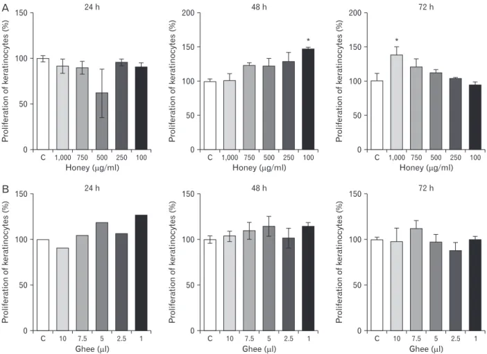
Fig. 1. Proliferative effect of different doses of the honey (A) and ghee (B) on keratinocytes during 24, 48, and 72 hours as indicated by MTT assay.
*P<0.05 in comparison with control (C, untreated group).
incubation with the test materials using a built-in camera in the microscope (40× magnification).
The captured images were then analyzed using T Scratch software (CSE Lab, ETH Zurich, Clausiusstrasse, Switzer- land). The area of the scratch in each image was estimated.
Scratch area on the 0th hour was considered 100% and the reduction in area was calculated for each set in 24, 48, and 72 hours and plotted on a graph.
Statistical analysis
The data were analyzed using Graph Pad Prism software (Microsoft, San Diego, CA, USA) and were expressed as mean±SEM. One-way ANOVA, followed by Dunnett’s post- hoc test was used to compare control group and treated group.
P-value of ≤0.05 was considered statistically significant.
Results
Evaluation of the role of the medicinal preparations in the survival and proliferation of the keratinocytes (HaCaT cells)
Cell proliferation/cytotoxicity assay
The HaCaT cells treated with different concentrations of honey showed no proliferative effects after 24 hours of exposure. After 48 hours, all the concentrations showed pro- liferation. However, the finding was significant only at a con- centration of 100 µg/ml (147.75±2.433, P<0.05). Treatment for 72 hours also showed an increase in the cells, which was significant at a concentration of 1,000 µg/ml (138.22±12.303, P<0.05) (Fig. 1A). Cells treated with ghee also showed better proliferative effects. The findings, however, were not signifi- cant statistically (Fig. 1B).
The aqueous extract of GG had a positive proliferative ef-
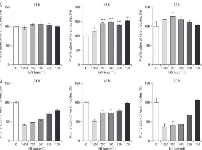
Fig. 2. Proliferative effect of different doses of Glycyrrhiza glabra (GG) (A) and Nerium indicum (NI) (B) on keratinocytes during 24, 48, and 72 hours as indicated by MTT assay. *P<0.05 and ***P<0.001 in comparison with control (C, untreated group).
150
100
50
Proliferationofkeratinocytes(%)
0
24 h 200
100
50
Proliferationofkeratinocytes(%)
0 150
48 h
100
50
Proliferationofkeratinocytes(%)
0
150 72 h
*
* A
*** *** *** ***
C 1,000 750 500 250 100 C 1,000 750 500 250 100 C 1,000 750 500 250 100
GG ( g/ml) GG ( g/ml) GG ( g/ml)
150
100
50
Proliferationofkeratinocytes(%)
0
24 h
100
50
Proliferationofkeratinocytes(%)
0
150 48 h
100
50
Proliferationofkeratinocytes(%)
0
150 72 h
B
C C C
NI ( g /ml)) NI ( g/ml)) NI ( g/ml)
1,000 750 500 250 100 1,000 750 500 250 100 1,000 750 500 250 100
fect on the HaCaT cells. All the doses of the extract showed increased survival and proliferation of the cells. The find- ings were statistically significant after 48 hours of the treat- ment, i.e., 1,000 µg/ml: 114.78±2.46, P<0.05; 750 µg/ml:
142.54±4.28, P<0.001; 500 µg/ml: 147.03±2.62, P<0.001;
250 µg/ml: 136.36±3.37, P<0.001; 100 µg/ml: 152.72±0.96, P<0.001.
At 72 hours, all the doses demonstrated a natural increase in the proliferation of the cells, however statistically signifi- cant observations were made only at a dose of 750 µg/ml (125.38±1.48, P<0.05) (Fig. 2A). The aqueous extract of NI, on the contrary, was cytotoxic to the keratinocytes in all the doses calculated. The survival rate of the HaCaT cells showed a gradual decrease with the increase in the concentration of NI (Fig. 2B).
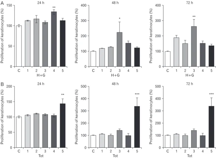
H+G also showed proliferative effects. The finding was significant at a dosage of 250 µg/ml at 24 hours (133.65±5.81, P<0.01) and 500 µg/ml in both 48 (223.34±68.44, P<0.5) and
72 hours (261.30±46.98, P<0.01) respectively (Fig. 3A). Tot also showed proliferative effect. But the finding was signifi- cant only at the dosage of 50 µg/ml during all the durations of treatment, i.e., 24 (142.70±16.38, P<0.01), 48 (337.49±70.97, P<0.001), and 72 hours (340.68±32.95, P<0.001) (Fig. 3B).
The higher doses of Tot failed to show a significant increase in the rate of proliferation. It could be because of the greater doses of NI present in them.
Wound scratch assay
All the experimental groups showed significant closure compared to control (Fig. 4). The scratch area was occupied by the migrating HaCaT cells. All the treated groups showed significant migration and narrowing of the scratch area by 24 and 48 hours (P<0.001). Among them, the groups treated with honey, H+G, and Tot showed significantly best results at 48 hours. Complete closure of the scratch area was observed in all the treated groups by 72 hours (Table 1, Fig. 5).
Fig. 3. Proliferative effect of different doses of honey+ghee (H+G) (A) and combination of all test materials (Tot) (B) on keratinocytes during 24, 48, and 72 hours as indicated by MTT assay. *P<0.05, **P<0.01, and ***P<0.001 in comparison with control (C, untreated group).
Discussion
Keratinocytes are the chief cellular constituents of the
epidermis and have numerous critical responsibilities in the process of wound healing. They are involved in the intricate mechanisms of initiation, maintenance, and completion of
Fig. 4. Analyzed images of Scratch assay of HaCaT cells treated with the different test materials compared to control (untreated). Yellow dotted line indicates the site of the scratch wound.
GG, Glycyrrhiza glabra; NI, Nerium indicum; H+G, honey+ghee; Tot, combination of all test materials.
Table 1. The percentage of wound closure in the experimental groups in the scratch assay using HaCaT cells
Time (h) Group (mean±SEM)
C H G GG NI H+G Tot
0 100 100 100 100 100 100 100
24 94.37±0.36 58.39±0.02a) 62.66±0.49a) 75.13±0.24a) 87.16±0.19a) 66.87±0.04a) 56.90±0.46a) 48 31.17±0.24 8.39.033b) 28.80±0.09b) 24.87±0.11b) 15.16±0.03b) 10.66±0.16b) 11.23±0.04b) C, control; H, honey; G, ghee; GG, Glycyrrhiza glabra; NI, Nerium indicum; H+G, honey+ghee; Tot, combination of all test materials. a)P<0.001 and b)P<0.001 in comparison with 24 hours and 48 hours’ control (untreated) groups respectively.
wound healing [1].
Owing to its significant role in wound healing, keratino- cytes (HaCaT cells) have been chosen in the current study to appreciate the healing benefits of the traditional medicines.
Measuring viability and growth of the cells, i.e., keratino- cytes when treated with the different concentrations of the test materials is a positive step to test their efficiency. The ac- curate method to evaluate the same is the use of MTT assay [12]. It provides a precise determination of the alterations in the cell proliferation rate [14].
The scratch wound healing assay is yet another important method used to portray the cell proliferation and migration [15]. Both the methods are therefore employed in the present study to understand the influence of the traditional medicines on the keratinocytes.
The exposure of honey was previously found to stimulate wound healing in the rat dermal fibroblasts (RDFs) as indi- cated by the scratch assay. Among the three kinds of honey used, i.e., Acacia (Robinia pseudacacia), Buckwheat (Fago- pyrum sp.), and Manuka (Leptospermum scoparium); Acacia and buckwheat honey showed significant results compared to Manuka honey. It indicates that the different types of honey may present different wound healing properties [16].
Our previous reports on the MTT assay using RDFs re- vealed that honey neither showed any proliferative nor cy- totoxic effect on the fibroblasts. The scratch assay revealed better migration of the fibroblasts treated with honey. But the findings were not statistically significant [9]. Honey-treated
keratinocytes, on the contrary, showed proliferative effects after 48 and 72 hours of treatment as indicated in the present study. The scratch assay conducted on honey-treated kera- tinocytes showed a significant narrowing of the wound. It denotes that honey has better proliferative effects on keratino- cytes compared to the fibroblasts.
Studies have confirmed that honey induces low cytotoxic- ity in keratinocytes. Further, the honey driven wound repair goes through activation of keratinocyte re-epithelization [17].
Greater the duration of exposure to honey, a greater rate of proliferation as was observed in the present study.
The proliferative benefits of honey could be attributed to the involvement of specific mechanisms that are still ob- scure. Recent studies have however shown that honey has a chemoattractant effect [17]. It could be responsible for the improved proliferation and migration of the keratinocytes as was observed in the present study.
Honey is responsible for the enhanced expression of syn- decan-4 (transmembrane heparin sulfate) and matrix metallo- proteinase (MMP)-9. MMPs are known to play essential roles in wound repair linked cell migration [17]. Honey also aids in the activation of proteins involved in the proliferation and cy- toskeletal rearrangement. Honey activates cyclin-dependent kinase 2, a cyclin dependent kinase known to be involved in G1-S transition and keratinocyte proliferation. Focal adhesion kinase and rasGAP SH3 binding protein 1, both involved in cell locomotion are also shown to be activated by honey. Va- sodilator-stimulated phosphoprotein, integrin-β3, CDC25C, and p42/44 mitogen activated protein kinase are among the other peptides related to cytoskeletal and cell cycle dynamics.
They have also shown variable amount of activation on expo- sure to honey [17]. Honey is also known to play a prime role in the induction of epithelial mesenchymal transition (EMT).
The EMT is essential for the keratinocyte re-epithelization as is observed in wound healing [17].
The abovementioned factors and mechanisms support the observations made in the present study. It further reaffirms the proliferative and migratory benefits of honey and rein- states the belief that honey increases the wound repair capa- bilities of keratinocytes.
The polyunsaturated fatty acid (PUFA) present in the ghee is capable of controlling cell to cell interaction and intracel- lular signal transduction [18]. The n-3 and n-6 PUFA also en- able the proliferation of epithelial cells in vitro which serves as an essential step in wound repair [19].
Ghee showed proliferative effects on the keratinocytes as Fig. 5. Graphical representation of the percentage of wound closure
in all the experimental groups in a scratch assay using HaCaT cells.
GG, Glycyrrhiza glabra; NI, Nerium indicum; H+G, honey+ghee;
Tot, combination of all test materials.***P<0.001 and ###P<0.001 in comparison with 24 and 48 hours’ control (untreated) groups respectively.
indicated in the current study. The cells were proliferative when exposed to ghee for shorter durations. The decrease in the proliferative implications of these cells on exposure to longer duration may be probably due to the immiscible nature of ghee which formed an oily layer of film on the cul- ture medium, thereby interfering with the CO2 and oxygen ratio in the culture medium, which has an adverse effect on cell survival and growth. Similar observations were made by the fibroblasts treated with ghee in previous reports [9]. The scratch assay, however, showed significant wound closure in ghee treated cells. It indicates that ghee can increase the wound closure abilities of the keratinocytes by influencing its migration. The actual mechanisms of action are however un- clear and further needs to be explored.
The H+G treated cells on prolonged exposure also showed better proliferative and migratory effects.
GG had a positive proliferative effect on the keratinocytes.
Greater the dose, higher was the proliferation rate. Further, the migration of the keratinocytes was also influenced by GG on exposure to longer duration as indicated by the scratch as- say. Research has revealed that GG inhibits the proliferation of abnormal cells, and is anti-carcinogenic in nature. How- ever, the exact mechanisms influencing these activities are still being investigated [20]. On the contrary, in the present study, GG has shown a positive influence on cell proliferation.
A similar observation was made in the previous study on fi- broblasts [9]. This indicates that GG has a variable influence on the normal and abnormal cells.
Although GG has shown to improve the rate of cell prolif- eration and migration of keratinocytes and thereby improve wound healing, the underlying mechanisms are obscure.
However, it could be suggested that GG aids in the activa- tion of proteins involved in the proliferation and cytoskeletal rearrangement, and thus improves healing. The increased duration of exposure to GG also has a positive impact on the increased activities of the keratinocytes. Longer the duration of exposure, greater could be the activation of specific growth factors/proteins aiding in healing that needs to be further in- vestigated. The antioxidant and rejuvenating effects of some of the constituents of GG such as glycyrrhizin and glabridin may also aid in improving the wound healing abilities of kera- tinocytes [21].
NI, on the contrary, was cytotoxic to keratinocytes in all the calculated higher doses as indicated by the MTT assay.
The lower doses of NI neither showed proliferative nor toxic effects. The migration of the keratinocytes treated with NI
was however significantly improved compared to control as shown by the scratch assay. It suggests that NI may have a var- ied influence on the activity of keratinocytes at the wound site at particular dose. Similar observation was made in the previ- ous study on fibroblasts [9]. Thus, it could be postulated that NI favors cellular migration, but is anti-proliferative in nature.
Further analytical studies are required to reaffirm the same.
The animal experiments conducted in the past revealed that although NI exhibited prolonged inflammatory re- sponses, but was however effective in quickening re-epithe- lialization and reducing fibrosis at the wound site. Further, the wounds treated with NI also showed increased tensile strength at the wound site compared to other test groups [3].
It indicates that NI is not entirely toxic. In our current study using the HaCaT cells, NI is found to be toxic indicating the many other intrinsic factors are triggered by the use of NI in- vivo setup which aid in the wound repair. Therefore, there is a need for further evaluation. Conducting the future experi- ments using lower doses of NI would show positive results.
The Tot treated keratinocytes showed proliferative effects.
Significant observations were made at the lower concentra- tions used, owing to the reduction in the amount of NI pres- ent in Tot. The scratch assay employing the keratinocytes also showed significant results.
Studies in the past have focused on the role of keratino- cyte–fibroblast interactions in the wound healing process.
There is ample evidence that keratinocytes stimulate fibro- blasts to synthesize growth factors, which in turn stimulates the keratinocyte proliferation in a double paracrine man- ner. Moreover, fibroblasts can also acquire a myofibroblast phenotype under the control of keratinocytes. This depends on a finely tuned balance between a pro-inflammatory or a transforming growth factor β–dominated environment [22].
The use of fibroblast and keratinocyte co-cultures to study the healing efficacy of these natural medicines may, therefore, be beneficial.
The preliminary results obtained from the current study reestablishes the benefits of traditional medicine in healing wounds in general and their influence on the keratinocytes in particular. The study indicates that the test materials used, both singly and in combination have a positive influence on the proliferation and migration of the keratinocytes, an im- portant factor required for wound closure and thereby better wound healing.
Honey and GG had a positive influence on the wound healing capabilities of keratinocytes. Ghee and NI although
showed better keratinocyte proliferation and migration, the findings were good only at lower concentrations and shorter durations of exposure owing to their cytotoxic effects. The observations made in the present study may further encour- age and instigate newer objectives that would identify and explain the process of wound healing, and thereby glorify the effectiveness of the use of these traditional medicines. Further evaluation is also required to understand the intricate cellular and molecular mechanisms of these test materials which may help us to design the appropriate combination of these test materials in treating medically challenging wounds.
ORCID
Sushma R. Kotian: https://orcid.org/0000-0003-0271-3568 Kumar M. R. Bhat: https://orcid.org/0000-0003-1805-3453 K. Sreedhara R. Pai: https://orcid.org/0000-0002-2017-9533
Author Contributions
Conceptualization: KMRB. Data acquisition: SRK, KSRP.
Data analysis or interpretation: DP, SRK. Drafting of the manuscript: SRK. Critical revision of the manuscript: SRK, KMRB. Approval of the final version of the manuscript: all authors.
Conflicts of Interest
No potential conflict of interest relevant to this article was reported.
References
1. Pastar I, Stojadinovic O, Tomic-Canic M. Role of keratinocytes in healing of chronic wounds. Surg Technol Int 2008;17:105-12.
2. Isaac C, Paggiaro AO, Aldunate JL, Herson MR, Altran SC, Mathor MB, Ferreira MC. Role of keratinocytes in wound con- traction: an impact assessment using a model of collagen matrix populated with fibroblasts. Rev Bras Cir Plast 2011;26:402-6.
3. Kotian SR, Pai KS, Nayak J, Bangera H, Prasad K, Bhat KM. Bio- mechanical, biochemical and histological evidences for wound healing properties of Indian traditional medicines. Int J Pharm Pharm Sci 2015;7:163-71.
4. Prasad V, Dorle AK. Evaluation of ghee based formulation for wound healing activity. J Ethnopharmacol 2006;107:38-47.
5. Molan PC, Betts JA. Clinical usage of honey as a wound dress- ing: an update. J Wound Care 2004;13:353-6.
6. Molan PC. The evidence supporting the use of honey as a wound
dressing. Int J Low Extrem Wounds 2006;5:40-54.
7. Oloumi MM, Derakhshanfar A, Nikpoor A. Healing potential of liquorice root extract on dermal wounds in rats. J Vet Res 2007;62:147-54.
8. Sravanthi KC, Manthri S, Srilakshmi S, Ashajyothi V. Wound healing herbs: a review. Int J Pharm Technol 2010;2:603-24.
9. Kotian SR, Padma D, Madhukar R, Bhat KM. Effect of natural medicines on dermal fibroblasts in wound healing: an in-vitro study. Adv Sci Lett 2017;23:1949-56.
10. Davis H. Bentley’s textbook of pharmaceutics. 6th ed. London:
Balliere, Tindall and Co:; 1956. p.272-30.
11. Schurer N, Kohne A, Schliep V, Barlag K, Goerz G. Lipid com- position and synthesis of HaCaT cells, an immortalized human keratinocyte line, in comparison with normal human adult kera- tinocytes. Exp Dermatol 1993;2:179-85.
12. Ali A, Kim MJ, Kim MY, Lee HJ, Roh GS, Kim HJ, Cho GJ, Choi WS. Quercetin induces cell death in cervical cancer by reducing O-GlcNAcylation of adenosine monophosphate-activated pro- tein kinase. Anat Cell Biol 2018;51:274-83.
13. Balekar N, Katkam NG, Nakpheng T, Jehtae K, Srichana T. Eval- uation of the wound healing potential of Wedelia trilobata (L.) leaves. J Ethnopharmacol 2012;141:817-24.
14. Maioli E, Torricelli C, Fortino V, Carlucci F, Tommassini V, Pa- cini A. Critical appraisal of the MTT assay in the presence of rot- tlerin and uncouplers. Biol Proced Online 2009;11:227-40.
15. Menon MB, Ronkina N, Schwermann J, Kotlyarov A, Gaestel M. Fluorescence-based quantitative scratch wound healing assay demonstrating the role of MAPKAPK-2/3 in fibroblast migra- tion. Cell Motil Cytoskeleton 2009;66:1041-7.
16. Ranzato E, Martinotti S, Burlando B. Honey exposure stimu- lates wound repair of human dermal fibroblasts. Burns Trauma 2013;1:32-8.
17. Ranzato E, Martinotti S, Burlando B. Epithelial mesenchymal transition traits in honey-driven keratinocyte wound healing:
comparison among different honeys. Wound Repair Regen 2012;20:778-85.
18. Ruthig DJ, Meckling-Gill KA. Both (n-3) and (n-6) fatty acids stimulate wound healing in the rat intestinal epithelial cell line, IEC-6. J Nutr 1999;129:1791-8.
19. Calder PC. N-3 polyunsaturated fatty acids, inflammation and immunity: pouring oil on troubled waters or another fishy tale?
Nutr Res 2001;21:309-41.
20. Liu W, Kato M, Akhand AA, Hayakawa A, Takemura M, Yo- shida S, Suzuki H, Nakashima I. The herbal medicine sho- saiko-to inhibits the growth of malignant melanoma cells by upregulating Fas-mediated apoptosis and arresting cell cycle through downregulation of cyclin dependent kinases. Int J On- col 1998;12:1321-6.
21. Wang ZY, Nixon DW. Licorice and cancer. Nutr Cancer 2001;39:
1-11.
22. Werner S, Krieg T, Smola H. Keratinocyte-fibroblast interactions in wound healing. J Invest Dermatol 2007;127:998-1008.