Key Words :
Fig. 1. Chest CT shows 1.3 cm sized calcified endobronchial nodule within the left main-stem bronchus, abutting posterior wall of left main bronchus. Both lower paratracheal LNs are detected, but less than 1cm in size.
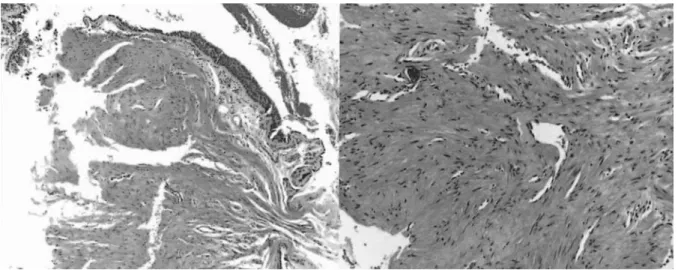
Fig. 3. The microphotograph shows squamous metaplasia of bronchial mucosal epithelium and underlying proliferation of neoplastic spindle cells. Left microphotograph: H & E stain, 100 Right microphotograph: H
& E stain, 200
Fig. 2. Bronchoscopy shows hyperemic irregular surfaced polypoid exophytic mass in left main bronchus, partially obstructing lumen.
1. Arrigoni MG, Woolner LB, Bernatz BE, Miller WE, Fontana RS: Benign tumors of the lung. A ten-year surgical experiences. J Thorac Cardiovasc Surg 1970;60:589-99.
2. White SH, Ibrahim NBN, Forrester-Wood CP, Jeyasinghan K. Leiomyomas of the lower respiratory tract. Thorax 1985;40:306-11.
3. Wolff M, Kaye G, Salva F. Pulmonary metastases ( with admixed epithelial tumor)from smooth muscle neoplasia. Report of nine cases, including three males. Am J Surg Pathol 1979;3:325-42.
4. Sakaguchi M, Nakamura T, Shimizu T, Koike S, Kumeda S, Shigematsu H, et al. Leiomyoma of the lung: a case report Kyobu Geka 1999;52(2):157-60.
5. Yelin A, Resenman Y, Leiberman Y. Review of smooth muscle tumours of the lower respiratory tract.
Br J Dis Chest 1984;78:337-50.
6. Sung DF. Complete endobronchial obstruction and left non-aerated hemithorax caused by a leiomyoma:
report of a case. Surg Today 1995;25:161-3.
7. Xiaogang Z, Huasheng W, Xingtao J. Carinal leiomyoma: report of a case treated by carinal resection and recontruction. Thorac Cardiovasc Surg 2001;49(4):235-7.
8. Ayabe H, Tsuji H, Tagawa Y, Tomita M, Tsuda N, Chen J. Endobronchialleiomyoma: report of a case treated by bronchoplasty and a review of the literature. Surg Today 1995;25(12):1057-60.
9. Yamada H, Katoh O, Yamaguchi T, Natsuaki M, Itoh Fig. 4. Bronchoscopy shows healed scars in left main
bronchus after LASER bronchoscopic resection.