수술전 관상피내암 환자의 수술후 침윤암 진단 예측인자
가톨릭대학교 의과대학 은평성모병원 외과, 1성균관대학교 의과대학 강북삼성병원 외과
이관호*, 한정우
1,
*, 김은영1
, 윤지섭1
, 박용래1
, 박찬흔1
Clinical Predictors of Upstaging to Invasive Cancer Postoperatively in Patients Diagnosed with Ductal Carcinoma In Situ before Surgery
Kwan Ho Lee*, Jeong Woo Han
1,*, Eun Young Kim
1, Ji Sup Yun
1, Yong Lai Park
1, Chan Heun Park
1Department of Surgery, Eunpyeong St. Mary’s Hospital, The Catholic University of Korea College of Medicine,
1
Department of Surgery, Kangbuk Samsung Hospital, Sungkyunkwan University School of Medicine, Seoul, Korea
Received October 24, 2019 Revised November 4, 2019 Accepted November 4, 2019
Purpose: Upstaging to invasive cancer (IC) is often found after surgery in those patients di-
agnosed with ductal carcinoma in situ (DCIS) and who underwent preoperative needle biopsy. This may change the post-surgical plans that include the re-operation, chemo- therapy, and/or radiotherapy. Yet, there are no clinically available factors to predict IC in preoperatively diagnosed DCIS patients. This study evaluated the clinical and pathological predictive risk factors for upgrading DCIS to IC.Methods: This study retrospectively evaluated those patients who were diagnosed with
DCIS preoperatively, and this diagnosis was followed by performing breast surgery between Jan 2005 and June 2018. Clinico-pathological factors were collected for the analysis be- tween the pure DCIS group and the IC group.Results: Of the 431 patients included in the study, 34 (7.9%) were upstaged to IC after sur-
gery, and 397 (92.1%) were diagnosed as having pure DCIS. The nuclear grade was the sole predictor of upstaging to IC on the analysis of the clinico-pathological factors (odds ratio [OR] = 2.39, 95% confidence interval [95% CI] = 1.05–5.42, P = 0.038 on the univariate analysis; aOR = 2.86, 95% CI = 1.14–7.14, P = 0.025 on the multivariate analysis). The mass’s size and characteristics, as determined by sonography, were not predictive of IC.Conclusion: The sonographic findings were not significant factors for predicting IC in pre-
operative DCIS patients. A high nuclear grade was the only statistically significant factor as- sociated with IC. Considering the variability of the gauge of biopsy needles or the method for needle biopsy, large-scale prospective studies that control these variables may well reveal available predictive factors of IC in patients with DCIS.Keywords: Carcinoma, Intraductal, Noninfiltrating, Breast, Carcinoma, Ductal, Risk factor
*These authors contributed equally to this work.
Correspondence to:
Chan Heun Park
Department of Surgery, Kangbuk Samsung Hospital, Sungkyunkwan University School of Medicine, 29 Saemunan-ro, Jongno-gu, Seoul 03181, Korea
Tel: +82-2-2001-1722 Fax: +82-2-2001-1883 E-mail: [email protected]
서 론
유방암은 전세계적으로 여성암 중에 가장 흔한 암이
며,(1) 이에 따른 건강검진으로 유방 촬영술(mammo- graphy)이 시행되고 있고, 치밀 유방이거나 고위험군 여 성에서는 유방 초음파(ultrasonography), 자기공명영상 J Surg Ultrasound 2019;6:38-45
https://doi.org/10.46268/jsu.2019.6.2.38
JSU Journal of Surgical Ultrasound
(magnetic resonance imaging; MRI) 등이 추가로 시행 되어져 왔다. 특히 아시아 여성에서는 치밀 유방의 빈도가 높아 초음파 검사의 중요성은 서구에 비해 높다고 할 수 있다.(2)
유방암의 병리학적 진단을 위해서는 중심부 바늘 생검 (Core needle biopsy; CNB) 또는 진공 흡인 보조 생검 (vacuum-assisted breast biopsy; VABB)이 많이 사용 되는데, 이러한 검사법은 관상피내암(ductal carcinoma in situ; DCIS) 내의 침윤암(invasive cancer; IC)의 유무 를 완벽히 판별할 수 없는 단점이 있다.(3-5) 관상피내암 은 유관 내에만 한정되어 암세포가 있는 것으로, 주변 조 직을 침윤하지 않는 특징이 있어 침윤암과는 치료 및 예후 에 차이가 있다. 수술 전 관상피내암으로 진단된 환자에서 수술 후 조직에서 침윤암이 발견되는 경우에는, 추가적인 감시림프절 생검술 및 방사선 치료, 보조항암화학요법 등 에 의한 삶의 질 저하 및 유방 재건술 동시 시행한 환자의 경우 재건 부위의 손상이 발생할 수 있다.(6)
수술 전 관상피내암이 진단된 환자에서 수술 후 침윤암 이 발견될 수 있는 임상 병리적 예측 인자들에 관한 연구들 이 있었으나(연령, 유방촬영 BI-RADS [breast imag- ing-reporting and data system], 촉진가능성, 유두 분 비물, 검체 개수, 유방촬영상 병변의 최대 크기, 초음파검 사상 병변의 최대 크기, 초음파상 mass 형성 여부(sono- graphic mass), 고등급 DCIS, 액와림프절 침윤, 다중심 병변, 반대쪽 병변, MRI상 종괴 유사 병변, MRI상 유두유 륜복합체 침윤, ADC area, MRI상 조영증강패턴, HER2 과발현 등), 결과들이 일치하지 않아 현재 정립된 결론이 없다.(7-12) 따라서 이 연구에서는 이러한 인자들에 대한 연구를 본 기관의 환자 정보를 이용하여 분석하였다.
방 법
1. 대상
2005년 1월부터 2018년 6월까지 성균관대학교 강북삼 성병원에서 유방암 수술을 받은 환자의 수술 전 병리결과 에서 관상피내암으로 진단된 439명의 환자를 대상으로 후향적 분석을 시행하였다. 임상-병리학적 종합 데이터 가 없는 환자들은 제외되었고, 결과적으로 431명의 환자
직검사 결과, 치료 정보(수술, 방사선치료, 항암치료), 에 스트로겐 수용체(estrogen receptor; ER), 프로게스테론 수용체(progesterone receptor; PR), 인간 표피 성장인 자 수용체 2 (human epithelial growth factor receptor 2; HER-2) 발현상태를 포함한 의무기록에서 얻었다. 이 연구는 2017년 3월 3일 성균관대학교 강북삼성병원 연구 윤리심의위원회(Institutional Review Board)에 의해 승 인되었다(KBSMC 2017-03-018). 연구윤리심의위원회 는 국가생명윤리심의위원회의 2017년 National Statement on Ethical Conduct in Human Research에 따라 이 프로 젝트가 저위험도 또는 무시할 수 있는 위험도 범주로 포함 되어 환자들로부터의 서면동의서는 받지 않았다.
2. 영상-유도 조직검사 기법
초음파는 중심부 바늘 생검시 고해상도 5-12 MHz 선 형 배열 탐측자(linear array transducer)를 이용하여 시 행되었다(Aixplorer Ultimate, Supersonic, Weston, FL, USA). 진찰 시, 환자의 유방은 양팔을 머리 위로 올린 상태에서 횡면과 방사면으로 검사되었다. 표준 유방촬영 유닛(Mammomat 3000, Nova Siemens, Pennsylvania, PA, USA)을 이용하여 양쪽 유방촬영상을 얻었다. 초음파 에서 확인된 검체는 16 또는 18 게이지 바늘을 이용하여 초음파 유도 중심부 바늘 생검을 통상 4-6회 시행하여 획 득하였고, 유방촬영상에서만 발견된 병변은 입체 정위 생 검 또는 바늘 국소화(needle localization)가 동반된 절제 생검을 통해 얻어졌다. Automated gun과 14 게이지 Starcut biopsy needle (22-mm-throw)을 사용했다 (TSK Laboratory Japan, Tochigi-Shi, Japan).
3. 병리학적 평가와 면역조직화학 분석
모든 환자의 표본들은 영상 유도 바늘 생검(중심부 바늘 생검 또는 진공흡인 보조 생검) 또는 10% 포름알데히드 용 액에 고정 후 파라핀 처리된 수술 검체로부터 얻어졌다.
검체의 절편은 종양의 크기, 조직학적 유형, 핵분화도, 절 제면 상태, 전이된 림프절의 존재 유무를 확인하기 위해 H&E (hematoxylin and eosin)에 의해 염색되었다. 면역 조직화학(immunohistochemical; IHC) 분석은 숙련된 유방 조직병리학자에 의해 ER, PR, HER-2의 상태와
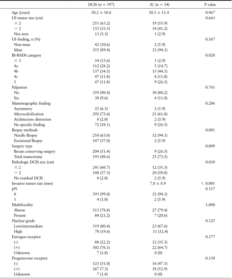
Table 1. Clinico-Pathological Features of 431 Patients with a Preoperative Histopathologic Diagnosis of DCIS
DCIS (n = 397) IC (n = 34) P value
Age (years) 50.2 ± 10.6 50.3 ± 11.4 0.967
US tumor size (cm) 0.663
≤ 2 251 (63.2) 19 (55.9)
> 2 133 (33.5) 14 (41.2)
Not seen 13 (3.3) 1 (2.9)
US finding, n (%) 0.567
Non-mass 42 (10.6) 2 (5.9)
Mass 355 (89.4) 32 (94.1)
BI-RADS category 0.028
≤ 3 54 (13.6) 1 (2.9)
4a 112 (28.2) 5 (14.7)
4b 137 (34.5) 15 (44.1)
4c 47 (11.8) 4 (11.8)
5 47 (11.8) 9 (26.5)
Palpation 0.761
No 359 (90.4) 30 (88.2)
Yes 38 (9.6) 4 (11.8)
Mammographic finding 0.286
Asymmetry 25 (6.3) 2 (5.9)
Microcalcification 292 (73.6) 21 (61.8)
Architecture distorsion 8 (2.0) 2 (5.9)
No specific finding 72 (18.1) 9 (26.5)
Biopsy methods 0.001
Needle Biopsy 250 (63.0) 32 (94.1)
Excisional Biopsy 147 (37.0) 2 (5.9)
Surgery type 0.009
Breast conserving surgery 204 (51.4) 9 (26.5)
Total mastectomy 193 (48.6) 25 (73.5)
Pathologic DCIS size (cm) 0.010
≤ 2 241 (60.7) 12 (35.3)
> 2 148 (37.3) 20 (58.8)
No residual DCIS 8 (2.0) 2 (5.9)
Invasive tumor size (mm) - 7.0 ± 8.9 < 0.001
pN 0.117
0 393 (99.0) 32 (94.1)
1 4 (1.0) 2 (5.9)
Multifocality 1.000
Absent 313 (78.8) 27 (79.4)
Present 84 (21.2) 7 (20.6)
Nuclear grade 0.125
Low/intermediate 319 (80.4) 23 (67.6)
High 78 (19.6) 11 (32.4)
Estrogen receptor 0.177
(-) 88 (22.2) 12 (35.3)
(+) 302 (76.1) 22 (64.7)
Unknown 7 (1.8) 0 (0)
Progesterone receptor 0.130
(-) 123 (31.0) 16 (47.1)
(+) 267 (7.3) 18 (52.9)
Unknown 7 (1.8) 0 (0)
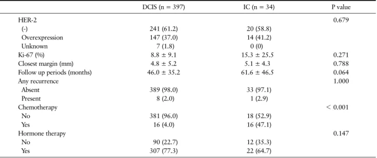
Table 1. Continued
DCIS (n = 397) IC (n = 34) P value
HER-2 0.679
(-) 241 (61.2) 20 (58.8)
Overexpression 147 (37.0) 14 (41.2)
Unknown 7 (1.8) 0 (0)
Ki-67 (%) 8.8 ± 9.1 15.3 ± 25.5 0.271
Closest margin (mm) 4.8 ± 5.2 5.1 ± 4.3 0.788
Follow up periods (months) 46.0 ± 35.2 61.6 ± 46.5 0.064
Any recurrence 1.000
Absent 389 (98.0) 33 (97.1)
Present 8 (2.0) 1 (2.9)
Chemotherapy < 0.001
No 381 (96.0) 18 (52.9)
Yes 16 (4.0) 16 (47.1)
Hormone therapy 0.147
No 90 (22.7) 12 (35.3)
Yes 307 (77.3) 22 (64.7)
Data are expressed as mean ± standard deviations or n (%).
DCIS = ductal carcinoma in situ; HER-2 = human epidermal growth factor receptor 2; IC = invasive component; US = ultrasonography.
4. 통계 분석
모든 통계 분석은 R version 3.3.2를 이용하였 다.(13,14) 관상피내암 환자와 침윤암 환자에서 범주형 변 수는 n (%)로 나타내었고, Fisher’s exact test 또는 Chi-square test를 이용하여 분석하였다. 연속형 변수는 mean ± standard deviation으로 나타내었고, Student’s t-test가 이용하여 검정하였다. 단변수와 다변수 분석을 위해 로지스틱 회귀분석을 사용하여 Odds ratio (95% CI) 를 추정하였다. 모든 검정은 양측 검정, P-value < 0.05 일 때 유의성이 있는 것으로 판단하였다.
결 과
1. 대상 환자의 특성
431명 환자들의 특성이 Table 1에 나타나 있다. 이러한 특성은 관상피내암을 진단 받은 환자군과 침윤암을 포함 하는 환자군에서 비교되었다. 수술 전 관상피내암을 진단 받은 환자 431명 중에서 92.1%인 397명은 수술 후 조직검 사에서 관상피내암으로 진단되었고, 7.9%인 34명의 환자 들은 관상피내암과 침윤암을 가지고 있다고 진단되었다.
RADS category (P = 0.028), 조직검사방법(needle bi- opsy or excisional biopsy, P = 0.001), 수술유형(breast- conserving surgery or total mastectomy, P = 0.009), 병리학적 관상피내암의 크기(≤ 2 cm, > 2 cm, or no re- sidual DCIS, P = 0.010)가 두 환자군에서 유의미하게 차 이가 있었다.
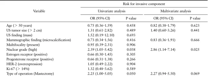
2. 수술 전 바늘 생검으로 관상피내암을 진단받은 환자에서 침윤암의 임상-병리학적 예측인자
수술 전 바늘 생검(needle biopsy)을 통해 관상피내암 을 진단 받은 282명의 환자들 중 절제생검(excisional bi- opsy)을 통해 수술 전 관상피내암을 진단 받은 149명의 환 자들은 제외되었다. 수술 중에 획득된 검체에서 침윤암을 예측할 수 있는 임상 병리학적 예측인자를 평가하였다. 단 변수분석과 다변수분석에서 핵분화도(OR = 2.39, 95%CI; 1.05-5.42, P = 0.038 in univariate analysis; aOR
= 2.86, 95% CI; 1.14-7.14, P = 0.025 in multivariate analysis)만이 유일하게 침윤암의 유의미한 예측 인자였 다(Table 2). 연령, 초음파상 종양의 크기, 초음파 소견 (mass
vs
. non-mass) (Fig. 1), 유방촬영상 소견, 다병소 성(multifocality), ER, PR, HER-2, Ki-67, 수술의 유Table 2. Clinico-Pathological Predictors of Invasive Component in Patients with Preoperative Needle Biopsy-Based Diagnosis of DCIS
Variable
Risk for invasive component
Univariate analysis Multivariate analysis
OR (95% CI) P value OR (95% CI) P value
Age (> 50 years) 0.75 (0.36–1.59) 0.458 0.82 (0.38–1.79) 0.623
US tumor size (> 2 cm) 1.31 (0.61–2.82) 0.489 1.40 (0.60–3.26) 0.441
US finding (mass) 1.52 (0.19–12.10) 0.693
Mammographic finding (microcalcification) 0.73 (0.34–1.56) 0.416 0.83 (0.36–1.91) 0.666
Multifocality (present) 0.95 (0.39–2.31) 0.906
Nuclear grade (high) 2.39 (1.05–5.42) 0.038 2.86 (1.14–7.14) 0.025
Estrogen receptor (positive) 0.66 (0.30–1.45) 0.305
Progesterone receptor (positive) 0.66 (0.31–1.38) 0.266
HER-2 (overexpression) 1.05 (0.49–2.22) 0.904
Ki-67 (> 14%) 1.32 (0.48–3.62) 0.589
Type of operation (Mastectomy) 2.25 (1.00–5.05) 0.050 2.27 (0.94–5.50) 0.069
DCIS = ductal carcinoma in situ; HER-2 = human epidermal growth factor receptor 2; US = ultrasonography.
Fig. 1. Non-mass vs. mass lesion on pre-operative breast sonography. (A) Non mass lesion on pre-operative breast US, left breast 10H, BI-RADS C4a, diagnosed of DCIS on CNB, (B) Mass lesion on pre-operative breast US, left breast 12H, subareolar area, BI-RADS C4a, diagnosed of DCIS on CNB. BI-RADS = breast imaging- reporting and data system; CNB = core needle biopsy; DCIS = ductal carcinoma in situ; US = ultraso- nography.
고 찰
이 연구는 수술 전에 관상피내암으로 진단된 환자에게 서 수술 후에 침윤성 유방암으로 변경 될 수 있는 예측인자 에 대해서 분석하였다. 이전의 연구에서는 나이, 체질량 지수, 유방촬영술상의 의심 소견, 촉지되는 소견, 유두 분 비물, 바늘 생검상 채취된 조직의 개수, 석회질의 범위, 종 양의 크기, 핵등급, 액와 림파선 전이, 다중심성 병소, 반 대측 유방의 병소, HER-2 과발현 등이 위험인자라고 보 고하였다.(3,6-12,15-29) 하지만 이러한 연구들은 대부 분 소규모를 대상자로 한 후향적 연구였으며, 결과들이 일 치하지 않아 현재 임상적으로 이용할 수 있는 예측인자는 없다. 따라서 비교적 많은 수를 대상으로 하는 이 연구를 통해 이러한 한계점을 극복하고자 하였다.
현재 예측인자로서의 나이에 대한 가치는 불분명하다.
Yen 등(15)은 55세 이하의 나이가 관상피내암에서 침윤암 으로 병기상승의 예측인자라고 하였다. 하지만 Nori 등 (11)은 반대의 결과를 보고하였는데, 55세이상의 나이가 오히려 침윤암의 예측인자라고 하였다. 이러한 상반된 결 과 및 다른 대부분의 연구들에서 나이를 유의한 예측인자 로 보고하지 않은 점을 종합했을 때, 나이는 예측인자로 보기 힘들며 이것은 우리의 연구 결과와도 일치한다.
핵등급의 예측적 가치에 대해서도 다양한 결과들이 보 였었다. 본 연구에서 핵등급은 침윤암을 예측하는 유일한 병리학적 인자였다(OR = 2.39, CI = 1.05-5.42, P = 0.038 in univariate analysis; OR = 2.86, CI = 1.14-7.14, P = 0.025 in multivariate analysis). Guillot 등(6)은 고 등급의 핵등급을 가진 관상피내암은 수술 후 침윤암으로 병기상승이 될 가능성이 높다고 하였다. Brennan 등(21) 도 고등급의 관상피내암은 수술 전 검사에서 침윤암이 관
상피내암으로 저평가되는 위험 요소라고 하였다. 그러나 다른 연구들에서는 핵등급을 예측인자로서의 가치가 없 다고 분석하였다.(3,15,30) 이러한 일관되지 않은 결과의 이유는 암 미세침윤(microinvasion)에 대한 병리 의사간 해석의 다양성 때문일 가능성이 크다. 따라서 이론적 암의 진화과정을 고려했을 때 핵등급의 예측가치는 있어 보이 며, 앞으로의 연구에서 면포상 괴사(comedo-necrosis) 와 같은 인자들과 함께 고려하여 분석 한다면 더 확실한 결과를 알 수 있을 것이다.
큰 종양의 크기는 침윤암으로의 병기상승의 예측인자 로 알려져 왔다. 유방촬영술상의 병변의 크기가 예측인자 로 작용한다는 연구도 있었고,(10,24) Lee 등(31)은 초음 파상 종양의 크기가 20 mm이상일 때 침윤암일 가능성이 높다고 하였다. Park 등(10)은 32 mm이상의 초음파상 종 양의 크기의 관상피내암이 수술 후 침윤암으로 병기 상승 의 위험성이 있다고 하였고, MRI와 유방 촬영술상의 크기 도 관련성이 있다고 보고하였다.(9) 이러한 이론적 배경 은 조직검사 대상이 되는 범위가 클수록 바늘 검사의 채취 량이 모든 범위를 반영하기 힘들어져, 결국 침윤성 암세포 를 채취하지 않게 될 개연성이 높아진다는 것이다. 하지만 우리의 연구에서는 초음파상 종양의 크기 및 침윤암의 위 험성은 통계적으로 유의한 연관성을 보이지 않았다. 이처 럼 다른 결과를 보이는 이유는, 생검 시 사용하는 바늘 종 류 및 채취 횟수의 차이, 술자 및 병리 의사의 숙련도, 불 충분한 연구 대상자 수가 원인일 수 있을 것이다. 본 연구 결과와는 다르게 초음파상의 mass 형성 여부(sonographic mass)와 높은BI-RADS 범주는 기존연구에서 침윤암의 유의미한 예측인자로 보고되고 있고, 초음파에서 병변의 여러 특징들(shape, orientation, margin, Echo pat- tern, posterior feature, calcification)은 병리학적 악성 병변(malignant lesion)을 예측하는 인자로서 평가의 대 상이 되고 있으나, 일관된 결과를 보이지는 않는 다.(32-34) 이는 초음파의 판독이 보이는 형태학적 특징 을 기반으로 이루어지기 때문에 병변의 병리를 정확하게 반영하는 데 한계점이 있기 때문으로 생각된다.(34) 형태 학적 특징을 기반으로 하는 기존 초음파의 판독에 보조적 인 정보를 제공하여 위양성율을 줄일 수 있는 횡탄성 초음 파의 사용은 이러한 초음파의 악성병변 예측에 대한 진단
최근 유방암의 탄성초음파(elastography)의 임상적 유 용성에 대한 연구가 보고되고 있다. Youn 등(35)은 많은 고형 병변들을 양성으로 판정하여 낮은 특이도(specifi- city)를 갖는 유방초음파의 제한점을 극복하는데 탄성초 음파(elastography)가 사용되고 있으며, 양성과 악성 병 변의 구별 뿐만 아니라 선행항암화학요법의 반응 예측, 유 방암 환자의 예후 예측에 임상적으로 매우 유용하다고 하 였다. Vinnicombe 등(36)은 정량적 전단파 탄성초음파 (quantitative SWE)에서 양성으로 분류된 유방암이 더 종양(tumor)의 크기가 작고(≤ 10 mm), 저등급이거나 순 수 관상피내암일 가능성이 높다고 하였다. 또한 전단파 탄 성초음파상 종양의 단단한 정도(stiffness)는 초음파 유 도 중심 바늘 생검으로 관상피내암이 진단된 환자에서 잠 재적 침윤(occult invasion)의 예측인자로 보고되고 있다 는 점에서,(37) 탄성초음파의 악성과 양성 감별을 위한 임 상적 유용성에 더하여 추후 침윤암의 예측인자로서의 추가 적인 연구들이 더욱 활발히 진행되어야 한다고 생각된다.
이 연구의 한계점은, 후향적 연구로서 생검을 시행한 술자가 다수여서 숙련도가 일치하지 않는다는 것이다. 또 한 진단력에 차이가 있을 수 있는 중심부 바늘 생검과 진공 흡인 보조 생검간의 바늘 굵기 차이나 생검 횟수의 차이가 반영이 되지 않았다는 점이다. 그러나 본 연구는 상대적으 로 이전 연구에 비해 많은 대상자를 분석하여, 불일치 된 결과에 대한 종합적인 결론 형성에 기여할 것으로 생각된다.
결 론
본 연구는 수술 전 조직검사에서 관상피내암이 진단되 었을 때 고등급의 핵등급만이 유일한 수술 후 침윤암으로 의 병기상승을 예측할 수 인자라는 것이다. 또한 이전의 연구들에서 관련이 있는 인자였던 초음파 상의 종양의 크 기는 관련이 없었다는 점에서 볼 때, 술자의 숙련도나 바 늘 생검의 종류 및 방법에 따라 많은 차이가 있을 수 있다 는 가정을 해 볼 수 있다. 앞으로의 더 많은 연구를 통해 침윤암으로의 병기 상승의 예측인자를 파악하여, 2차 수 술을 피하고 방사선 치료 및 항암화학요법의 예측하지 못 했던 시행으로 인한 유방 재건술의 합병증의 빈도를 낮추기 위한 수술 전 계획 수립을 효과적으로 할 수 있을 것이다.
CONFLICT OF INTEREST
The authors declare that they have no conflict of interest.
AUTHORS’ CONTRIBUTIONS
JWH, EYK, YLP and JSY collected and interpreted the patient data, and KHL was a major contributor in writing the manuscript. CHP inspired the topic se- lection and led the authors. All authors read and ap- proved the final manuscript.
REFERENCES
1. World Cancer Research Fund, American Institute for Cancer Research. Breast cancer statistics: breast cancer is the most common cancer in women world- wide [Internet]. London: World Cancer Research Fund 2019 [cited 2019 Oct 30]. Available at: https://
www.wcrf.org/dietandcancer/cancer-trends/breast- cancer-statistics.
2. Youn HJ, Ahn HR, Kang SY, Jung SH. Efficacy of ul- trasonography in breast cancer screening. J Surg Ultrasound 2019;6:1-7.
3. Dillon MF, McDermott EW, Quinn CM, O’Doherty A, O’Higgins N, Hill AD. Predictors of invasive disease in breast cancer when core biopsy demonstrates DCIS only. J Surg Oncol 2006;93:559-63.
4. Maffuz A, Barroso-Bravo S, Nájera I, Zarco G, Alvarado-Cabrero I, Rodríguez-Cuevas SA. Tumor size as predictor of microinvasion, invasion, and ax- illary metastasis in ductal carcinoma in situ. J Exp Clin Cancer Res 2006;25:223-7.
5. Meijnen P, Oldenburg HS, Loo CE, Nieweg OE, Peterse JL, Rutgers EJ. Risk of invasion and axillary lymph node metastasis in ductal carcinoma in situ di- agnosed by core-needle biopsy. Br J Surg 2007;94:
952-6.
6. Guillot E, Vaysse C, Goetgeluck J, Falcou MC, Couturaud B, Fitoussi A, et al. Extensive pure ductal carcinoma in situ of the breast: identification of pre- dictors of associated infiltrating carcinoma and lymph node metastasis before immediate reconstructive surgery. Breast 2014;23:97-103.
7. Goto M, Yuen S, Akazawa K, Nishida K, Konishi E, Kajihara M, et al. The role of breast MR imaging in pre-operative determination of invasive disease for ductal carcinoma in situ diagnosed by needle biopsy.
Eur Radiol 2012;22:1255-64.
8. Mori N, Ota H, Mugikura S, Takasawa C, Tominaga J, Ishida T, et al. Detection of invasive components in cases of breast ductal carcinoma in situ on biopsy by using apparent diffusion coefficient MR parameters.
Eur Radiol 2013;23:2705-12.
9. Wisner DJ, Hwang ES, Chang CB, Tso HH, Joe BN, Lessing JN, et al. Features of occult invasion in biop- sy-proven DCIS at breast MRI. Breast J 2013;
19:650-8.
10. Park AY, Gweon HM, Son EJ, Yoo M, Kim JA, Youk JH. Ductal carcinoma in situ diagnosed at US-guided 14-gauge core-needle biopsy for breast mass: pre- operative predictors of invasive breast cancer. Eur J Radiol 2014;83:654-9.
11. Nori J, Meattini I, Giannotti E, Abdulcadir D, Mariscotti G, Calabrese M, et al. Role of preoperative breast MRI in ductal carcinoma in situ for prediction of the presence and assessment of the extent of occult invasive component. Breast J 2014;20:243-8.
12. Lee CW, Wu HK, Lai HW, Wu WP, Chen ST, Chen DR, et al. Preoperative clinicopathologic factors and breast magnetic resonance imaging features can pre- dict ductal carcinoma in situ with invasive com- ponents. Eur J Radiol 2016;85:780-9.
13. R Core Team. R: a language and environment for statistical computing [Internet]. Vienna: R Foundation for Statistical Computing 2016 [cited 2019 Feb 20].
Available at: https://www.R-project.org/.
14. Therneau TM. A package for survival analysis in S.
version 2.38. [Internet]. Vienna: R Foundation for Statistical Computing 2015 [cited 2019 Feb 20].
Available at: https://CRAN.R-project.org/package=
survival.
15. Yen TW, Hunt KK, Ross MI, Mirza NQ, Babiera GV, Meric-Bernstam F, et al. Predictors of invasive breast cancer in patients with an initial diagnosis of ductal carcinoma in situ: a guide to selective use of sentinel lymph nodebiopsy in management of ductal carcinoma in situ. J Am Coll Surg 2005;200:516-26.
16. Rutstein LA, Johnson RR, Poller WR, Dabbs D, Groblewski J, Rakitt T, et al. Predictors of residual invasive disease after core needle biopsy diagnosis of ductal carcinoma in situ. Breast J 2007;13:251-7.
17. Go EM, Chan SK, Vong JS, Lui PC, Chan AW, Ma TK, et al. Predictors of invasion in needle core biopsies of the breast with ductal carcinoma in situ. Mod Pathol 2010;23:737-42.
18. Chan MY, Lim S. Predictors of invasive breast cancer in ductal carcinoma in situ initially diagnosed by core biopsy. Asian J Surg 2010;33:76-82.
19. Huang YT, Cheung YC, Lo YF, Ueng SH, Kuo WL, Chen SC. MRI findings of cancers preoperatively di- agnosed as pure DCIS at core needle biopsy. Acta Radiol 2011;52:1064-8.
20. Han JS, Molberg KH, Sarode V. Predictors of invasion and axillary lymph node metastasis in patients with a
core biopsy diagnosis of ductal carcinoma in situ: an analysis of 255 cases. Breast J 2011;17:223-9.
21. Brennan ME, Turner RM, Ciatto S, Marinovich ML, French JR, Macaskill P, et al. Ductal carcinoma in situ at core-needle biopsy: meta-analysis of under- estimation and predictors of invasive breast cancer.
Radiology 2011;260:119-28.
22. Kim J, Han W, Lee JW, You JM, Shin HC, Ahn SK, et al. Factors associated with upstaging from ductal carcinoma in situ following core needle biopsy to in- vasive cancer in subsequent surgical excision. Breast 2012;21:641-5.
23. Hollingsworth AB, Stough RG. Multicentric and con- tralateral invasive tumors identified with pre-op MRI in patients newly diagnosed with ductal carcinoma in situ of the breast. Breast J 2012;18:420-7.
24. Trentin C, Dominelli V, Maisonneuve P, Menna S, Bazolli B, Luini A, et al. Predictors of invasive breast cancer and lymph node involvement in ductal carcino- ma in situ initially diagnosed by vacuum-assisted breast biopsy: experience of 733 cases. Breast 2012;
21:635-40.
25. Nadrljanski M, Maksimović R, Plešnac-Karapandžć V, Nikitović M, Marković-Vasiljković B, Milošvić Z.
Positive enhancement integral values in dynamic con- trast enhanced magnetic resonance imaging of breast carcinoma: ductal carcinoma in situ vs. invasive duc- tal carcinoma. Eur J Radiol 2014;83:1363-7.
26. Deurloo EE, Sriram JD, Teertstra HJ, Loo CE, Wesseling J, Rutgers EJ, et al. MRI of the breast in patients with DCIS to exclude the presence of invasive disease. Eur Radiol 2012;22:1504-11.
27. Wilkie C, White L, Dupont E, Cantor A, Cox CE. An update of sentinel lymph node mapping in patients with ductal carcinoma in situ. Am J Surg 2005;
190:563-6.
28. Goyal A, Douglas-Jones A, Monypenny I, Sweetland H, Stevens G, Mansel RE. Is there a role of sentinel lymph node biopsy in ductal carcinoma in situ?: anal- ysis of 587 cases. Breast Cancer Res Treat 2006;98:
311-4.
29. Lee JM, Kaplan JB, Murray MP, Mazur-Grbec M, Tadic T, Stimac D, et al. Underestimation of DCIS at MRI-guided vacuum-assisted breast biopsy. AJR Am J Roentgenol 2007189:468-74.
30. Santamaría G, Velasco M, Farrús B, Zanón G, Fernández PL. Preoperative MRI of pure intraductal breast carcinoma--a valuable adjunct to mammog- raphy in assessing cancer extent. Breast 2008;17:
186-94.
31. Lee JW, Han W, Ko E, Cho J, Kim EK, Jung SY, et al.
Sonographic lesion size of ductal carcinoma in situ as a preoperative predictor for the presence of an in- vasive focus. J Surg Oncol 2008;98:15-20.
32. Kim S, Kim J, Park HS, Kim HY, Lee K, Lee J, et al.
An updated nomogram for predicting invasiveness in preoperative ductal carcinoma in situ of the breast.
Yonsei Med J 2019;60:1028-35.
33. Si J, Yang B, Guo R, Huang N, Quan C, Ma L, et al.
Factors associated with upstaging in patients pre- operatively diagnosed with ductal carcinoma in situ by core needle biopsy. Cancer Biol Med 2019;16:312-8.
34. Kim YS, Park WS, Min SY. Diagnostic usefulness of shear-wave elastography for an evaluation of BI-RADS category 4a breast lesions detected by US. J Surg Ultrasound 2017;4:48-54.
35. Youn HJ, Kang SY, Jung SH. Clinical application of shear wave elastography in patients with breast cancer. J Surg Ultrasound 2018;5:33-8.
36. Vinnicombe SJ, Whelehan P, Thomson K, McLean D, Purdie CA, Jordan LB, et al. What are the character- istics of breast cancers misclassified as benign by quantitative ultrasound shear wave elastography? Eur Radiol 2014;24:921-6.
37. Evans A, Purdie CA, Jordan L, Macaskill EJ, Flynn J, Vinnicombe S, et al. Stiffness at shear-wave elastog- raphy and patient presentation predicts upgrade at surgery following an ultrasound-guided core biopsy diagnosis of ductal carcinoma in situ. Clin Radiol 2016;71:1156-9.