1 순천향대학교 의과대학 이비인후과학교실 , 2 연세대학교 의과대학 이비인후과학교실
박진수1, 박무균1, 이종대1, 이원상2
1 Department of Otolaryngology and Head & Neck Surgery, College of Medicine, Soonchunhyang University Korea
2 Department of Otolaryngology and Head & Neck Surgery, College of Medicine, Yonsei University, Korea
Jin Su Park, MD1, Moo Kyun Park, MD1, Jong Dae Lee, MD1, Won-Sang Lee, MD, PhD2
청신경종양에서 Vascular Endothelial Growth Factor의 발현
Expression of Vascular Endothelial Growth Factor in Acoustic Neuroma
J Korean Skull Base Society 7권 2호 : 17~20, 2012
Objectives : VEGF(vascular endothelial growth factor) is a potent key mediator of angiogenesis, which is necessary during tumor growth. The goal of this study was to determine expression of VEGF in acoustic neuromas and to examine a correlation with clinical characteristics.
Methods : The VEGF expression was evaluated by immunohistochemical staining with monoclonal anti-VEGF antibody in 25 cases of the acoustic neuromas.
Results : All tumors showed expression of VEGF in cytoplasm of schwannoma cell. The staining density revealed an insignificant correlation between VEGF expression and tumor size, symptom duration.
Conclusions : This study suggests that VEGF in acoustic tumor has a role of both angiogenesis and mitogenesis in the growth of schwannoma.
논문 접수일 : 2012년 11월 10일 심사 완료일 : 2012년 11월 30일 주소 : 420-767 순천향대학교 부천병원
이비인후과
경기도 부천시 원미구 중동 1174 Tel : 82-32-621-5054
Fax : 82-32-621-5016 E-mail : [email protected]
이 종 대
교신저자
Acoustic neuroma, Vascular endothelial growth factor
Key Words
17
청신경종양에서 Vascular Endothelial Growth Factor의 발현
종설1 증례1 증례2 증례3 증례4
18 JOURNAL OF KOREAN SKULL BASE SOCIETY DECEMBER |Vol. 7 |No. 2
▒
서 론청신경종양은 8번째 뇌신경의 전정신경분지의 신경초세포 (schwann cell)에서 유래하는 종양으로 비교적 느리게 성장한다.
청신경종양의 약 30~90%가 다양한 속도로 성장한다고 보고되고 있다.1)따라서 성장에 영향을 미치는 성장인자(growth factor)가 있을 것으로 생각되며 몇몇 성장인자에 대한 연구가 진행되어 왔 다.2, 3)
종양의 성장에 있어 신생혈관생성(angiogenesis)은 필수적인 과 정으로, 그 중 VEGF(vascular endothelial growth factor)는 혈관 생성에 중요한 매개체로 알려져 있어 종양의 성장에 중요한 인자로 생각된다.4)VEGF는 다양한 종양에서 발현되며 또한 일부 종양세포 에서는 세포증식을 일으키는 인자(mitogen)로도 알려져 있다.5)
본 연구는 청신경종양에서 VEGF의 발현여부를 알아보고 종양의 크기, 증상기간 등의 임상적 특성과 비교하였다.
▒
대상 및 방법2006년부터 2010년까지 청신경종양으로 진단받고 수술치료를 시행받은 25예를 대상으로 하였다. 남녀 비율은 2:3으로 여성에서
많았으며 환자군의 연령은 29세에서 65세까지 다양하였고 평균 연 령은 47.8세였다.
각 환자의 종양조직에서 VEGF에 대한 면역조직화학염색을 시행 하였다. 조직의 파라핀블록에서 xylene으로 탈 파라핀을 시행하고 알콜로 함수과정을 거친 후 pH 7.6의 TBS(tris buffered saline)로 세척하였다. 항원성 회복을 위해 0.1M citrate 용액에 담궈 20분간 가열한 뒤 실온에서 천천히 식혀주었다. 내인성 peroxidase를 억제 하기 위하여 3% 과산화수소와 메탄올에 10분간 처리한 후 TBS로 세척하였다. 항체에 대한 비특이적 반응을 억제하기 위하여 0.5%
토끼 혈청(Dako, CA, USA) 용액으로 1시간 실온에서 처리하였다.
슬라이드에서 혈청을 제거한 뒤 VEGF의 경우 rabbit polyclonal antibody(Santa Cruz biotechnology, Inc, USA)을 1:100으로 희 석한 후 4°C에서 16시간 처리하였다. 후에 TBS에 세척 후 이차항체 로 anti-rabibit immunoglobulin(Envision kit, Glostrup, Denmark)을 가하여 실온에서 30분간 반응시킨 후 TBS로 세척하 였다. DAB(3,3′-diaminobenzidien-tetra-hydrochloride) 용액 으로 현미경하에서 발색을 확인한 후 증류수로 세척하고, 대조염색 은 Hematoxylin을 사용한 후 현미경하에서 발색부위를 관찰하였 다. 음성대조염색은 일차항체 대신에 TBS를 사용하여 시행하였다.
양성대조는 전립선암 조직을 대상으로 하였다. 염색된 표본은 고배
Immunohistochemical stain for VEGF in acoustic tumors
A. Negative control
B. Prostate cancer tissue as positive control showing dark brown stain
C. Weak positive expression D. Strong postive expression(×200)
A
D B
C Fig. 1
19
청신경종양에서 Vascular Endothelial Growth Factor의 발현
율 시야(200배)에서 염색된 부분을 기준으로 약(weak)양성과 강 (strong)양성으로 분류하였다(Fig.1).
본 연구는 SPSS 통계프로그램(14.0 version)을 이용하여 분석하 였고, 면역조직화학 염색 결과와 임상적 지표들과의 비교는 Pearson Chi-Square test를 사용하였으며, 유의수준은 5% 이내로 하였다.
▒
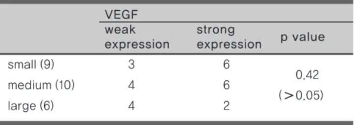
결 과면역조직화학검사상 25개의 종양에서 모두 VEGF에 발현하였으 며 VEGF에서 강양성을 나타낸 경우는 13례(52%), 약양성을 나타 낸 경우는 12례(48%)였다. 종양의 크기를 최대직경을 기준으로 10mm 이하를 소형으로, 11~25mm를 중형으로, 26mm 이상을 대 형으로 분류할 때 각각 9 : 10 : 6 예였으며, VEGF의 발현강도와 종 양의 크기와는 통계적인 유의성을 보이지 않았다(Table 1). 환자는 청력감소, 이명, 어지러움을 주 증상으로 하였으며 증상의 지속기간 을 1년 미만과 1년 이상으로 분류하였을 때 각각 8례와 17례였으며, 증상의 지속기간과 VEGF의 발현강도와 상관 관계에 있어서 통계적 인 유의성을 보이지 않았다(Table 2).
▒
고 찰청신경종양의 종양의 성장은 매우 다양한 형태로 나타나며 transforming growth factor, fibroblast growth factor 등의 성 장인자에 대한 연구가 보고되어 왔으나2)정확한 기전은 밝혀져 있지 않다.
혈관신생은 기존의 혈관으로부터 새로운 혈관을 만드는 과정으로 고형종양이 약 2~3mm3)의 크기 이상으로 성장하기 위해서는 혈관 신생이 반드시 필요하다.6)다양한 혈관신생인자 중에 매우 강력한 것으로 알려진 VEGF는 34~46kD의 당단백으로 혈관내피세포에 특이적으로 작용하며 121, 145, 165, 189, 206 등의 5가지 이형체 중
VEGF121, VEGF165가 세포에서 분비되어 기능을 하는 것으로 알 려져 있다.4, 5)VEGF가 혈관신생에 관여하는 기전으로는 혈관내피 세포에 존재하는 VEGFR(vascular endothelial growth factor receptor)-1(Flt-1, film-like tyrosine kinase-1)과 VEGFR- 2(KDR, kinase insert domain containing receptor)에 결합하여 세포분열을 촉진시켜 강력한 혈관신생인자로 작용하며, 미세혈관의 투과도를 증가시키는 매개자 역할을 하여 혈장단백이 주변 조직으 로 배출되어 세포외 기질을 변화시켜 혈관신생에 관여한다.
VEGF는 폐암7), 유방암8), 소화기암9)등의 매우 다양한 암에서 발 현됨이 알려졌고 몇몇 두개내 종양에서도 발현이 보고되었다.10)이 는 종양의 성장에 필수적인 혈관신생에 있어서 VEGF가 중요한 매 개체로 작용하고 있음을 알 수 있다. 청신경종양에서의 VEGF 발현 에 대한 연구도 발표되어, Caye-Thomasen 등11)은 18명의 청신경 종양에서 모두 VEGF가 발현되며 특히 VEGF의 발현양상이 청신경 종양의 성장속도와 밀접한 관련이 있음을 보고하였다. 하지만 Brieger 등12)은 34명의 청신경종양에서 단 1명만이 VEGF가 발현되 어 청신경종양의 성장에 있어서 혈관신생은 중요한 기전이 아님을 보고하였다. 본 연구의 결과는 전자의 결과와 일치하여 25명의 환자 에서 모두 VEGF가 발현됨을 알 수 있었으나 발현양상과 종양의 크 기, 증상지속기간과의 관계에서 통계적인 의의는 없었다.
VEGF는 주로 내피세포(endothelial cell)에서 세포증식을 일으키 지만 췌장분비세포13), 망막색소세포14), 유방암세포15)등의 내피세포 외에서도 세포증식을 일으키는 인자(mitogen)로도 작용한다고 알 려져 있다. Sondell 등16)은 말초신경계의 세포에도 신경영양작용 (neutrophic)과 세포증식인자(mitogen)로 작용한다고 보고하였다.
현재 암을 치료하는 데 있어서 VEGF를 표적으로 하여 혈관생성 및 증식을 억제함으로써 암을 치료하는 다양한 치료가 시행되고 있 다.17)현재 청신경종양이 주 표현형인 신경섬유종 2형 환자에서도 VEGF를 표적으로 하는 Bevacimumab이 일부 환자에서 효과가 있 다고 보고된 후 2형 신경섬유종증 환자에서 Bevacimumab를 이용 한 임상시험이 몇몇 국가에서 진행중이다.18, 19)
small (9) 3 6
medium (10) 4 6 0.42
( 0.05)
large (6) 4 2
VEGF
weak strong
p value expression expression
Table 1. The relationship between expression of VEGF and tumor size
symptom 9 1yr (8) 3 5 0.86
symptom ≥ 1yr (17) 7 10 ( 0.05)
VEGF
weak strong
p value expression expression
Table 2. The relationship between expression of VEGF and symptom duration
20 JOURNAL OF KOREAN SKULL BASE SOCIETY DECEMBER |Vol. 7 |No. 2
▒
결 론본 연구를 통해 종양의 성장에 있어 필수적인 강력한 혈관신생인 자인 VEGF를 청신경종양에서 면역조직화학염색을 통해 그 발현을 알아본 결과 종양에서 모두 발현됨을 알 수 있었다. 따라서 청신경 종양세포에서 VEGF가 분비되어 이는 종양의 성장에 있어서 필요한 혈관생성에 관여함을 추론할 수 있었다.
References
1. Stangerup SE, Caye-Thomasen P, Tos M, Thomsen J. The natural history of vestibular schwannoma. Otol Neurotol 2006;27 :547-52 2. Weerda HG, Gamberger TI, Siegner A, Gjuric M, Tamm ER. Effects of
transforming growth factor-beta1 and basic fibroblast growth factor on proliferation of cell cultures derived from human vestibular nerve schwannoma. Acta otolaryngol (Stockh) 1998;118:337-43
3. Adams EF, Rafferty B, Mower J, Ward H, Petersen B, Fahlbusch R.
Human acoustic neuromas secrete interleukin-6 in cell culture:
possible autocrine regulation of cell proliferation. Neurosurgery 1994;35:434-8
4. Ferrara N. Molecular and biological properties of vascular endothelial growth factor. J Mol Med. 1999;77:527-43
5. Ferrara N. Vascular endothelial growth factor: Basic science and clinical progress. Endocrine Reviews 2005;25:581-611
6. Folkman J. What is the evidence that tumors are angiogenesis- dependent? J Natl Cancer Inst 1990;82:4-6
7. Volm M, Koomagi R, Mattern J. Prognostic value of vascular endothelial growth factor and its receptor Flt-1 in squamous cell lung cancer. Int J Cancer 1997; 74:64-68
8. Yoshiji H, Gomez DE, Shibuya M, Thorgeirsson UP. Expression of vascular endothelial growth factor, its receptor, and other angiogenic factors in human breast cancer. Cancer Res 1996;56:2013-16 9. Brown LF, Berse B, Jackman RW, Tognazzi K, Manseau EJ, Senger
DR, et al. Expression of vascular permeability factor (vascular endothelial growth factor) and its receptors in adenocarcinomas of the gastrointestinal tract. Cancer Res 1993;53:4727-35
10. Berkman RA, Merrill MJ, Reinhold WC, Monacci WT, Saxena A, Clark
WC, et al. Expression of the vascular permeability factor /vascular endothelial growth factor gene in central nervous system neoplasms.
J Clin Invest 1993;91:153-159
11. Cay -Thomasen P, Baandrup L, Jacobsen GK, Thomsen J, Stangerup SE. Immunohistochemical demonstration of vascular endothelial growth factor in vestibular schwannomas correlates to tumor growth rate. Laryngoscope 2003;113:2129-34
12. Brieger J, Bedavanija A, Lehr HA, Maurer J, Mann WJ. Expression of angiogenic growth factors in acoustic neurinoma. Acta Otolaryngol.
2003;123:1040-5
13. Oberg-Welsh C, Sandler S, Andersson A, Welsh M. Effects of vascular endothelial growth factor on pancreatic duct cell replication and the insulin production of fetal islet-like cell clusters in vitro. Mol Cell Endocrinol 1997;126:125-132
14. Guerrin M, Moukadiri H, Chollet P, Moro F, Dutt K, Malecaze F, et al.
Vasculotropin/vascular endothelial growth factor is an autocrine growth factor for human retinal pigment epithelial cells cultured in vitro. J Cell Physiol 1995;164:385-394
15. Liang Y, Brekken RA, Hyder SM. Vascular endothelial growth factor induces proliferation of breast cancer cells and inhibits the anti- proliferative activity of anti-hormones. Endocr Relat Cancer.
2006;13:905-19
16. Sondell M, Lundborg G, Kanje M. Vascular endothelial growth factor has neurotrophic activity and stimulates axonal outgrowth, enhancing cell survival and Schwann cell proliferation in the peripheral nervous system. J Neurosci 1999;19:5731-40
17. Toi M, Matsumoto T, Bando H. Vascular endothelial growth factor: its progostic, predictive, and therapeutic implications. Lancet Oncol 2001;2:667-73
18. Mautner VF, Nguyen R, Kutta H, Fuensterer C, Bokemeyer C, Hagel C, et al. Bevacizumab induces regression of vestibular schwannomas in patients with neurofibromatosis type 2. Neuro Oncol. 2010;12(1):14-8 19. Plotkin SR, Stemmer-Rachamimov AO, Barker FG 2nd, Halpin C,
Padera TP, Tyrrell A,et al. Hearing improvement after bevacizumab in patients with neurofibromatosis type 2. N Engl J Med. 2009;361(4) :358-67