INTRODUCTION
Middle ear adenoma is a rare benign epithelial tumor derived from middle ear mucosal cells and can present both a neuroen- docrine and an epithelial differentiation (1). Preoperative diag- nosis is not possible in most cases, because the clinical features and otoscopic findings are not typical. Although a few cases fo- cusing the imaging features of middle ear adenoma have been reported in the English literature (2, 3), and a few case reports fo- cusing the clinical aspects of middle ear adenoma have appeared in the Korean otolaryngology literature (4-6), imaging features of this rare entity at high-resolution CT and gadolinium (Gd)- enhanced MRI altogether in a single case have rarely been pre-
sented in the literature (2). We report herein a case of middle ear adenoma of neuroendocrine differentiation in a 36-year-old man along with CT, MRI and pathologic features.
CASE REPORT
A 36-year-old man presented with aggravated hearing loss of the left ear for the last 5 months. He had experienced mild hear- ing losses of the left ear without specific management since 10 years prior. Approximately 5 months prior, he had visited local medical clinic with aggravated hearing losses and intermittent otalgia of the left ear, and had medicated under the impression of otomastoiditis without significant symptomatic improve-
J Korean Soc Radiol 2013;69(3):187-191 http://dx.doi.org/10.3348/jksr.2013.69.3.187
Received April 27, 2013; Accepted June 14, 2013 Corresponding author: Sang Kwon Lee, MD Department of Radiology, Keimyung University School of Medicine, 56 Dalseong-ro, Jung-gu, Daegu 700-712, Korea.
Tel. 82-53-250-7735 Fax. 82-53-250-7766 E-mail: [email protected]
This is an Open Access article distributed under the terms of the Creative Commons Attribution Non-Commercial License (http://creativecommons.org/licenses/by-nc/3.0) which permits unrestricted non-commercial use, distri- bution, and reproduction in any medium, provided the original work is properly cited.
Middle ear adenoma is a rare benign epithelial tumor. We report the CT and mag- netic resonance imaging findings of a case of middle ear adenoma of neuroendo- crine differentiation in a 36-year-old man. On high-resolution CT, the mass was found to fill the middle ear, in which the ossicles were embedded, but not de- stroyed, with outward bulging of the intact tympanic membrane. On MRI, the mass, which was intensely enhanced on 3-dimensional (3D) gadolinium (Gd)-enhanced spoiled gradient-recalled (SPGR) sequence, involved the middle ear, aditus ad an- trum and a portion of mastoid antrum. Histological and immunohistochemical findings of the specimen obtained by surgical excisions were consistent with middle ear adenoma of neuroendocrine differentiation. Middle ear adenoma of neuroendo- crine differentiation should be included in the differential diagnosis of an intensely enhancing mass filling the middle ear/mastoid antrum without ossicular destruc- tions. The extent of the mass can be excellently assessed with 3D Gd-enhanced SPGR sequence.
Index terms Adenoma Middle Ear
Tomography, X-Ray Computed Magnetic Resonance Imaging
CT and Magnetic Resonance Imaging Features of Middle Ear Adenoma of Neuroendocrine Differentiation: A Case Report
1중이에서 발생한 신경내분비 분화 선종의 전산화단층촬영 및 자기공명영상 소견: 증례 보고1
Sang Kwon Lee, MD
1, Mi Sun Choe, MD
2Departments of 1Radiology, 2Pathology, Keimyung University School of Medicine, Daegu, Korea
findings were consistent with middle ear adenoma of neuroen- docrine differentiation. The postoperative audiometric assess- ment revealed improved hearing. His postoperative course was unremarkable without evidences of recurrence until 14 months after operation.
DISCUSSION
Two separate adenomatous tumors have been identified in the temporal bone: middle ear adenoma and aggressive papil- lary tumor (7). The histogenesis of the middle ear adenoma re- mains controversial, although the consensus tends toward a plu- ripotential stem cells of the middle ear mucosa as the origin of the lesion. It has been believed that neuroendocrine tumor and middle ear adenoma were separate entities. Recent studies sug- gest that middle ear adenoma with either epithelial or neuroen- docrine differentiation represents opposite ends of a spectrum of differentiation (8). The middle ear adenoma may express syn- aptophysin, chromogranin, and various polypeptides which are typical for neuroendocrine differentiations (9). Another adeno- matous lesion distinct from middle ear adenoma is an aggres- sive papillary tumor. It is a histologically benign tumor but it clinically shows aggressive growth patterns (10). It expresses cy- tokeratins, epithelial membrane antigen, and S100.
The middle ear adenoma, a rare benign tumor, is often mis- taken for more commonly chronic otomastoiditis or cholestea- toma clinically, as in our case. The overwhelming majority of middle ear adenoma does not invade the temporal bone. Metas- tasis does not occur (11). The architectural patterns of middle ear adenoma with neuroendocrine differentiation may be glan- dular, trabecular, or solid. The cells are cuboidal or columnar, and nuclei are round to oval with a “salt and pepper” chromatin pattern and inconspicuous nucleoli. Clinically, the most com- mon symptoms consist of conductive hearing loss, tinnitus and vertigo. Other symptoms include a sense of fullness in the af- fected ear, otalgia, and, rarely, the facial nerve palsy. The clinical presentation and otoscopic findings are not typical. Thus, the definitive diagnosis is based on histological and immunohisto- chemical examinations. The imaging features of middle ear ade- noma of neuroendocrine differentiation have not yet been clear- ly defined, because of its relative rarity. Maintz et al. (2) reported CT and/or MR imaging findings of three adenomatous tumors ments. He denied otorrhea, dizziness or vertigo and his facial
expression was intact. Otoscopic examination revealed a pro- truding mass behind the intact tympanic membrane (TM).
High resolution CT, performed using Sensation 64 scanner (Sie- mens Healthcare, Forchheim, Germany) with a slice thickness of 0.6 mm, showed a mass filling the middle ear, in which the ossicles were embedded, but not destroyed (Fig. 1A-C). Also be- ing noted were outward bulging of the intact TM (Fig. 1B, C) and complete opacification of the mastoid antrum and air cells (Fig. 1A-C). On the basis of CT findings, we suggested neoplas- tic conditions or cholesteatoma with associated otomastoiditis.
MRI was performed for better evaluations and for better assess- ments for the extent of the lesion by using a 3.0-T unit (Signa Excite; GE Medical System, Milwaukee, WI, USA). The mass was isointense to gray matter on axial fast spin echo T2-weight- ed [repetition time (TR)/echo time (TE) = 4800/111; echo train length = 19; matrix number/number of excitation = 512 × 224/3.0; slice thickness = 3.0 mm] (Fig. 1D) and 3-dimensional (3D) spoiled gradient-recalled (SPGR) sequences (TR/TE = 7.9/3.1; flip angle = 20; matrix number/number of excitation = 256 × 224/2; slice thickness = 1.6 mm) (Fig. 1E). In contrast, the opacified mastoid antrum and air cells were hyperintense on both sequences (Fig. 1D, E). Homogeneous and intense en- hancement of the mass, involving the entire middle ear cavity, aditus ad antrum, and a portion of mastoid antrum, was noted on Gd-enhanced 3D SPGR sequence (TR/TE = 7.9/3.1; flip angle = 20; matrix number/number of excitation = 256 × 224/2; slice thickness = 1.6 mm) (Fig. 1F, G). In view of CT and magnetic resonance (MR) imaging features, our tentative diagnoses were glomus tympanicum paraganglioma and other neoplastic con- ditions. He underwent tympanoplasty, canal wall up mastoidec- tomy, and ossiculoplasty with complete excision of tumor in the middle ear cavity, aditus ad antrum, and mastoid antrum. Intra- operatively, the malleus, incus and stapes superstructures were embedded in the yellowish mass, but they were not destroyed.
The incudostapedial articulation was subluxated. The mass did not invade the walls of tympanic cavity nor did it expose the fa- cial nerves. Histologically, the tumor displayed a trabecular growth pattern (Fig. 1H). The tumor cells were cuboidal, and the nuclei were round and uniform. Immunohistochemically, the tumor cells were positive for synaptophysin and chromo- granin (not shown). The histological and immunohistochemical
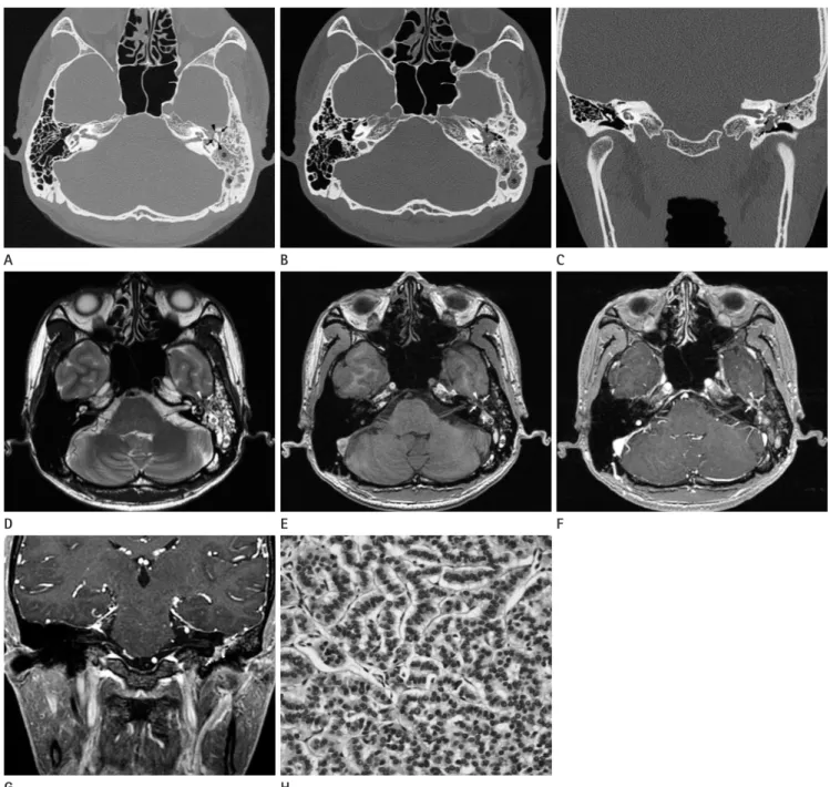
Fig. 1. CT, MRI, and pathologic features of middle ear adenoma of neuroendocrine differentiation in a 36-year-old man.
A, B. Axial high-resolution CT images show a soft tissue mass filling the middle ear (arrows), in which the ossicles (arrowheads) are embedded, but not destroyed. Also noted are outward bulging of the intact tympanic membrane (open arrow) and complete opacification of the mastoid antrum and air cells (asterisks).
C. Coronal high-resolution CT reveals the mass occupying entire middle ear (arrows). The incus and stapes (arrowheads) are embedded within the mass, but not destroyed.
D, E. Axial fast spin echo T2-weighted (D) and 3-dimensional (3D) spoiled gradient-recalled (SPGR) (E) sequences reveal the mass (arrows), isointense to the gray matter, filling the entire middle ear cavity, aditus ad antrum, and a portion of the mastoid antrum. In contrast, the fluid re- tention in the mastoid antrum and air cells (asterisks) is hyperintense on both sequences. Note that the hypointense malleus and incus (arrow- head) are embedded within the mass.
F. Axial gadolinium (Gd)-enhanced 3D SPGR sequence demonstrates homogeneous and intense enhancement of the mass (arrows) involving the entire middle ear cavity, aditus ad antrum, and a portion of the mastoid antrum. Fluid retention in the mastoid antrum and air cells (asterisks) shows varying signal intensities and the ossicles (arrowhead) are hypointense.
G. Coronal Gd-enhanced 3D SPGR sequence shows intact incus (arrowhead) embedded within the enhancing middle ear mass (arrows).
H. Histologically, the tumor demonstrates a trabecular growth pattern. The tumor cells are cuboidal, and nuclei are round and uniform (hematox- ylin-eosin, × 400).
B
E
H A
D
G
C
F
case of middle ear adenoma whose initial radiologic diagnosis favored glomus tympanicum paraganglioma versus cholesteato- ma. Bierry et al. (13) have noticed that a vascular blush was pres- ent on angiography in glomus tympanicum paraganglioma, but was absent in middle ear adenoma. A close relationship be- tween the tumor and the Jacobson’s nerve or its branches and pulsatile tinnitus were identified in glomus tympanicum para- ganglioma, but were absent in middle ear adenoma. Other dif- ferential diagnoses include schwannoma of the facial nerves and meningioma.
Complete surgical removal via the external auditory canal or radical mastoidectomy is the treatment of choice.
In summary, middle ear adenoma of neuroendocrine differen- tiation, a rare entity, should be included in the differential diagno- sis when managing inflammatory diseases that does not respond to conservative treatments, or in cases with unclear expansile pro- cesses of the middle ear, clinically, and an intensely enhanced mass filling the middle ear/mastoid antrum without ossicular destructions on high-resolution CT and MRI. The extent of the middle ear adenoma of neuroendocrine differentiation can be excellently assessed with high resolution 3D Gd-enhanced SPGR sequences.
REFERENCES
1. Torske KR, Thompson LD. Adenoma versus carcinoid tumor of the middle ear: a study of 48 cases and review of the literature. Mod Pathol 2002;15:543-555
2. Maintz D, Stupp C, Krueger K, Wustrow J, Lackner K. MRI and CT of adenomatous tumours of the middle ear. Neu- roradiology 2001;43:58-61
3. Zan E, Limb CJ, Koehler JF, Yousem DM. Middle ear adeno- ma: a challenging diagnosis. AJNR Am J Neuroradiol 2009;
30:1602-1603
4. Shim MJ, Song CI, Yoon TH. A case of middle ear adenoma.
Korean J Audiol 2012;16:27-30
5. Shin BS, Kim JR, Jeong HW, Kim EK. Two cases of middle ear adenoma with neuroendocrine differentiation (carcinoid tumor). Korean J Otorhinolaryngol-Head Neck Surg 2011;
54:573-577
6. Jun BH, Han YH, Yoon SP, Park SY. A case of the middle ear adenoma. Korean J Otolaryngol-Head Neck Surg 2000;43:
of the middle ear, one adenoma, one adenocarcinoma, and one aggressive papillary tumor, respectively. In all cases, they found a small intratympanic mass in which the ossicles were embed- ded on high-resolution CT. On MRI, the tumors were isointense or slightly hyperintense compared with white matters on T1- weighted images, and isointense to the gray matters on T2- weighted images, and all cases revealed contrast enhancement.
Notably, irrespective of biological behavior of the tumors, none showed destruction of the ossicles or walls of the tympanic cavi- ty. Zan et al. (3) reported high-resolution CT features of a case of middle ear adenoma. They also noticed a well-defined, lobu- lated, homogeneous, soft tissue-attenuation mass in which the ossicles were embedded, but not destroyed. Despite of fairly large volume of the middle ear adenoma that occupied entire middle ear cavity and a portion of mastoid antrum, the middle ear ossi- cles were not destroyed but embedded within the mass in our case. The reason why the ossicles are not destroyed but embed- ded within the tumor in overwhelming majority of the middle ear adenomas is not fully discussed in the literature (2-7). We retrospectively speculated and hypothesized that the soft fragile texture, un-encapsulated with well-delineated outline, and slow growth rate of the middle ear adenoma might be responsible for these findings (3, 5, 7). These typical imaging features of the middle ear adenoma may be helpful in the differentiation of the middle ear adenoma from cholesteatoma, paragangliomas, and malignant tumors of the middle ear cavity.
The middle ear adenoma may present diagnostic dilemma because chronic otomastoiditis and cholesteatoma, which com- prise the majority of middle ear diseases, may masquerade mid- dle ear adenoma on high-resolution CT, the most commonly used imaging modality for middle ear diseases (3, 12). Gd-en- hanced MRI may be useful in this context. In our case, the ex- tent of the tumor cannot be assessed with high-resolution CT, because the mass and fluid retention in the mastoid antrum and air cells had similar attenuation value, thus could not be distin- guished. 3D Gd-enhanced SPGR sequence enabled clear dis- tinction between the intensely enhanced masses in the middle ear, aditus ad antrum and a portion of mastoid antrum, and non- enhancing fluid retention of the mastoid antrum and air cells.
Glomus tympanicum paragangliomas may also mimic mid- dle ear adenoma (3, 13) in that both tumors can show intense enhancements on Gd-enhanced MRI. Zan et al. (3) reported a
Aggressive papillary middle-ear tumor. A clinicopathologic entity distinct from middle-ear adenoma. Am J Surg Pathol 1988;12:790-797
11. Saliba I, Evrard AS. Middle ear glandular neoplasms: ade- noma, carcinoma or adenoma with neuroendocrine dif- ferentiation: a case series. Cases J 2009;2:6508
12. Tomazic PV, Beham A, Lackner A, Ropposch T, Stockreiter U, Walch C. Neuroendocrine adenoma of the middle ear (NAME) mimicking as chronic otitis media with an episode of facial nerve palsy. B-ENT 2011;7:121-125
13. Bierry G, Riehm S, Marcellin L, Stierlé JL, Veillon F. Middle ear adenomatous tumor: a not so rare glomus tympani- cum-mimicking lesion. J Neuroradiol 2010;37:116-121 95-98
7. Duderstadt M, Förster C, Welkoborsky HJ, Ostertag H. Ade- nomatous tumors of the middle ear and temporal bone:
clinical, morphological and tumor biological characteristics of challenging neoplastic lesions. Eur Arch Otorhinolaryn- gol 2012;269:823-831
8. Devaney KO, Ferlito A, Rinaldo A. Epithelial tumors of the middle ear--are middle ear carcinoids really distinct from middle ear adenomas? Acta Otolaryngol 2003;123:678-682 9. Wassef M, Kanavaros P, Polivka M, Nemeth J, Monteil JP,
Frachet B, et al. Middle ear adenoma. A tumor displaying mucinous and neuroendocrine differentiation. Am J Surg Pathol 1989;13:838-847
10. Gaffey MJ, Mills SE, Fechner RE, Intemann SR, Wick MR.
중이에서 발생한 신경내분비 분화 선종의 전산화단층촬영 및 자기공명영상 소견: 증례 보고1
이상권
1· 최미선
2중이 선종은 드문 양성 상피성 종양이다. 저자들은 36세 남자의 중이에서 발생한 신경내분비 분화 선종 1예의 CT 및 MRI 소견을 보고하고자 한다. 고해상능 CT에서 종괴는 중이를 채웠으며, 내부에 골 파괴를 동반하지 않은 이소골이 묻혀 있었 고, 천공되지 않은 고막의 외측 돌출이 동반되어 있었다. 종괴는 조영증강 후 3차원 spoiled gradient-recalled (SPGR) 자 기공명영상에서 강한 조영증강을 보였으며, 중이, 유돌동구 및 유양동의 일부를 침범하였다. 수술적 절제에 의해 얻어진 검체의 조직학적 및 면역조직화학적 소견은 신경내분비 분화 중이 선종에 합당하였다. 신경내분비 분화 중이 선종은 중 이/유양동을 채우며, 이소골의 파괴를 동반하지 않으며, 강한 조영증강을 보이는 종괴의 감별진단에 포함되어야 할 것으 로 생각되며, 조영증강 후 3차원 SPGR 자기공명영상은 종괴의 범위를 평가하는 데 있어서 우수한 방법으로 생각된다.
계명대학교 의과대학 1영상의학교실, 2병리학교실