Address reprint requests to Kyung-Soo Suk, M.D.
Department of Orthopaedic Surgery, College of Medicine, Kyung Hee University
#1 Hoeki-Dong, Dongdaemun-ku, Seoul, Korea 130-702
Tel : 822-958-8345, Fax : 822-964-3865, E-mail : [email protected]
경추부 측괴나사못 고정술을 위한
경추부 측괴의 자기공명영상을 이용한 측정
석경수・김기택・이상훈・류경남*
경희대학교 의과대학 정형외과학교실, 진단방사선과학교실*
Measurements of Lateral Mass of Cervical Spine Using MRI for Lateral Mass Screw Fixation
Kyung-Soo Suk, M.D., Ki-Tack Kim, M.D., Sang-Hoon Lee, M.D., Kyung-Nam Ryu, M.D.*
Department of Orthopaedic Surgery, Diagnostic Radiology*, School of Medicine, Kyung Hee University, Seoul, Korea – Abstract –
Study design : Lateral mass was measured using MRI for lateral mass screw fixation
Objectives : To measure the lateral mass of cervical spine using MRI for lateral mass screw fixation and find out the ideal entry point and insertion angle and length of lateral mass screws.
Summary of Literature Review : Two methods of screw placement are in common use. The original technique, described by Ray- mond Roy-Camille, places the screw in a more or less straight sagittal direction and angling the screw laterally 10 to 20 degrees.
Margerl technique involves placing the screw parallel to the facet joint and angling the screw laterally 25 to 30 degrees.
Materials and Methods : Axial MR images of the cervical spine parallel to the facet joints were obtained from C3 to C6 of 10 patients.
The mean age of the patients were 48.0 years. The patients consisted of 6 male and 4 female patients. Ideal entry points, insertion angle and length of the lateral mass for lateral mass screw fixation were measured on the axial MR images using PACS digital measuring i n s t r u m e n t .
Results : Ideal entry point of a lateral mass screw was center of lateral mass in sagittal plane, 16mm lateral to the midline of the cervical spine, ideal direction of the lateral mass screw was parallel to the facet joint and angling the screw laterally 25.3 degrees, and ideal length of lateral mass screw was 17.9mm.
Conclusions : Based on the results of the study, there were some differences of measurements depending on the patients and the level of the cervical spine. Therefore, a preoperative measurement of lateral mass was recommended in each patient and each level of the cervical spine.
Key Words : Lateral mass screw fixation, Magnetic resonance image
Journal of Korean Spine Surg.
Vol. 9, No. 2, pp 121~126, 2002
서 론
경추부 측괴 나사못 및 금속판 고정술은 후방 감압술로
경추의 후궁 및 극돌기가 없거나 골절 등으로 결손이 있 는 경우와 같이 기존의 강선을 이용한 유합술이 불가능한 경우에 있어서 유용하게 이용할 수 있는 술식이다2 , 3 , 6 , 8 , 1 0 )
.
그러나 측괴 나사못 고정술은 척수, 신경근, 척추 동맥의 손상을 초래할 수 있는 위험성이 있다. 따라서 정확한 위
치, 삽입 각도 및 나사못의 길이 가 중요하며 특히 신경근 및 척추 동맥의 손상을 주지 않도록 측괴의 해부학에 대 한 완전한 이해가 필요하겠다. 본 연구에서는 안전한 양 측 피질골 고정 측괴 나사못 고정술에 도움을 주고자 경 추 측괴를 자기공명영상을 이용하여 측정하고자 하였다.
연구대상 및 방법
10명의 환자를 대상으로 경추부 자기공명영상을 시행
하였다. 자기공명영상은 제 3경추에서 제 6경추까지 경 추의 후관절에 평행한 T1 강조 축상면 영상(T1-weightedaxial image) 을 얻었다(Fig. 1). 대상 환자는 남자 6명, 여
자 4명이었으며 이들의 평균 연령은 4 8 . 0세 이었다. 상 기한 40개의 축상면 영상에서 좌우 80개의 측괴에서 측 괴 나사못의 삽입 위치, 삽입 각도 및 적절한 나사못의 길이를 PACS를 이용한 digital 계측을 이용하여 측정하 였다. 각 부위별 측정값을 전체 평균과 비교하여 O n e -sample T test로 통계 분석하였으며 성별 및 연령에 따른
측정값을 Mann-whitney U test로 통계 분석하였다.
결 과
제 3경추에 적절한 삽입 위치는 상하로는 측괴의 중앙 에 위치하고 좌우로는 정중선에서 15.5 mm에 위치하였 다. 삽입 각도는 시상면상 후관절면에 평행하고 외측을 향하여 평균 24.3도이었다. 적절한 측괴 나사못의 길이 는 평균 16.2 mm 이었다.
제 4경추에 적절한 삽입 위치는 상하로는 측괴의 중앙 에 위치하고 좌우로는 정중선에서 16.0 mm에 위치하였 다. 삽입 각도는 시상면상 후관절면에 평행하고 외측을 향하여 평균 25.2도이었다. 적절한 측괴 나사못의 길이 는 평균 17.1 mm 이었다.
제 5경추에 적절한 삽입 위치는 상하로는 측괴의 중앙 에 위치하고 좌우로는 정중선에서 15.9 mm에 위치하였 다. 삽입 각도는 시상면상 후관절면에 평행하고 외측을 향하여 평균 26.6도이었다. 적절한 측괴 나사못의 길이
Fig. 1. Axial MR images of the cervical spine parallel to the facet joints were obtained from C3 to C6.
는 평균 18.6 mm 이었다.
제 6경추에 적절한 삽입 위치는 상하로는 측괴의 중앙 에 위치하고 좌우로는 정중선에서 16.4 mm에 위치하였 다. 삽입 각도는 시상면상 후관절면에 평행하고 외측을 향하여 평균 24.9도이었다. 측괴 나사못의 길이는 평균
19.8 mm 이었다.
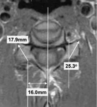
제 3경추에서 제 6경추의 평균적인 적절한 삽입 위치 는 경추의 정중선에서 16.0 mm 거리에 있었다. 삽입 각 도는 시상면상 후관절에 평행하고 외측을 향하여 2 5 . 3 도이었다. 적절한 측괴 나사못의 삽입 길이는 17.9 mm 이었다(Fig. 2).
각 경추의 부위에 따른 삽입 위치, 삽입 각도 및 삽입 길이를 제 3경추에서 제 6경추의 전체 평균 값과 비교하 여 보면 삽입 위치 및 삽입 각도는 전체 평균과 의의 있
는 차이는 없었으나 삽입 길이는 제 3경추의 경우 전체 평균보다 1.7 mm가 의의 있게 짧았으며(P=0.000), 제 6 경추의 경우 전체 평균보다 1.9 mm가 의의 있게 길었다
(P=0.018). 제 4, 5경추는 통계적으로 의의 있는 차이는
없었다(Table 1).성별에 따른 각 경추 부위의 삽입 위치, 각도 및 길이를 분석하여 보면 삽입 위치 및 각도는 성별에 따른 통계적 인 차이는 없으나 삽입 길이는 의의 있는 차이가 있었다.
삽입 길이는 제 3경추에서 남자 16.9 mm, 여자 14.8 mm
(Mann-Whitney U= 6.000, P=0.003), 제 4경추에서 남자 18.1 mm, 여자 14.7mm(Mann-Whitney U= 7.000, P=0.004),
제 5경추에서 남자 20.5 mm, 여자 14.1 mm(Mann-WhitneyU= 2.000, P=0.001), 제 6 경추에서 남자 21.4 mm, 여자 16.1 mm(Mann-Whitney U=4.000, P=0.002)로 모든 경추부
위에서 성별에 따른 삽입 길이에 의의 있는 차이가 있었 다(Table 2).연령에 따른 측정치를 60세 이상인 군과 60세 이하인 군을 나누어 비교하여 보았을 때 연령에 따른 통계적인 차이는 없었다.
고 찰
경추 측괴 나사못 및 금속판 고정술은 경추 후궁 및 극 돌기의 결손이 있어 기존의 강선 고정술을 시행할 수 없 는 경우에도 시행할 수 있는 유용한 술식으로 최근에는 강선 고정술에 비하여 우수한 고정력으로 경추의 골절, 탈구 등 경추 불안정성의 치료에 점차 널리 사용되고 있
Fig. 2. Ideal entry points, insertion angle and length of the lat- eral mass for lateral mass screw fixation were measured on the axial MR images using PACS digital measuring i n s t r u m e n t .
Table 1. Measurements of lateral mass depending on the level Entry Angle Length Significance
(mm) (˚) (mm) (P)
C3 15.5 24.3 16.2* 0.000*
C4 16.0 25.2 17.1 0.197
C5 15.9 26.6 18.6 0.463
C6 16.4 24.9 19.8† 0.018†
Mean 16.0 25.3 17.9 1.000
Table 2. Measurements of lateral mass depending on sex of the patients
Male Female
Entry Angle Length* Entry Angle Length* Sig(P)*
C3 16.1 23.8 16.9 14.2 25.3 14.8 0.003
C4 16.4 25.8 18.1 15.3 24.0 14.7 0.004
C5 16.0 26.7 20.5 15.8 26.4 14.1 0.001
C6 16.7 24.8 21.4 15.7 25.1 16.1 0.002
다. 또한 여러 분절의 고정도 용이하게 시행할 수 있으 며 강성고정술에 비하여 필요한 이식골의 양도 줄일 수 있는 장점이 있다.
경추 측괴 나사못 고정술은 처음 R o y - C a m i l l e에 의하여 광범위하게 사용되었으며1 3 ) 이후 많은 술자들이 사용하 게 되었다1 , 3 - 5 , 7 , 9 , 1 1 , 1 3 - 1 5 )
. Roy-Camille의 연구에 의하면 굴곡-
신전 부하에 대한 안정성은 강선고정술을 시행하였을 경 우 3 3 %가 증가하는데 비하여 측괴 나사못 고정술을 시행 하였을 경우 9 2 %가 증가하였다고 보고한 바 있다1 3 ).
경추 측괴 나사못 고정술의 방법은 두가지 방법이 가 장 널리 사용되고 있는데 그 하나는 Roy-Camille 방법이 고 다른 하나는 Magerl 방법이다. Roy-Camille방법은 측 괴의 중앙부에 나사못을 삽입하며 삽입 방향은 상하 방 향으로 측괴에 대하여 직각, 내외측 방향으로는 외측으 로 10도의 방향으로 삽입하는 것이다. 반면에 Magerl방 법은 측괴의 중앙에서 내측으로 1 mm 지점에서 삽입하 여 상방으로 후관절에 평행하고 외측으로 25~30도 각도 로 삽입하는 것이다. 많은 저자들이 Roy-Camille의 방법 과 Magerl의 방법을 비교한 연구를 시행하였는데 Errico 등은 나사못의 방향이 금속판의 안정성에 중요하며
R o y - C a m i l l e의 방법과 M a g e r l의 방법을 비교하여 p u l l - out강도가 Magerl의 방법이 더 우수함을 보고한 바 있다 (471N versus 607N)
5).
M o n t e s a n o등은 R o y - C a m i l l e의 방법에 비하여 M a g e r l의
방법이 삽입할 수 있는 나사못을 더 길게 할 수 있으며( 1 4mm versus 20 mm) 또한 실패 부하(failure load)도 우수하
고(585 Nm versus 152 Nm) 강도도(223 Nm versus 34 Nm) 우수함을 보고하였으며 나사못의 방향이 R o y - C a m i l l e의 방법은 후관절에서 전방에 있는 신경근 및 척추 동맥을 향하고 있으나 M a g e r l의 방법은 나사못이 신경 및 혈관에 서 멀리가는 방향을 향하고 있음 보고하였다1 2 ).
상기한 바와 같이 측괴 나사못 삽입에서 중요한 점은 측괴의 전방에 위치하는 신경근 및 척추 동맥의 손상은 주지 않으면서 보다 강한 고정력을 얻기 위하여 가능하 면 긴 나사못을 삽입하여야 하며 또한 측괴 나사못의 삽 입은 한쪽 피질골만을 고정하는 방법(unicortical fixation) 보다는 양쪽 피질골을 고정하는 방법(bicortical fixation) 을 사용하여야 할 것이다. 이와 같은 개념으로 본 연구에 서는 한국인의 경추를 대상으로 측괴 나사못 삽입을 위 한 측정을 하였다. 측정 방법은 후관절에 평행한 방향으 로 얻은 T1 강조 축상면에서 삽입위치, 삽입 각도 및 삽입 길이를 측정하였다. 본 연구에서 일반적인 자기공명영상 에서 시행하는 추간판에 평행한 방향으로 얻은 축상면 대신 후관절에 평행한 방향으로 얻은 축상면에서 측정한 이유는 시상면상의 삽입 각도가 후관절에 평행한 방향으
더 많아지고 신경근 손상을 피할 가능성이 더 높기 때문 이었다. 따라서 본 연구에서 시상면상의 각도는 후관절 에 평행한 방향으로 삽입하는 것으로 정하고 나머지 요 소인 삽입 위치, 방향, 길이에 대하여 측정하였다.
본 연구 결과 삽입 각도는 시상면상 후관절에 평행하 고 외측을 향하여 2 5 . 3도이었으며 적절한 측괴 나사못 의 삽입 길이는 17.9 mm로 이는 Magerl의 방법과 유사 한 결과이었으며 삽입 위치도 M a g e r l의 연구와 비교하 여 보았을 때 측괴의 중앙에서 1~2 mm 내측에 존재함을 알 수 있었다. 그러나 삽입 길이는 17.9 mm로 Montesano 등12)이 보고한 20 mm 보다 약간 짧음을 알 수 있었으며 이는 서양인과 한국인의 체격의 차이로 인한 것으로 생 각되었다. 본 연구에서 시상면상 후관절과 평행한 삽입 각도를 측정하지 않은 이유는 실제 수술시 환자의 자세 에 따라 후관절면이 향하는 방향은 변화하게 되며 이러 한 후관절면은 수술시 촉진하여 그 방향을 알 수 있기 때문이었다. 본 연구에서 삽입 위치는 상하로는 측괴의 중앙이었으며 내외측으로는 극돌기에서 16.0 mm에 위 치하였다. 다른 연구에서는 삽입 위치의 내외측을 측괴 의 중앙에서 얼마나 떨어져 있는 가로 측정하였으나 실 제 수술시에 측괴와 후궁의 경계를 명확하게 구분하기 힘든 경우가 있으며 이러한 경우 삽입 위치를 파악하기 어려울 수 있다. 따라서 본 연구에서는 가장 명확한 해 부학적 구조물인 극돌기에서의 거리를 측정하였다. 이 는 실제 수술시 Caliper를 이용하여 극돌기에서의 거리 로 삽입위치를 찾을 수 있게 하고자 함이었다.
본 연구 결과 삽입 위치 및 삽입 방향은 경추의 각 부 위에 따른 차이는 없었으나 삽입 나사못의 길이는 제 3 경추에서 의의 있게 짧았고 제 6 경추는 의의 있게 길었 다. 또한 성별에 따른 측정값도 삽입 위치 및 삽입 방향 은 차이가 없었으나 삽입 길이는 통계적으로 의의 있게 남자에서 길었다.
결 론
본 연구 결과 경추 측괴 나사못 삽입의 이상적인 위치 는 상하로는 측괴의 중앙에 있으며 내외측으로는 경추 의 정중선에서 16.0 mm 거리에 있었으며 삽입 각도는 시상면상 후관절에 평행하고 외측을 향하여 25.3도이었 고 측괴 나사못의 삽입 길이는 17.9 mm이었다. 그러나 각각의 환자에 따른 삽입 위치, 각도, 길이의 차이가 있 으므로 수술전 각각의 환자의 측괴에 대한 측정을 하는 것이 안전할 것으로 생각되었다.
REFERENCES
01) Anderson PA, Henley MB, Grady MS, Montesano PX and Winn HR : Posterior cervical arthrodesis with AO reconstruction plates and bone graft. Spine, 16(S):S72-9, 1991.
02) Coe JD, Warden KE, Sutterlin CE and McAfee PC : Biomechanical evaluation of cervical spinal stabilization methods in a human cadaveric model. Spine, 14(10):1122- 31, 1989.
03) Cooper PR, Cohen A, Rosiello A and Koslow M : Pos - terior stabilization of cervical spine fractures and sublux - ations using plates and screws. Neurosurgery, 23:300-6, 1988.
04) Ebraheim NA, An HS, Jackson WT and Brow JA : In- ternal fixation of the unstable cervical spine using posteior Roy-Camille plates: preliminary report. J Orthop Trauma, 3:23-8, 1989.
05) Errico T, Uhl R, Cooper P, Casar R and McHenry T : Pullout strength comparison of two methods of orienting screw insertion in the lateral masses. J Spinal Dis, 7:429- 38, 1992.
06) Gill K, Paschal S, Corin J, Asham R and Bucholz R : Posterior plating of the cervical spine: a biomechanical comparison of different posterior fusion technique. Spine, 13:813-6, 1988.
07) Graham AW, Swank ML, Kinard RE, Lowery GL and Dials BE : Posterior cervical arthrodesis and stabilization
with a lateral mass plate: clinical and computed tomo - graphic evaluation of lateral mass screw placement and associated complications. Spine, 21:323-8, 1996.
08) Grob D and Magerl F : Dorsal spondylodesis of the cer - vical spine using a hooked plate. Orthopade, 16:55-61, 1987.
09) Lowery GL, Swank ML and McDonough RF : Surgical revision for failed anterior cervical fusions: articular pil - lar plating or anterior revision? Spine, 20:2436-42, 1995.
10) Magerl F : Posterior fusion using hook plates. Bulletin 61, Paoli, PA: Synthes.
11) Magerl F and Grob D : Stable dorsal fusion of the cervical spine(C2-T1) using hook plates. In Kehr P, Weidner A, eds.
Cervical spine. Vienna: Springer-Verlag, 217-21, 1987.
12) Montesano PX, Jauch E, Anderson PA, Benson DR and Hanson B : Biomechanics of cervical spine internal fixation. Spine, 16(suppl.):S10-6, 1991.
13) Roy-Camille RR, Sailant G and Mazel C : Internal fixa - tion of the unstable cervical spine by posterior osteosynthe - sis with plate and screws. In Cervical Spine Research Soci - ety, The cervical spine, 2nd ed. Philadelphia: JB Lippin - cott, 390-404, 1989.
14) Savini R, Parisini P and Cevellati S : The surgical treat - ment of late instability of flexion-rotation injuries in the lower cervical spine. Spine, 12:178-82, 1987.
15) Swank ML, Sutterlin CE III, Bossons CR and Dials BE : Rigid internal fixation with lateral mass plates in multi - level anterior and posterior reconstruction of the cervical spine. Spine, 22:274-82, 1997.
연구계획 : 자기공명영상을 이용하여 측괴나사못 고정술을 위한 측괴의 계측을 시행하였다.
연구목적 : 경추부 측괴를 자기공명영상을 이용하여 측정하여 경추부 측괴 나사못 고정술에 도움을 주고자 하였다.
대상 및 방법 : 10명의 환자를 대상으로 제 3, 4, 5, 6 경추의 후관절에 평행한 방향으로 얻은 자기공명영상의 축상면 에서 80개의 측괴를 대상으로 적절한 측괴나사못의 삽입 길이, 방향 및 그 위치를 측정하여 통계 분석하였다. 각각의 측정값은 PACS를 이용한 digital 계측을 이용하였다. 연구대상 환자의 평균 나이는 48.0세 이었고 남자 6명 여자 4명 이었다.
결과 : 적절한 측괴 나사못의 삽입 길이는 평균 17.9 mm, 적절한 삽입 각도는 외측을 향하여 평균 25.3도, 적절한 삽입 위치는 정중선에서 평균 16.0 mm 거리에 있었다.
결론 : 각각의 환자 및 부위에 따라 삽입 위치, 삽입 각도, 삽입 길이에 차이가 있으므로 수술전 각각의 환자 및 부위 에 따라 측괴나사못 삽입을 계획하는 것이 좋을 것으로 생각되었다.
색인단어 : 자기공명영상, 측괴나사못 고정술 국 문 초 록
※ 통신저자 : 석 경 수
서울특별시 동대문구 회기동 1 경희대학교 의과대학 정형외과학교실
Tel : 82-2-958-8345, Fax : 82-2-964-3865, E-mail : [email protected]