submit.radiology.or.kr J Korean Soc Radiol 2012;67(4):241-244
241 INTRODUCTION
Progressive systemic sclerosis (PSS) is a chronic, multisys- temic disease, which is characterized by vascular changes, fi- brosis, and inflammation of the skin and visceral organs. Pul- monary involvement occurs frequently in patients with PSS and is now considered as the main cause of death among these patients (1). Lung abnormalities are often subtle, most fre- quently occurring at the lung bases, and progressively expand- ing to involve the lower two-thirds of the lungs (2). High reso- lution CT (HRCT) findings of interstitial fibrosis in PSS include fine reticular opacities, ground-glass opacity, consolidation, traction bronchiectasis, honeycombing, and coarse or irregular reticulations.
The presence of faint parenchymal micronodules on CT scans is not unexpected, as focal lymphoid hyperplasia (follicu-
lar bronchiolitis) has been reported as a frequent histologic fea- ture. Subpleural micronodules may reflect lung involvement at the pleural parenchymal interface, as small lymphoid aggre- gates adjacent to the pleura have been observed in lower lobe biopsies of patients with PSS (3). However, the radiologic de- tection of progressive intrapulmonary lymph nodes (IPLNs) with PSS has not yet been reported.
In this review, we report the serial chest radiologic findings in one case of PSS with multiple growing lung nodules, which turned out to be IPLNs on video-assisted thoracoscopic lung biopsy (VATS).
CASE REPORT
A 40-year-old female visited the internal medicine outpatient department for the evaluation of Raynaud’s phenomenon for a
Case Report
pISSN 1738-2637
J Korean Soc Radiol 2012;67(4):241-244
Received April 19, 2012; Accepted July 24, 2012 Corresponding author: Seon Mun Kim, MD Department of Radiology, Seoul St. Mary’s Hospital, College of Medicine, The Catholic University of Korea, 222 Banpo-daero, Seocho-gu, Seoul 137-701, Korea.
Tel. 82-2-2258-1455 Fax. 82-2-599-6771 E-mail: [email protected]
Copyrights © 2012 The Korean Society of Radiology
A 40-year-old female visited the clinic for evaluation of Raynaud’s phenomenon for a period of four years. The initial chest radiograph showed a fine reticular density and ground glass opacity with lower lobe predominance. These findings are consis- tent interstitial fibrosis. Additionally, high resolution CT showed multiple, small, co- existing nodular opacities, ranging from 3 to 7 mm in size in both lungs. These nod- ules grew up to 1.5 cm and showed moderate enhancement. Because of the rareness of intrapulmonary lymph node in patient of progressive systemic sclerosis, we couldn’t exclude the possibility of malignancy. These nodules are turned out to be intrapulmonary lymph nodes on video-assisted thoracoscopic lung biopsy.
Index terms
Progressive Systemic Sclerosis Intrapulmonary Lymph Node
Gradual Progression of Intrapulmonary Lymph Nodes Associated with Usual Interstitial Pneumonia in Progressive Systemic Sclerosis on Chest Radiographs and CT
1진행성 전신경화증에서 간질폐렴과 동반하여 점진적 진행을 보이는 폐내 림프절: 증례 보고1
Seon Mun Kim, MD
1,2, Joon Young Ohm, MD
1, Myung Hee Chung, MD
1, Yong Hyun Kim, MD
31Department of Radiology, Bucheon St. Mary’s Hospital, College of Medicine, The Catholic University of Korea, Bucheon, Korea
2Department of Radiology, Seoul St. Mary’s Hospital, College of Medicine, The Catholic University of Korea, Seoul, Korea
3Department of Internal Medicine, Bucheon St. Mary’s Hospital, College of Medicine, The Catholic University of Korea, Bucheon, Korea
Intrapulmonary Lymphnode in Progressive Systemic Sclerosis
submit.radiology.or.kr
J Korean Soc Radiol 2012;67(4):241-244
242
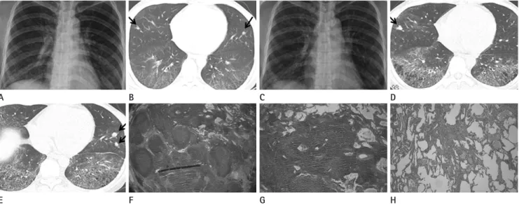
low-up enhanced chest CT and CO diffusion test. HRCT showed interval increase in size of the multiple small nodules (7-11 mm in size) and an aggravation in areas of honeycomb cystic change in the right middle lobe and both lower lobes (Fig. 1D, E). Nodules were found more than 1 cm away from the pleura. Finally, she was admitted for a VATS lung biopsy of small nodules in the right middle lobe. The macroscopic find- ings revealed that the nodules were 10 × 10 mm in size, round, marginated, white and black in color. Histologic examination (Fig. 1F, G) revealed a lymph node with lymphoid follicles in the lung parenchyma. The lung tissue obtained by wedge resec- tion of the right middle lobe showed organizing pneumonia with lymphocyte aggregation with follicle formation. Lung tis- sue from the right lower lobe showed the usual interstitial pneumonia pattern (Fig. 1H). The findings were compatible with interstitial pneumonia due to lung involvement in PSS.
The activity was of a moderate degree. Two years later, the last follow-up nonenhanced chest CT was performed, and it re- vealed a slight regression or no definite aggravation of diffuse lung disease, and no overall interval change in the larger pe- period of four years (from 2004 to 2008). She had symptoms of
dry cough, skin pigmentation, skin thinning, scleroderma-like change below both elbows, and gastroesophageal reflux.
Initial chest radiograph showed the pattern of interstitial lung disease in the lung bases (Fig. 1A). These findings were consistent with lung involvement in PSS. HRCT performed one month later showed diffuse ground-glass opacities and fine reticular densities in the posterior portions of both lower lobes (Fig. 1B). In addition, multiple, small, coexisting nodular opac- ities, ranging from 3 to 7 mm in size, were detected in the both upper, right middle and both lower lobes. The authors thought that HRCT findings were consistent with interstitial lung in- volvement, as seen in the usual interstitial pneumonia or non- specific interstitial pneumonia in patients with PSS. However, larger nodules were overlooked during interpretation of the initial HRCT scan. After three months, pulmonary function test showed mild restrictive patterns.
Follow-up chest radiograph showed minimally increased ex- tent of coarse reticulation in both lower lung zones (Fig. 1C).
However, she had no respiratory symptoms at the time of fol-
F B
G C
H D
E A
Fig. 1. A 40-year-old female with history of Raynaud’s phenomenon.
A. The initial chest radiograph shows diffuse fine reticular densities and ground-glass opacities in both lung fields with basal lung predominance.
B. HRCT shows fine reticulonodular densities and diffuse ground-glass opacities in the right middle lobe, both lower lobes and left lingular seg- ment. Findings are compatible with interstitial lung disease due to PSS. Nodules in the right middle lobe and left lingular segments (arrows) were overlooked at this point of time.
C. Six years later, follow-up chest radiograph shows irregular reticulonodular opacities bilaterally. The lung volume was slightly decreased.
D, E. Follow-up HRCT shows interval increase in size of the nodules in the right middle lobe (arrow) and left lingular segment (arrow). The nodules in the right middle lobe become lobulated. Nodules are found more than 1 cm away from the pleura. There are increased in the honeycomb cysts.
F, G. Histologic examination of the nodules reveals a lymph node with lymphoid follicles in the lung parenchyma (Hematoxylin and Eosin stain, x 100).
H. Lung tissue obtained by wedge resection of the right lower lobe shows interstitial pneumonia compatible with the usual interstitial pneumo- nia pattern, due to the moderate activity of PSS (Hematoxylin and Eosin stain, x 100).
Note.-HRCT = high resolution CT, PSS = progressive systemic sclerosis
Seon Mun Kim, et al
submit.radiology.or.kr J Korean Soc Radiol 2012;67(4):241-244
243
4) they may cause pleural indentations; and 5) the border may sometimes be rough, like that in lung cancer. These findings are not necessarily specific for IPLNs. Nevertheless, they suggest that when nodular shadows with a sharp border are found just under the pleura, IPLNs should be suspected. Peripheral small lung cancer does not always have specific features, and the CT features of IPLNs and small lung cancer sometimes overlap (8).
These IPLNs have been occasionally found to coexist with sev- eral diseases, such as silicosis (9) and diseases affecting African miners (10).
In PSS, IPLNs are very rare, and hence, we should be able to differentiate them from cancerous conditions, infected nodules, and nodules of other origin.
REFERENCES
1. Arroliga AC, Podell DN, Matthay RA. Pulmonary manifes- tations of scleroderma. J Thorac Imaging 1992;7:30-45 2. Silver RM, Metcalf JF, Stanley JH, LeRoy EC. Interstitial
lung disease in scleroderma. Analysis by bronchoalveolar lavage. Arthritis Rheum 1984;27:1254-1262
3. Harrison NK, Myers AR, Corrin B, Soosay G, Dewar A, Black CM, et al. Structural features of interstitial lung disease in systemic sclerosis. Am Rev Respir Dis 1991;144(3 Pt 1):706- 713
4. Howling SJ, Hansell DM, Wells AU, Nicholson AG, Flint JD, Müller NL. Follicular bronchiolitis: thin-section CT and his- tologic findings. Radiology 1999;212:637-642
5. Szekanecz E, Szamosi S, Gergely L, Keszthelyi P, Szekanecz Z, Szucs G. Incidence of lymphoma in systemic sclerosis: a retrospective analysis of 218 Hungarian patients with sys- temic sclerosis. Clin Rheumatol 2008;27:1163-1166 6. Yoshitomi A, Sato A, Toyoshima M, Suganuma H, Imokawa
S, Tamura R, et al. [Two cases of intrapulmonary lymph node associated with either progressive systemic sclerosis or idiopathic pulmonary fibrosis]. Nihon Kyobu Shikkan Gakkai Zasshi 1995;33:1003-1008
7. Ohtsuka T, Nomori H, Horio H, Naruke T, Suemasu K. Radio- logical examination for peripheral lung cancers and benign nodules less than 10 mm. Lung Cancer 2003;42:291-296 8. Yokomise H, Mizuno H, Ike O, Wada H, Hitomi S, Itoh H.
Importance of intrapulmonary lymph nodes in the differ- ripheral nodules.
DISCUSSION
Most reports of lung pathology in PSS describe abnormali- ties, including extensive interstitial fibrosis with cystic changes, bronchiolectasis, and pleural thickening. Focal lymphoid hy- perplasia (follicular bronchiolitis) is more common in the PSS patient than in the lone cryptogenic fibrosing alveolitis (3). Fo- cal lymphoid hyperplasia manifest as faint parenchymal mi- cronodules on a CT scan, although, the nodules of focal lym- phoid hyperplasia in rheumatoid arthritis patient measured about 3 and 10 mm in diameter (4). Recent results suggest that the B cells may have multiple pathogenic roles in PSS and there may be increased incidence of B cell lymphomas in PSS (5).
The association of PSS and non-Hodgkin’s lymphoma may be a rather uncommon feature. The other report was about a soli- tary lung nodule in PSS, which was an IPLN in patients with PSS (6). In each patient, a follow-up chest CT scan showed a pulmonary nodule, which could not be seen on a chest radio- graph, as in this case. In these cases, the nodules had an irregu- lar margin and a linear shadow resembling pleural indentation.
Histological examination of the open lung biopsy specimens revealed that these nodules were IPLNs surrounded by an in- terstitial pneumonia and lymphoid hyperplasia. In our case, the nodules were less than 5 mm in diameter, without definite pleu- ral indentation, on an initial CT scan, and were grown up to 1.5 cm for a period of five years. Our patient had longer follow up time and serial CT findings. In general, ill-defined margins, in- volvement of bronchi or vessels, and nodule enlargement visu- alized by CT are important signs of malignancy, even for less than 10 mm in size (7). All nodules in our patient grew in size, the largest nodule in the right middle lobe increased in size from 7 mm to 1.5 cm in diameter.
It has been generally believed that IPLNs exist in the vicinity of the hilar region of the lung and the central bronchi, but not after the fourth branching point. Nevertheless, it has been known for a long time that IPLNs sometimes show up on chest radio- graphs as solitary nodular shadows. The following points of the CT findings in IPLNs were confirmed; 1) the border was sharp in most cases; 2) IPLNs were found within several millimeters from the pleura; 3) sometimes, more than one node were seen;
Intrapulmonary Lymphnode in Progressive Systemic Sclerosis
submit.radiology.or.kr
J Korean Soc Radiol 2012;67(4):241-244
244
nia]. Nihon Kyobu Shikkan Gakkai Zasshi 1993;31:117-122 10. Honma K, Nelson G, Murray J. Intrapulmonary lymph nodes
in South African miners--an autopsy survey. Am J Ind Med 2007;50:261-264
ential diagnosis of small pulmonary nodular shadows.
Chest 1998;113:703-706
9. Yoshii C, Hamada M, Tao Y, Sasaki M, Okamoto T, Obata H, et al. [A case of intrapulmonary lymph node with silicotic nodules in a patient with idiopathic interstitial pneumo-
진행성 전신경화증에서 간질폐렴과 동반하여 점진적 진행을 보이는 폐내 림프절: 증례 보고1
김선문
1,2· 엄준영
1· 정명희
1· 김용현
340세 여자 환자가 4년 이상의 레이노 증상을 주소로 내원하였다. 내원시 단순흉부촬영 소견에서 망상 음영 소견과 간유 리음영이 주로 양 폐하에 있었다. 이는 사이질 섬유화에 합당한 소견이다. 고해상도 흉부전산화단층촬영에서 3~7 mm 크기의 다수의 작은 결정형 음영들이 양 폐에 있었다. 이 결절들은 추적 관찰 중 1.5 cm까지 성장하였고 조영증강이 잘 되었다. 진행성 전신경화증 환자에서의 폐내 림프절은 매운 드문 일이기에 진단시 악성 종양의 가능성을 배제할 수 없었 다. 환자는 생검을 통하여 폐내 림프절로 조직학적 확진을 받았다.
1가톨릭대학교 의과대학 부천성모병원 영상의학과, 2가톨릭대학교 의과대학 서울성모병원 영상의학과,
3가톨릭대학교 의과대학 부천성모병원 내과