Legg-Calve-Perthes(LCP) 질환은 소아의 대퇴골두의 원 발성 골괴사로서 무혈성 괴사기, 분절기와 재생기, 잔여기 등 의 진행경로를 거쳐 자연 치유되나 때로는 고관절 기형을 가 져오기도 하며 이로 말미암아 퇴행성 고관절 질환의 발생률이 높다(1-3). LCP 질환은 반복된 경색(infarct)과정이 필수적이 며 반복적인 골세포의 죽음에 이어 신생골 형성을 한다. 이는 대퇴골두의 혈류장애가 있는 상태에서 불충분한 재혈관화에 기 인하는 것으로 생각된다(1). LCP질환의 영상진단에 이용되는 자기공명영상은 단순촬영에서 병변이 나타나기 전에 골수의 신 호강도 변화를 보이는 민감도가 높은 검사이고 골괴사의 범위 를 측정하는데 유용하다(4, 5). 그러나 질병의 초기에 고식적 인 스핀에코법을 이용한 자기공명 영상에서 위음성 결과가 보 고되어 있고 이는 골단의 허혈상태를 보여주지 못한 것으로 생 각된다(6-8). 조영증강 자기공명영상을 이용하여 골관류를 반 영하여 조기에 골단의 허혈상태와 재혈관화를 알아보고자 한 연구가 있다(9, 10).
저자들은 소아의 LCP 질환의 조영증강 자기공명영상 소견 을 분석하고 진단과 예후 판정에서 조영증강 자기공명영상의 유용성을 알아보고자 하였다.
대상과 방법
본원에서 LCP질환으로 진단 받은 환아 중 조영증강 자기공 명영상을 시행한 12명의 환아 14예를 대상으로 하였다(양측 성 병변 2예). 연령분포는 3세에서 9세로 평균연령은 6.2 세 였다. 남녀 비는 11:1로 남아가 많았다.
모든 환아에서 고관절 전후 및 개구리 다리(frog leg) 단순 촬영과 자기공명영상을 시행하였다. 자기공명영상 기기는 1.5 Tesla Magnetom Vision과 Vision Plus(Siemens, Erlangen Germany)를 이용하였고 스핀에코기법으로 축상면, 관상면의 T1 강조영상(TR/TE=450/14 msec)과 T2 강조영상 (TR/TE=3800/99 msec)을 얻었다. 환아의 몸무게당 0.2 cc 의 gadopentetate dimeglumine(Magnevist, Shering, Berlin, Germany)을 정맥 주사하여 조영증강 영상을 얻었다. 대퇴골 단 정상 지방골수는 T1강조영상에서 고신호강도를 보이므로 조영증강 여부를 알려면 지방억제 T1 강조영상이 필수적이므 로 축상면, 관상면의 조영증강 지방억제 T1 강조영상을 얻었 다. 절편두께와 간격은 각각 3 mm, 0.3 mm로 하였고 체부코 일을 이용하였다.
단순촬영을 Waldenstrom분류법에 따라 분석하였다. 2예가 1기(초기: 대퇴골두는 정상소견이고 관절간격이 넓어보임), 10
Legg-Calve-Perthes 질환의 Gadolinium 조영증강 자기공명영상의 유용성
1김 지 은・김 지 혜2・김 형 식
목적: 소아의 Legg-Calve-Perthes(LCP) 질환에서 조영증강 자기공명영상의 유용성을 알아보 고자 하였다.
대상과 방법: LCP 질환으로 진단받은 환아 중 조영증강 자기공명영상을 시행한 12명의 환아
14고관절을 대상으로 하였다. 자기공명영상을 후향적으로 분석하였고 골괴사의 범위, 골단의 재혈관화 기전과 골간단의 변화를 분석하였다.
결과: 자기공명영상에서 LCP질환의 모든 환아는 조영증강 결손을 보였다. 9예에서 미만성 조 영증강 결손을 보였다. 이중 2예에서는 T1, T2 강조영상에서 골수에 정상신호강도를 보였다.
5예에서 국소적 조영증강 결손을 보였다. 골단의 재혈관화는 외측부에 우세한 경우 5예, 외측 부와 내측부 양측에서 보인 경우가 4예, 성장판을 통한 재혈관화를 보인 경우가 3예 있었다.
골간단의 이상소견은 2예에서 보였다.
결론: 조영증강 자기공명영상은 LCP질환의 모든 환아에서 조영증강 결손을 보여 LCP질환의 진단에 유용하였고 재혈관화 기전을 보여 주어 예후 판정에 유용한 정보를 제공하는 좋은 검 사이다.
1가천의과학대학교 길병원 영상의학과
2성균관대학교 삼성서울병원 영상의학과
이 논문은 2007년 11월 13일 접수하여 2008년 2월 13일에 채택되었음.
예가 2기(무혈성 괴사기: 대퇴골두의 음영증가, 연골하 골음영 감소, 대퇴골두 편평화), 2예가 3기(재생기 혹은 분절화기: 골 음영의 감소와 분절화)였고 4기(잔여기: 해면골로 대치, 다양 한 정도의 변형이 남음)는 없었다. 자기공명영상을 후향적으로 분석하였고 골괴사의 범위(extent of necrosis), 골단의 재혈 관화 기전(epiphyseal revascularization pathway)과 골간단의 변화(metaphyseal change)를 분석하였다. 골단의 재혈관화 기 전은 대퇴골두를 관상면에서 3등분 하여 내측, 중간, 외측으로 분류하고 어느쪽에 우세한 재혈관화를 보이는지 분석하였고 성 장판을 통한 재혈관화(transphyseal revascularization)가 보이 는지 분석하였다.
결 과
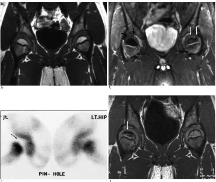
정상 대퇴골두의 골단의 조영증강은 대퇴골두의 바깥쪽 둘 레와 성장판(physis)을 따라 강한 조영증강으로 대퇴골두 주 변의 테두리 조영증강과 성장판을 따라 선상의 조영증강을 보 였다(Fig. 1). 정상 대퇴골두의 조영증강과 비교하여 조영증강 결손을 판독하였고 그 형태에 따라 미만성과 국소적 조영증강 결손으로 나누었다.
조영증강 자기공명영상에서 14예 모두에서 대퇴골두에 조영 증강 결손을 보였다. 이중 9예에서 미만성 조영증강 결손을 보 였고 5예에서는 국소적 조영증강 결손을 보였다. 미만성 조영 증강 결손을 보인 경우 중 Waldenstrom 분류1기였던 2예에서 T1, T2 강조영상에서 정상신호강도를 보여 고식적인 스핀에
코방법으로는 이상소견을 발견할 수 없었으나 조영증강 영상 에서는 뚜렷한 조영증강 결손을 보여 LCP질환을 진단할 수 있 었다. 6개월 후의 추적검사상 T1 강조영상에서 전형적인 골괴 사의 소견이 나타났다(Fig. 2). 미만성 조영증강 결손을 보인 나머지 7예는 Waldenstrom 분류 2기였고 T1 강조영상에서 저신호강도, T2 강조영상에서 4예에서 저신호강도, 3예에서 비 균질성 고신호강도를 보였다(Fig. 3). 국소적 조영증강 결손을 보인 5예 모두 T1 강조영상에서 저신호강도, T2 강조영상에 서 비균질성 고신호강도를 보였다.
Waldenstrom 분류 1기였던 2예를 제외한 나머지 12예에서 재혈관화를 평가할 수 있었는데 조영증강 영상에서 국소적으 로 강한 조영증강을 보이고 T2 강조영상에서 고신호강도를 보 였다. 외측부에 우세한 재혈관화를 보인 예가 5예, 외측과 내 측 모두에서 보인 경우가 4예 있었다(Fig. 3). 성장판을 통한 재혈관화를 보인 예가 3예 있었다(Fig. 4).
골간단의 이상소견은 2예에서 보였고 T1강조영상에서 저신 호강도, T2강조영상에서 고신호강도를 보이고 약한 변연부 조 영증강을 보였다(Fig. 4) (Table 1).
고 찰
LCP 질환은 동맥이 차단되거나 대퇴골두 내의 압력이 높아 져서 결국 대퇴골두로의 혈행에 장해를 받고 궁극적으로는 골 수의 괴사와 대퇴 골두의 변형이 유발하는 질환이다(11, 12).
주로 4세에서 12세 사이에 발생하고 6-8세에 잘 발생한다.
A B
Fig. 1. Normal femoral head enhance- ment pattern
A, B. Coronal T1-weighted enhanced MR image shows rim-like epiphyseal (white arrows) and thin linear physeal (black arrows) enhancement of the normal femoral head.
양측성 질환이 10-12% 보고되고 있다(1). 자기공명영상은 LCP 질환의 진단에 민감도가 높은 검사이다. 단순촬영상 정상 인 환아에서 조기 발견이 가능하며(13-15) 골괴사의 범위 측 정, 관절연골, 대퇴골두의 모양, 관절삼출액과 관절막 비후 그 리고 대퇴골두 유치의 정도(quality of containment)를 평가하 는데 유용하다(5, 14). 또한, 자기공명영상에서 보이는 골수의 신호강도 변화는 조직학적으로는 지방괴사, 염증성 침윤, 섬유 세포와 골 복구(fibrocyte and bone repair)에 의한 것으로 알 려졌다(16). 그러나 LCP질환의 초기에 고식적인 스핀에코법 을 이용한 자기공명영상에서 위음성(false negative)이 보고되 어 있다(6-8). Vande Berg 등에 의하면 위음성이 나타나는
원인으로 조기 골수 괴사 시기에 지방조직이 건사(mummified) 되어 지방과 같은 신호강도를 유지하여 T1 강조영상에서 정 상 신호강도를 보일 수 있다고 하였다(6). 이러한 문제점을 해 결하고자 동물실험을 통해 대퇴골두의 허혈상태를 조영증강 자 기공명영상으로 조기진단 할 수 있다는 연구가 있다(17). 또 한, 최근에 조영증강 자기공명영상을 이용하여 골관류를 반영 하여 조기에 골단의 허혈 상태와 재혈관화를 알아보고자 한 연 구가 있다(9, 10). 정상 고관절에서 역동적 조영증강 자기공명 영상에 관한 연구에 의하면 대퇴골두와 골간단 그리고 비구 (acetabulum)에 조기(0-2분)에 강한 조영증강이 이루어지고 2-5분에 천천히 감소한다. 대퇴골두의 바깥쪽 둘레와 성장판
A B
C D
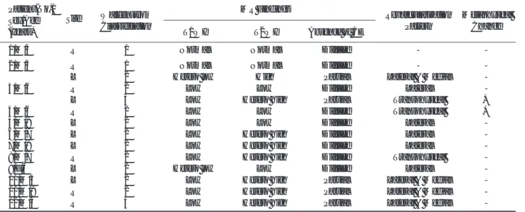
Fig. 2. Right side early LCP in a 4-year-old boy with right hip pain
A. Coronal T1-weighted image shows normal homogeneous fatty marrow signal of both femoral heads without any abnormality.
B. Complete absence of enhancement of right femoral epiphysis (black arrows) is only shown on the enhanced fat suppressed T1- weighted image. Normal rim-like epiphyseal (white arrows) and thin linear physeal enhancement of the normal contralateral hip is shown for comparison.
C. Pinhole bone scan shows cold defect of right femoral epiphysis (arrow) and normal perfusion of the left femoral epiphysis.
D. Follow up T1-weighted MR image (after 6 months) shows the linear hypointensity of subchondral fracture and decreased height of the right femoral epiphysis (arrow).
(physis)을 따라 가장 강한 조영증강이 이루어지고 이는 대퇴 골두 주변의 테두리 조영증강과 성장판을 따라 선상의 조영증 강을 보인다. 대퇴골두의 중심부는 상대적으로 조영증강이 잘 되지 않아 낮은 신호강도를 보이고 골간단 보다 낮은 신호강 도를 보인다(10). 본 연구에서 단순촬영과 고식적 자기공명영 상에서 정상소견을 보이는 환아에서 조영증강 영상에서 미만 성 조영증강 결손을 보여 LCP 질환의 초기에 진단할 수 있었 다. 이는 조영증강 자기공명영상이 골관류를 반영하여 골단의 허혈상태를 보여주는 것으로 생각된다.
LCP 질환의 예후에 관한 중요한 요소 중 하나가 재혈관화 과정인데. 외측부를 통한 재혈관화가 보이는 경우 예후가 좋은 것으로 보고되고 있다. 이는 이미 있던 혈관의 재관통 (recanalization of the existing vessels)에 의해 재혈관화가 이 루어지는 경로로 수분에서 수일 내로 이루어진다고 한다(18, 19). 또 다른 재혈관화 경로로는 신생혈관형성을 통한 것이 있 는데, 이는 수개월에서 수년에 걸쳐 이루어지고 특히 이 중에 서 성장판을 경유한 재혈관화가 이루어지면 성장장애와 기형 등의 나쁜 예후를 시사하는 소견이 된다(19). 본 연구에서 조
A B
Fig. 3. Right sided LCP disease in a 9-year-old boy presented with right hip pain.
A. Coronal T1-weighted image shows diffuse low signal intensity area of the right femoral epiphysis.
B. Coronal enhanced T1-weighted image shows central focal absence of enhancement (white arrow) and reperfusion with the me- dial and lateral columns of increased enhancement (black arrows).
Table 1. Data of 14 Affected Hips in Twelve Patients with LCP disease Patient(No)/
Waldenstrom MR Findings
Revascularization Metaphyseal Sex/Age Site
Classification Pattern Change
(years) T1WI T2WI Absence of CE
1/M/4 R 1 Normal Normal Diffuse - -
2/M/5 R 1 Normal Normal Diffuse - -
L 2 Hetero low High Partial Lateral & Medial -
3/M/6 R 2 Low Low Diffuse Lateral -
L 3 Low Hetero high Partial Transphyseal +
4/M/6 R 2 Low Low Diffuse Transphyseal +
5/M/8 L 2 Low Low Diffuse Lateral -
6/M/7 L 2 Low Hetero high Diffuse Lateral -
7/M/8 L 2 Low Hetero high Diffuse Lateral -
8/M/7 R 2 Low Hetero high Diffuse Transphyseal -
9/F/6 L 2 Hetero low Low Diffuse Lateral -
10/M/5 L 2 Low Hetero high Partial Lateral & Medial -
11/M/9 R 2 Low Hetero high Partial Lateral & Medial -
12/M/3 R 3 Low Hetero high Partial Lateral & Medial -
Hetero; heterogeneously, CE; enhancement - ; negative finding, + ; positive finding
영증강 자기공명영상에서 재혈관화가 강한 조영증강으로 뚜렷 하게 보였는데 이는 혈강관 확장, 혈관 분포의 증가, 증가된 모 세혈관 투과성 등에 의한 것으로 생각된다.
결론적으로 조영증강 자기공명영상은 LCP 질환의 가진 모 든 환아에서 조영증강 결손을 보이는 특징적인 소견을 보이며 특히 고식적 자기공명영상에서 위음성을 보이는 초기 질환의 진단에도 유용하였고 재혈관화 기전을 예측할 수 있어 예후 판 정에 유용한 정보를 제공하는 좋은 검사이다.
참 고 문 헌
1. 이덕용, 최인호, 정진엽, 조태준. 소아정형외과학 요람. 최신의학사,
2002:291-314
2. 국신호, 강흥식, 김인원, 연경모. Legg-Calve-Perthes 질환의 자기공 명영상소견. 대한방사선의학회지 1992;28:297-302
3. 문행진, 나재범, 심창민, 유진종, 정성훈. Legg-Calve-Perthes 질환 에서 골간단의 자기공명영상소견. 대한방사선의학회지 2001;44:
727-732
4. Ranner G, Ebner F, Fotter R, Linhart.W, Justich E. Magnetic reso- nance imaging in children with acute hip pain. Pediatr Radiol 1989;20:67-71
5. 정재동, 나재범, 정희영, 박일순, 송해룡, 유진종 등. Legg-Calve- Perthes 질환에서 대퇴골두의 괴사정도의 측정: Catterall 분류법과 MRI를 이용한 부피측정분류의 비교. 대한방사선의학회지 1998;
38:339-344
6. Vande Berg B, Malghem J, Labaisse MA, Noel H, Maldague B.
Avascular necrosis of the hip: comparison of contrast-enhanced
A B
C D
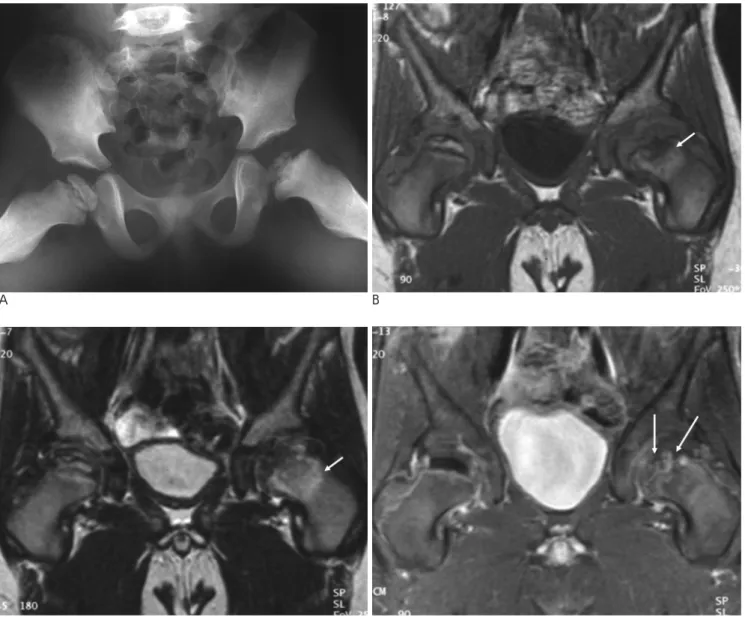
Fig. 4. Bilateral LCP disease in a 6-year-old boy
A. Radiography shows stage 2 subchondral fracture in the right femoral head and stage 3 fragmentation in the left side.
B, C. Metaphyseal change shows heterogeneous low signal intensity on T1-weighted image and increased signal intensity on T2- weighted image (arrow).
D. Coronal enhanced T1-weighted image shows absence of enhancement and reperfusion with lateral column on the right side and transphyseal revascularization on the left side (arrows) is indicative of a poor prognosis.
and nonenhanced MR imaging with histologic correlation.
Radiology 1992;182:445-450
7. Elsig JP, Exner GU, Von schulthess GK, Weitzel M. False-negative magnetic resonance imaging in early stage of Legg-Calve-Perthes disease. J Pediatr Orthop 1989;9:231-235
8. Uno A, Hattori T, Noritake K, Suda H. Legg-Calve-Perthes disease in the evolutionary period comparison of magnetic resonance imaging with bone scintigraphy. J Pediatr Orthop 1995;15:362-367 9. Sebag G, Ducou Le Pointe H, Klein I, Maiza D, Mazda K, Bensahel
H, et al. Dynamic gadolinium-enhanced subtraction MR imaging - a simple technique for the early diagnosis of Legg-Calve-Perthes disease: preliminary results. Pediatr Radiol 1997;7:216-220 10. Lamer S, Dorgeret S, Khairouni A, Mazda K, Brillet P-Y, Bacheville
E, et al. Femoral head vascularisation in Legg-Calve-Perthes dis- ease: comparison of dynamic gadolinium-enhanced subtraction MRI with bone scintigraphy. Pediatr Radiol 2002;32:580-585 11. 신용문, 강흥식, 김주완, 김희중, 김영민. 대퇴골두 무혈성 괴사: 조
영증강 자기공명 영상 소견. 대한방사선의학회지 1995;32:953-958 12. 오태경, 심재찬, 이기재, 전정동, 방선우, 김호균. 대퇴골두 무혈성
괴사의 지방억제 조영증강 자기공명영상소견. 대한방사선의학회 지 2000;42:327-331
13. Pinto MR, Peterson HA, Berquist TH. Magnetic resonance imaging in early diagnosis of Legg-Calve-Perthes disease. J Pediatr Orthop 1989;9:19-22
14. Toby EB, Koman LA, Bechtold RE. Magnetic resonance imaging of pediatric hip disease. J Pediatr Orthop 1985;5:665-671
15. Scoles PV, Yoon YS, Makley JT, Kalamchi A. Nuclear magnetic resonance imaging in Legg-Calve-Perthes disease. J Bone Joint Surg Am 1984;66:1357-1363
16. Brody AS, Strong M, Babikian G, Sweet DE, Seidel FG, Kuhn JP.
Avascular necrosis: early MR imaging and histologic finding in a canine model. AJR Am J Roentgenol 1991;157:341-345
17. Jaramillo D, Villegas-Medina OL, Doty DK, Dwek JR, Ransil BJ, Mulkern RV, et al. Gadolinium-enhanced MR imanging demon- strates abduction-caused hip ischemia and its reversal in piglets.
Pediatr Radiol 1995;25:578-587
18. Tsao AK, Dias LS, Conway JJ, Straka P. The prognostic value and significance of bone scintigraphy in Legg-Calve-Perthes disease. J Pediatr Orthop 1997;17:230-239
19. Conway JJ. A scintigraphic classification of Legg-Calve-Perthes dis- ease. Semin Nucl Med 1993;23:274-295
J Korean Radiol Soc 2008;58:417-423
Address reprint requests to : Jee-Eun Kim, M.D., Department of radiology, Gachon University of Medicine and Science, Gil Medical center, 1198 Kuwol-dong, Namdong-gu, Incheon 405-220, Korea.
Tel. 82-32-460-3060 Fax. 82-32-460-3065 E-mail: [email protected]
Efficacy of Gadolinium Enhanced MR Imaging for the Diagnosis of Legg-Calve-Perthes Disease
1Jee-Eun Kim, M.D., Ji Hye Kim, M.D.2, Hyung Sik Kim, M.D.
1Department of Radiology, Gachon University of Medicine and Science, Gil Medical Center
2Samsung Medical Center, Sungkyunkwan University School of Medicine
Purpose: The purpose of this study was to evaluate the efficacy of gadolinium enhanced MR imaging for mak- ing the diagnosis of Legg-Calve-Perthes (LCP) disease.
Materials and Methods: We studied the gadolinium enhanced MR images of 14 hips in 12 children who had the diagnosis of LCP disease. We retrospectively analyzed the extent of necrosis, the epiphyseal revasculariza- tion pathways and the metaphyseal changes.
Results: The absence of enhancement on gadolinium enhanced MRI was noted in all cases of LCP disease.
Diffuse absence of enhancement was observed in 9 femoral epiphyses. Two of them showed normal bone marrow signal intensity on the T1 and T2-weighted images. Focal absence of enhancement was observed in 5 femoral epiphyses. Enhanced MRI showed better epiphyseal revascularization in the lateral column (five cas- es), in the lateral and medial columns (four cases) and in the transphyseal pathway (three cases). Metaphyseal change was observed in two cases.
Conclusion: Gadolinium enhanced MRI allows detection of LCP disease and an accurate analysis of the differ- ent revascularization patterns, and this helpful for predicting the prognosis.
Index words :Child Hip
Legg-Perthes disease Magnetic resonance (MR) Femur head necrosis