https://doi.org/10.4093/jkd.2021.22.1.38
pISSN 2233-7431 · eISSN 2714-0547 Vol.22, No.1, 2021
Abstract
The prevalence of non-alcoholic fatty liver disease (NAFLD), the hepatic manifestation of metabolic syndrome, is rapidly increasing in Korean populations and is estimated to reach 39% among Korean adults. NAFLD is one of the strongest risk factors of type 2 diabetes mellitus (T2DM), and the presence of NAFLD in T2DM patients is associated with a worse natural course of diabetic complications. The gold standard for diagnosing NAFLD is liver biopsy, which involves sampling error, high cost, and other disadvantages related to its invasive nature. We summarize non-invasive diagnostic NAFLD algorithms including biomarkers and four major imaging tools (ultrasonography, computed tomography, magnetic resonance imaging, and liver FibroScan
Ⓡ) for diagnosing NAFLD including hepatic fibrosis. To prevent NASH (nonalcoholic steatohepatitis) or liver fibrosis, a combination of lifestyle modification with a low caloric diet and exercise, anti-diabetic agents, and non-anti-diabetic agents should be applied.
Keywords:
Diabetes mellitus; Fatty liver; Non-alcoholic fatty liver disease
당뇨병과 비알코올성 지방간: 진단 및 치료에 대한 고찰
이숙정, 이병완
연세대학교 의과대학 내과학교실
Diabetes Mellitus and Non-Alcoholic Fatty Liver Disease: Diagnosis and Treatment
Sook Jung Lee, Byung-Wan Lee
Division of Endocrinology and Metabolism, Department of Internal Medicine, Yonsei University College of Medicine, Seoul, Korea
Corresponding author: Byung-Wan Lee
Division of Endocrinology and Metabolism, Department of Internal Medicine, Yonsei University College of Medicine, 50-1 Yonsei-ro, Seodaemun-gu, Seoul 03722, Korea, E-mail: BWANLEE@yuhs.ac
Received: Dec. 2, 2020; Accepted: Dec. 4, 2020
This is an Open Access article distributed under the terms of the Creative Commons Attribution Non-Commercial License (http://creativecommons.org/licenses/by- nc/4.0/) which permits unrestricted non-commercial use, distribution, and reproduction in any medium, provided the original work is properly cited.
Copyright ⓒ 2021 Korean Diabetes Association
서론
당뇨병의 위험요소로 여겨지고 있는 비알코올성 지방간질 환(non-alcoholic fatty liver disease, NAFLD)은 우리나라 에서도 비만의 증가와 함께 유병률이 늘어나고 있다. 인슐린 저항성과 관련이 있는 것으로 여겨지는 NAFLD는 진단의 중 요성이 커지고 있으며, 이에 따른 치료법도 지속적으로 발표 되고 있다[1]. 본 글에서는 당뇨병과 함께 동반되는 NAFLD 의 진단 방법 및 치료법에 대해 논의해보고자 한다.
역학 및 정의
최근 7년간 우리나라의 당뇨병 유병률(2018년 현재 15.9%) 은 성별에 관계없이 지속적으로 증가하고 있다. 제2형 당뇨병 은 인슐린 저항성, 비만과 연관되는 NAFLD, 심혈관질환, 대 사증후군과 깊은 관련이 있다[1]. NAFLD는 제2형 당뇨병의 위험인자이며, 당뇨병환자의 NAFLD 유병률 또한 높다. 최 근 한국인의 비만 정도가 심해지면서 NAFLD 유병률은 30%
정도 되는 것으로 추측하고 있다. 특히 당뇨병환자와 당뇨 병이 없는 일반인과의 NAFLD 유병률은 2배의 차이를 보였 다(39% vs 19%) [2,3]. 지방의 분해로 혈액 내 지방산의 농 도가 높아짐으로써 이는 각 장기에 축적되어 염증을 일으키 게 되는데[4], 췌장의 베타세포에도 유리지방산이 축적되어 인 슐린 분비능을 감소시킴으로써 당뇨병 발생에 영향을 준다.
NAFLD는 지방이 간조직의 5% 초과 축적되고, 알코올을 남 성은 하루 30 g, 여성은 20 g 미만 마실 때로 정의된다[2].
NAFLD는 염증이나 섬유화 반응이 없이 지방만 침착되는 단 순 지방간(simple steatosis)에서 간세포의 ballooning 변성 및 apoptosis, 간엽 내 염증침윤 소견을 보이며, 섬유화를 보일 수도 있고 아닐 수도 있는 비알코올성 지방간염(nonalcoholic steatohepatitis, NASH), 그리고 비가역적 섬유화에 의한 간 경화로 진행되는 간질환을 포괄하는 질병이다. 단순 지방간 의 경우 대부분 치료 없이 저절로 호전되지만, 일부는 NASH 로 발전한다[1].
진단
NAFLD의 예후는 지방간염의 정도 또는 섬유화의 정도에 따라 달라지며 이를 확인하는 가장 좋은 방법은 간 생검이다.
NASH의 경우 침습적인 간 생검이 아닌 다른 검사 방법을 이 용하여 단순 지방간과 감별하는 것은 힘들지만, 간 생검은 합병증의 발생 가능성 및 비용상의 문제로 현실적으로 시행 하기가 쉽지 않다. 간 생검 시에는 표본오차가 존재할 수 있 으므로 충분한 양의 조직을 두 군데 이상에서 채취하는 것을 권고한다. 다만 검사자 사이에, 혹은 같은 검사자라고 하더 라고 진단이 불일치하는 경우도 있을 수 있다[5].
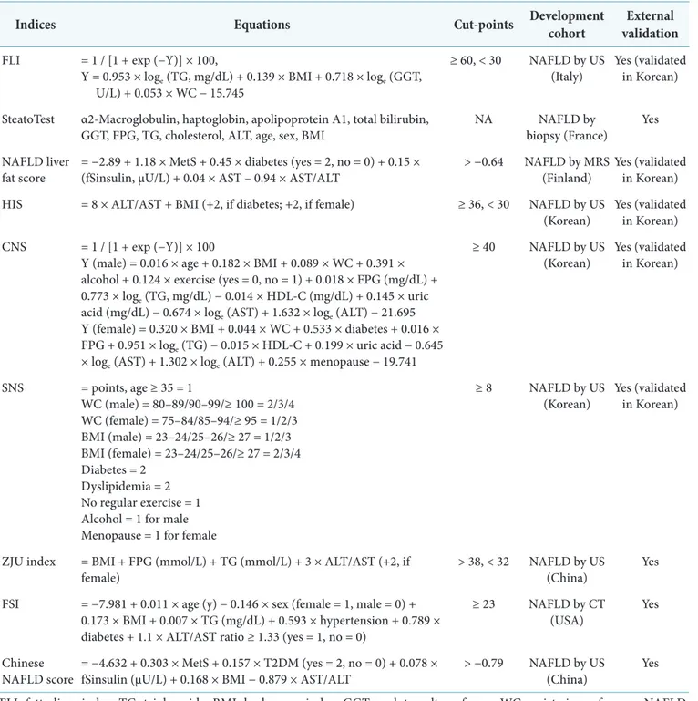
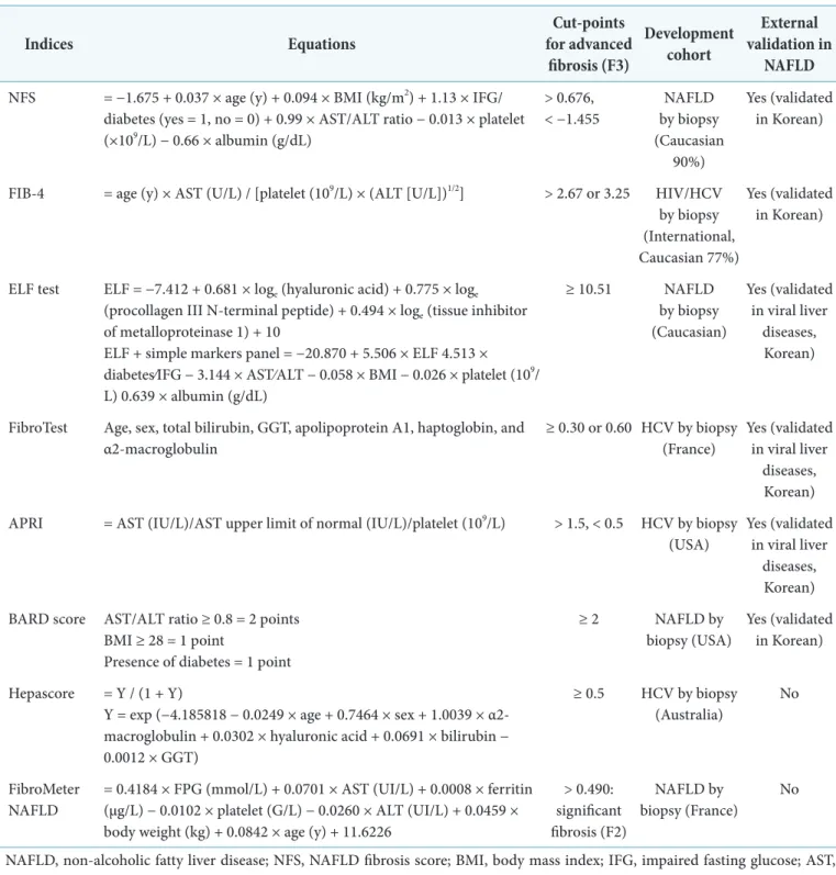
단순 지방간 및 간섬유화 진단을 위한 비침습적인 다양한 방법을 표로 정리하였다(Tables 1, 2) [2,5].
NASH 진단 방법으로 aminotransferase 측정이 있을 수 있으나 정확도는 상당히 낮다. 사이토카인 중 간세포 apoptosis를 반영하는 바이오마커인 cytokeratin-18 분절 (fragment)은 NASH 진단을 할 수 있는 유일한 marker로 밝혀진 바 있으나, 유의하게 진단을 위한 바이오마커로 쓰기 에는 여전히 무리가 있다[5].
영상학적으로 지방간(steatosis)을 진단하는 방법으로는 초음파가 가장 좋은 방법이나 간섬유화 측정에는 제한점 이 있어, 지방간과 간섬유화를 동시에 측정하는 제어감쇠매 개변수(controlled attenuation parameter, CAP)와 magnetic resonance imaging (MRI) 측정이 권고된다[2,6]. 초음파, computed tomography (CT)와 같은 영상학적인 방법으로 NASH 진단을 시도해보고자 하는 시도가 있었으나 이 역시 정확도는 떨어진다. 첫째, 초음파의 경우 비교적 쉽게 시행할 수 있는 장점이 있으나 중등도 이하의 경우 재현성이 낮고 정 량화가 어려운 단점이 있어 초기 진단 시에 제한적으로 시행 하게 될 가능성이 높다. 둘째, CT의 경우에는 MRI에 비해 비 용은 저렴하고 초음파와 다르게 정량화가 일부 가능한 장점 이 있으나 방사선 조사량에 대한 고려가 필요하여 임상에서 는 간내 지방량의 측정을 위해 단독으로 CT 검사를 시행하 는 경우보다는 건강검진이나 다른 질환의 진단 목적으로 CT 를 시행할 경우 부가적으로 간내 지방량의 측정을 시행하는
Table 1. Summary of biomarker-based prediction models to assess hepatic steatosis
Indices Equations Cut-points Development cohort External
validation FLI = 1 / [1 + exp (−Y)] × 100,
Y = 0.953 × loge (TG, mg/dL) + 0.139 × BMI + 0.718 × loge (GGT, U/L) + 0.053 × WC − 15.745
≥ 60, < 30 NAFLD by US
(Italy) Yes (validated in Korean) SteatoTest α2-Macroglobulin, haptoglobin, apolipoprotein A1, total bilirubin,
GGT, FPG, TG, cholesterol, ALT, age, sex, BMI NA NAFLD by
biopsy (France) Yes NAFLD liver
fat score = −2.89 + 1.18 × MetS + 0.45 × diabetes (yes = 2, no = 0) + 0.15 ×
(fSinsulin, μU/L) + 0.04 × AST – 0.94 × AST/ALT > −0.64 NAFLD by MRS
(Finland) Yes (validated in Korean) HIS = 8 × ALT/AST + BMI (+2, if diabetes; +2, if female) ≥ 36, < 30 NAFLD by US
(Korean) Yes (validated in Korean) CNS = 1 / [1 + exp (−Y)] × 100
Y (male) = 0.016 × age + 0.182 × BMI + 0.089 × WC + 0.391 × alcohol + 0.124 × exercise (yes = 0, no = 1) + 0.018 × FPG (mg/dL) + 0.773 × loge (TG, mg/dL) − 0.014 × HDL-C (mg/dL) + 0.145 × uric acid (mg/dL) − 0.674 × loge (AST) + 1.632 × loge (ALT) − 21.695 Y (female) = 0.320 × BMI + 0.044 × WC + 0.533 × diabetes + 0.016 × FPG + 0.951 × loge (TG) − 0.015 × HDL-C + 0.199 × uric acid − 0.645
× loge (AST) + 1.302 × loge (ALT) + 0.255 × menopause − 19.741
≥ 40 NAFLD by US
(Korean) Yes (validated in Korean)
SNS = points, age ≥ 35 = 1
WC (male) = 80–89/90–99/≥ 100 = 2/3/4 WC (female) = 75–84/85–94/≥ 95 = 1/2/3 BMI (male) = 23–24/25–26/≥ 27 = 1/2/3 BMI (female) = 23–24/25–26/≥ 27 = 2/3/4 Diabetes = 2
Dyslipidemia = 2 No regular exercise = 1 Alcohol = 1 for male Menopause = 1 for female
≥ 8 NAFLD by US
(Korean) Yes (validated in Korean)
ZJU index = BMI + FPG (mmol/L) + TG (mmol/L) + 3 × ALT/AST (+2, if
female) > 38, < 32 NAFLD by US
(China) Yes
FSI = −7.981 + 0.011 × age (y) − 0.146 × sex (female = 1, male = 0) + 0.173 × BMI + 0.007 × TG (mg/dL) + 0.593 × hypertension + 0.789 × diabetes + 1.1 × ALT/AST ratio ≥ 1.33 (yes = 1, no = 0)
≥ 23 NAFLD by CT
(USA) Yes
Chinese
NAFLD score = −4.632 + 0.303 × MetS + 0.157 × T2DM (yes = 2, no = 0) + 0.078 ×
fSinsulin (μU/L) + 0.168 × BMI − 0.879 × AST/ALT > −0.79 NAFLD by US
(China) Yes
FLI, fatty liver index; TG, triglyceride; BMI, body mass index; GGT, γ-glutamyltransferase; WC, waist circumference; NAFLD, non-alcoholic fatty liver disease; US, ultrasonography; FPG, fasting plasma glucose; ALT, alanine aminotransferase; NA, not available; MetS, metabolic syn¬drome; fSinsulin, fasting serum insulin; AST, aspartate aminotransferase; MRS, magnetic resonance spectroscopy; HSI, hepatic steatosis index; CNS, comprehensive NAFLD score; HDL-C, high density lipoprotein cholesterol; SNS, simple NAFLD score; ZJU, Zhejiang University; FSI, Framingham steatosis index; CT, computed tomography; T2DM, type 2 diabetes mellitus.
Adapted from the article of Lee et al. (Diabetes Metab J 2019;43:31-45) [2] in accordance with the Creative Commons Attribution Non-Commercial (CC BY-NC 4.0) license.
Table 2. Summary of biomarker-based prediction models to assess hepatic fibrosis
Indices Equations Cut-points
for advanced fibrosis (F3)
Development cohort
External validation in
NAFLD NFS = −1.675 + 0.037 × age (y) + 0.094 × BMI (kg/m2) + 1.13 × IFG/
diabetes (yes = 1, no = 0) + 0.99 × AST/ALT ratio − 0.013 × platelet (×109/L) − 0.66 × albumin (g/dL)
> 0.676,
< −1.455 NAFLD by biopsy (Caucasian
90%)
Yes (validated in Korean)
FIB-4 = age (y) × AST (U/L) / [platelet (109/L) × (ALT [U/L])1/2] > 2.67 or 3.25 HIV/HCV by biopsy (International, Caucasian 77%)
Yes (validated in Korean)
ELF test ELF = −7.412 + 0.681 × loge (hyaluronic acid) + 0.775 × loge
(procollagen III N-terminal peptide) + 0.494 × loge (tissue inhibitor of metalloproteinase 1) + 10
ELF + simple markers panel = −20.870 + 5.506 × ELF 4.513 × diabetes⁄IFG − 3.144 × AST⁄ALT − 0.058 × BMI − 0.026 × platelet (109/ L) 0.639 × albumin (g/dL)
≥ 10.51 NAFLD
by biopsy (Caucasian)
Yes (validated in viral liver
diseases, Korean)
FibroTest Age, sex, total bilirubin, GGT, apolipoprotein A1, haptoglobin, and α2-macroglobulin
≥ 0.30 or 0.60 HCV by biopsy (France)
Yes (validated in viral liver
diseases, Korean) APRI = AST (IU/L)/AST upper limit of normal (IU/L)/platelet (109/L) > 1.5, < 0.5 HCV by biopsy
(USA) Yes (validated in viral liver
diseases, Korean) BARD score AST/ALT ratio ≥ 0.8 = 2 points
BMI ≥ 28 = 1 point
Presence of diabetes = 1 point
≥ 2 NAFLD by
biopsy (USA) Yes (validated in Korean)
Hepascore = Y / (1 + Y)
Y = exp (−4.185818 − 0.0249 × age + 0.7464 × sex + 1.0039 × α2- macroglobulin + 0.0302 × hyaluronic acid + 0.0691 × bilirubin − 0.0012 × GGT)
≥ 0.5 HCV by biopsy (Australia)
No
FibroMeter
NAFLD = 0.4184 × FPG (mmol/L) + 0.0701 × AST (UI/L) + 0.0008 × ferritin (μg/L) − 0.0102 × platelet (G/L) − 0.0260 × ALT (UI/L) + 0.0459 × body weight (kg) + 0.0842 × age (y) + 11.6226
> 0.490:
significant fibrosis (F2)
NAFLD by
biopsy (France) No NAFLD, non-alcoholic fatty liver disease; NFS, NAFLD fibrosis score; BMI, body mass index; IFG, impaired fasting glucose; AST, aspartate aminotransferase; ALT, alanine aminotransferase; FIB-4, fibrosis-4; HIV, human immunodeficiency virus; HCV, hepatitis C virus; ELF, enhanced liver fibrosis; GGT, γ-glutamyltransferase; APRI, AST to platelet ratio index; FPG, fasting plasma glucose.
Adapted from the article of Lee et al. (Diabetes Metab J 2019;43:31-45) [2] in accordance with the Creative Commons Attribution Non-Commercial (CC BY-NC 4.0) license.
경우가 보다 많을 것으로 생각된다. 간과 비장의 Hounsfield unit (HU)의 비율 또는 간과 비장의 HU 차이를 측정하여 HU가 측정 위치에 따라 달라지는 단점을 보완할 수 있다.
간/비장 HU 비율이 0.9 이하인 경우 간내 지방침착이 있다고 간주하면 된다. 셋째, magnetic resonance를 이용한 MRI와 MRS (magnetic resonance spectroscopy)가 있다. 간접적으 로 간 내부의 지방량을 측정하는 초음파, CT와 다르게 MR 은 직접적으로 간 내부의 지방량을 측정한다. 높은 민감도 와 예민도로 치료 전후 간실질의 지방량을 측정할 수 있어 당 뇨병환자에게도 사용할 수 있으나, 고가의 비용, 숙련된 의 료진이 필요하다는 한계가 있다. 넷째, 제어감쇠매개변수는 transient elastography (FibroScanⓇ)를 이용하여 간실질에
지방 침착이 많을수록 초음파의 진폭이 감소하는 정도가 심 해진다는 점을 이용하여 간 내부의 지방 침착 정도를 측정한 다. Steatosis의 정도에 따라 S0의 경우 CAP 205 dB/m, S1 의 경우 245 dB/m, S2는 299 dB/m, S3는 321 dB/m의 중간 값을 보였다. CAP 250 dB/m 이상인 경우 지방간이 있음을 의미하며, 300 dB/m 이상인 경우 중등도의 지방간이 있음을 의미한다. CAP가 인슐린 저항성과 연관이 있다는 사실과 당 뇨병이 있는 환자의 CAP 수치가 당뇨병이 없는 환자의 수치 보다 높다는 점, 정량화 가능하여 검사자에 의존적이지 않다 는 점, 비용이 저렴하다는 점 때문에 FibroScanⓇ이 최근 많이 사용되고 있다. 간 섬유화는 FibroScanⓇ을 이용하여 LSM (liver stiffness measurement)을 통해 정량적으로 측정할 수
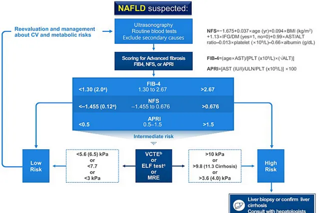
Fig. 1. Algorithm for non-alcoholic fatty liver disease (NAFLD) evaluation. Adapted from the article of Lee et al.
(Diabetes Metab J 2020;44:382-401) [1] in accordance with the Creative Commons Attribution Non-Commercial (CC BY-NC 4.0) license.
CV, cardiovascular; NFS, NAFLD fibrosis score; BMI, body mass index; IFG, impaired fasting glucose; DM, diabetes mellitus; AST, aspartate aminotransferase; ALT, alanine aminotransferase; FIB-4, fibrosis-4; PLT, platelet; APRI, AST to platelet ratio index; ULN, upper limit of normal; VCTE, vibration-controlled transient elastography; ELF, enhanced liver fibrosis; MRE, magnetic resonance elastography.
있다.
Fig. 1은 대한당뇨병학회 지방간연구회에서 NAFLD를 동 반한 제2형 당뇨병환자에서의 진단을 위한 접근 단계를 요약 한 권고안이다[1].
치료
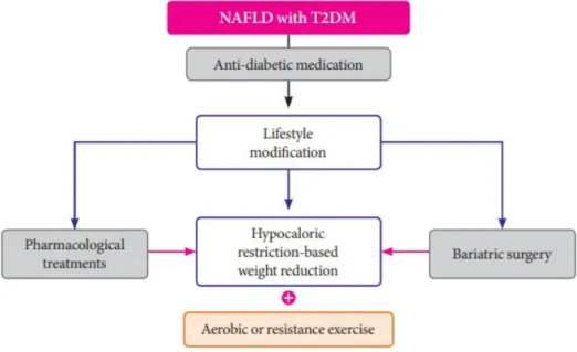
NAFLD와 당뇨병이 함께 동반되어 있을 경우 심혈관계 관 련 질환의 위험도를 낮추고 유리 지방산의 간 실질 침착을 줄 여 단순 지방간이 지방간염으로 진행하지 않도록 하는 것이 치료의 첫 번째 목표이고, NASH가 간섬유화로 진행하는 것 을 줄임으로써 간암으로 진행하는 위험을 낮추는 것이 이들 을 치료하는 목적이라 할 수 있겠다[1,7]. Fig. 2는 NAFLD 를 동반한 제2형 당뇨병환자에서의 관리 및 치료를 위한 접 근 단계를 요약한 권고안이다[1].
1. 생활습관을 통한 체중 감량
체중 감량은 식이, 운동과 같은 생활습관 개선과 bariatric surgery와 같은 수술적인 방법을 통해 이루어질 수 있다. 과
체중 또는 비만한 제2형 당뇨병환자가 체중을 감량한다는 것은 심혈관계 동반질환을 줄인다는 것 이외에 간 실질의 지 방 축적량을 감소시켜 염증과 인슐린저항성을 개선하는 의미 가 있다. 체중을 10% 이상 감량하면 NAFLD activity score 의 감소, 지방간염의 해소 및 fibrosis regression을 기대할 수 있다. 식단 조절과 유산소운동의 병행이 선호되고 있으나, weight regain에 대한 장기간 효과에 대해서는 현재로서는 알 려진 바가 없다[7].
2. 혈당강하제
Peroxisomal proliferator-activated receptor-γ (PPAR- γ) agonist인 thiazolidinedione은 인슐린 감수성 효과를 지니 며 간의 조직학적 소견을 향상시킨다고 보고되었다[8]. 생검 으로 NASH 진단을 받은 당뇨병 전단계 또는 제2형 당뇨병 환자들에게 하루 칼로리 소모를 제한하고 pioglitazone을 18 개월 투약하였을 경우 NASH의 호전율이 51%에 달했다는 연 구 결과도 있었다[9]. 위험성과 이득을 고려하여 pioglitazone 을 NASH와 제2형 당뇨병이 함께 동반되었을 때 일차 약제
Fig. 2. Suggested algorithm for the management of patients with non-alcoholic fatty liver disease (NAFLD) and type 2 diabetes mellitus (T2DM). Adapted from the article of Lee et al. (Diabetes Metab J 2020;44:382-401) [1] in accordance with the Creative Commons Attribution Non-Commercial (CC BY-NC 4.0) license.
로 쓰는 것을 고려해볼 수 있겠다. Glucacon-like peptide-1 receptor agonist (GLP-1RA)는 insulin 분비를 촉진시키고, 포만감을 주어 체중을 감량시킴으로써 NAFLD와 NASH의 치료제로 대두되고 있으나 연구가 더 필요한 상황이다[10].
Sodium-glucose co-transporter 2 (SGLT2) inhibitors는 체중감소를 유발함으로써 NAFLD의 치료를 위한 효과적인 약제로 부상하고 있다. SLGT2 inhibitors는 간효소 수치를 향상시키며, 간의 지방량, 간의 stiffness 정도를 호전시킨다 는 여러 연구가 있었다[10]. 제2형 당뇨병환자에게 광범위하게 사용되고 있는 1차 치료제인 metformin, 그리고 dipeptidyl peptidase-4 (DPP-4) 억제제의 경우 NAFLD와 당뇨병 사이 의 효과에 대해서는 데이터가 충분하지 않다[11].
3. 비혈당강하제
산화스트레스는 지방간염의 진행에 영향을 끼친다. 항산화 제인 vitamin E가 간손상을 방지하므로 생검으로 NASH 진 단을 받은 비당뇨병환자들을 대상으로 vitamin E를 투여하 였을 때 조직소견의 향상을 보이기는 하였으나, 당뇨병환자 들을 대상으로 한 데이터는 없으므로 권고하지는 않는다[1].
Statin, ezetimibe, fibrate, niacin, omega-3 polyunsaturated fatty acid는 NAFLD 진단을 받은 제2형 당뇨병환자들에 게는 효과가 있다는 증거가 없다[12]. 항산화, 항염증 작용 이 있는 것으로 알려 있는 ursodeoxycholic acid (UDCA) 는 NAFLD 진단을 받은 환자의 경우 간기능 향상 및 지 방간염의 향상을 보인다는 보고도 있었으나, NASH의 경 우 조직학적인 호전을 보이지는 않았다. TNF-α blocker인 pentoxyfylline은 NASH로 확인된 환자들의 조직학적인 호 전 소견을 보였으나, 당뇨병환자가 9.1%밖에 포함되지 않은 연구의 한계성이 있어 현재는 추천되지 않고 있다. Nuclear hormone인 farnesoid X receptor는 bile acid 합성에 주요한 역할을 하며 지방의 대사와 insulin sensitivity에 영향을 끼 친다. Farnesoid X receptor activator인 obeticholic acid는 chenodeoxycholic acid의 합성 형태이며, FLINT trial에서 간 생검으로 NASH 진단을 받은 환자에게 obeticholic acid 투약
군의 간 조직학적 소견이 향상되었다. 특히 이 연구의 52.7%
가 당뇨병환자였다는 데에서 의의가 있다. 미토콘드리아에서 fatty acid transport의 조절자 역할을 하는 carnitine은 당뇨 병과 NAFLD 진단을 받은 환자들을 대상으로 한 CORONA trial에서 12주 투약 이후 ALT의 호전과 CT로 판단한 지방 간염의 호전을 보였다[10,11].
결론
당뇨병의 유병률과 함께 대사성 증후군의 유병률 역시 증 가하고 있다. 당뇨병의 위험요인인 NAFLD의 유병률은 30%
로 알려져 있으며 NAFLD의 유병률은 당뇨병의 유무에 따 라 2배의 차이가 난다. 간조직에 지방이 축적되어 염증이 발 생하고 심한 간섬유화까지도 일어나므로 NAFLD 치료의 필 요성이 대두되었다. 간 생검, 생화학적인 방법, 영상학적인 방 법을 통해 단순 지방간, NAFLD, NASH, 진행된 간섬유화 까지 진단을 내릴 수 있게 되었다. 당뇨병이 동반되어 있는 NAFLD를 치료함으로써 심혈관계 질환 발생률을 낮춤과 동 시에 지방간염 또는 간섬유화로의 진행을 막는 데 목적이 있 다. 체중 감량을 포함한 생활습관 교정이 가장 중요하며, thiazolidinedione이 인슐린 감수성을 회복시켜주어 간의 조직 학적 소견의 호전을 가져온다고 알려져 있다. 당뇨병 약제가 아닌 여러 약제가 NAFLD, 지방간염을 호전시킨다는 여러 보 고가 있지만, 아직 연구가 더 진행되어야 할 필요가 있다.
REFERENCES
1. Lee BW, Lee Y, Park CY, Rhee EJ, Lee WY, Kim NH, et al. Non-alcoholic fatty liver disease in patients with type 2 diabetes mellitus: a position statement of the Fatty Liver Research Group of the Korean Diabetes Association.
Diabetes Metab J 2020;44:382-401.
2. Lee Y, Cho Y, Lee BW, Park CY, Lee DH, Cha BS, et al. Nonalcoholic fatty liver disease in diabetes. Part I: epidemiology and diagnosis. Diabetes Metab J
2019;43:31-45.
3. Rhee EJ. Clinical characteristics of non-alcoholic fatty liver disease based on analyses from the Kangbuk Samsung Health Study. J Korean Diabetes 2017;18:81-7.
4. Song YM, Lee Y, Kim JW, Ham DS, Kang ES, Cha BS, et al. Metformin alleviates hepatosteatosis by restoring SIRT1-mediated autophagy induction via an AMP- activated protein kinase-independent pathway. Autophagy 2015;11:46-59.
5. Lee Y. Diagnosis of non-alcoholic fatty liver disease based on clinical and laboratory data. J Korean Diabetes 2017;18:102-8.
6. Kim KJ, Kim SU, Chung YE, Kim CO. Radiologic evaluation of non-alcoholic fatty liver disease in diabetic patient. J Korean Diabetes 2017;18:88-101.
7. Lee BW. Management of patients with nonalcoholic fatty liver disease with lifestyle modification. J Korean Diabetes 2018;19:82-7.
8. Yang H, Park CY. Glucose-lowering agents in the management of nonalcoholic fatty liver disease. J Korean Diabetes 2018;19:88-96.
9. Cusi K, Orsak B, Bril F, Lomonaco R, Hecht J, Ortiz- Lopez C, et al. Long-term pioglitazone treatment for patients with nonalcoholic steatohepatitis and prediabetes or type 2 diabetes mellitus: a randomized trial. Ann Intern Med 2016;165:305-15.
10. Kim KS, Lee BW. Beneficial effect of anti-diabetic drugs for nonalcoholic fatty liver disease. Clin Mol Hepatol 2020;26:430-43.
11. Kim KS, Lee BW, Kim YJ, Lee DH, Cha BS, Park CY.
Nonalcoholic fatty liver disease and diabetes: part II:
treatment. Diabetes Metab J 2019;43:127-43.
12. Kim SU. Pharmacologic treatment using non- hypoglycemic agents for nonalcoholic fatty liver disease. J Korean Diabetes 2018;19:97-100.