蛇毒이 세포자멸사와 관계있는 Death Receptor를 통한 인간 대장암 세포 성장억제에 미치는 영향
오명진ᆞ송호섭
경원대학교 한의과대학 침구학교실
목적 : 이 연구는 Vipera lebetina turanica 사독(蛇毒)이 인간 대장암 세포주인 HCT116 세포에서 세포주 기진행, death receptor 의존적 세포자멸사 경로 관련단백질 발현 및 NK-κB와 STAT3 활성에 미치는 영향 을 규명함으로써 대장암 세포 성장에 대한 억제와 그 기전에 대하여 살펴보고자 하였다.
방법 : 사독을 처리한 후 HCT116의 세포주기를 분석하기 위해서 FACS analysis를 시행하였고, apoptosis 평가에는 TUNEL assay를 시행하였으며 death receptor 의존적 세포자멸사 경로 관련단백질 및 NF-κB와 STAT3 활성 변동 관찰에는 RT-PCR 및 western blot analysis를 시행하였다.
결과 : 1. 0.1, 0.5 및 1 ㎍/㎖ 등의 사독을 처리한 결과 농도 의존적으로 HCT116 대장암 세포활성의 억 제가 나타났다.
2. 0.1, 0.5 및 1 ㎍/㎖ 등의 사독을 처리한 결과 농도의존적으로 세포자멸사 활성세포의 증가가 나타났고, SVT 1 ㎍/㎖에서는 60-70%의 대장암세포 억제 효과가 나타났다.
3. 0.1, 0.5 및 1 ㎍/㎖ 등의 사독을 처리한 결과 약한 G1 arrest와 강한 G2/M arrest가 나타났고, G0/G1 또는 G2/M 관련 cyclin D, E 및 B1의 증가가 나타났다.
4. 0.1, 0.5 및 1 ㎍/㎖ 등의 사독을 처리한 결과 death receptor4, 5의 발현증가와 그에 따른 세포자멸사
1)
Inhibitory Effect of Snake Venom on Colon Cancer Cell Growth Through Induction of Death
Receptor Dependent Apoptosis
Oh Myung-jin and Song Ho-sueb
Dept. of Acupuncture & Moxibustion, College of Oriental Medicine, Kyungwon University
* This research was supported by the Kyungwon University Research Fund in 2012
․Acceptance : 2012. 1. 17. ․Adjustment : 2012. 1. 24. ․Adoption : 2012. 1. 26.
․Corresponding author : Song Ho-sueb, Kyungwon Gil Oriental Medical Hospital, 1200-1 Guwal-dong Namdong-gu Incheon Republic of Korea
Tel. 82-70-7120-5012 E-mail : [email protected]
국문초록
Original Article
촉진 Bax, PARP, caspase-3, -8, -9 발현 증가 및 세포자멸사 억제의 Bcl-2의 발현 감소 등이 나타났다.
6. 0.1, 0.5 및 1 ㎍/㎖ 등의 사독을 처리한 결과 NF-κB와 STAT3의 활성변동은 관찰되지 않았다.
결론 : 이상의 연구에서 사독은 death receptor 의존적인 세포자멸사를 촉진하여 대장암의 화학치료 내성 을 극복할 수 있는 하나의 대안이 될 것으로 생각되지만 보다 심화된 연구가 필요할 것으로 사료된다.
핵심 단어 : 사독(蛇毒), Vipera lebetina turanica, 대장암, 세포자멸사, 세포주기, death deceptor
Ⅰ. Introduction
Colon cancer is a major malignancy with a worldwide cumulative incidence rate of 9.4% and the second leading cause of cancer1). It develops in the cecum, colon and rectum2), which confined within the intestinal wall is treatable by enucleation of the local lesions. However, in the advanced colon cancer metastasizing into the deeper regions or other organ such as liver, surgical treatment alone has the limitations2). Although chemotherapy with anticancer drugs, such as 5-fluorouracil, leucovorin, irinotecan, and oxaliplatin is usually adopted for inhibiting the cancer growth and prolonging survival, it remains unsatisfactory due to current status of comparatively poor cancer drug development and cancer stem cell related highly chemoresistance in colon cancer3-6). Anti-cancer drug treatment generally results in apoptosis, programmed cell death through the activation of caspase cascade systems via triggering Death Receptor (DR) dependent apoptotic pathway at the cell surface, activating caspase-8 or cytochrome C dependent pathway at the mitochondria, characterized by loss of mitochondrial permeability transition, release of mitochondrial cytochrome C into the cytoplasm, consecutive activation of caspase-8 or -9, and caspase-37-9). However, it is usual for the activated apoptotosis related pathway to represent only some of the characteristics of the above classical apoptotic pathways in anti-cancer agents, depending on cancer cell type10,11). According to the previous reports12,13), acquired resistance to chemotherapeutics is closely related to the lower
apoptosis rate. Therefore, a new remedy enhancing it for colon cancer is urgently needed.
A snake venom toxin (SVT) from Vipera lebetina turanica, is a group of basic peptides, and important factor V activator made up of 236 amino acids with six disulfide bonds formed by twelve cysteins14). A few researchers revealed that SVT exerts its effect on cellular proteins such as Bax, Bcl-2, caspases in the classical apoptotic way, and that SVT inhibits cancer growth through the induction in cancers such as neuroblastoma and prostate cancer15-17). However, experiments demonstrating the molecular mechanisms of the anti-cancer effects of SVT in colon cancer cells have not been reported. Thus, in the present study, I investigated apoptosis related anti-cancer effects of SVT and confirmed whether increase of DR expression, cell cycle arrest, and inhibition of activity of NF-κB signal molecules or STAT3 pathway in the human colon cancer, HCT116 cells.
Ⅱ. Materials and methods
A. Materials
SVT from Vipera lebetina turanica was purchased from Sigma Chemical Co. (Saint Louis, USA). DR3, DR4, and DR6 siRNA were purchased from Santa Cruz Biotechnology Inc. (Santa Cruz, CA). All of the secondary antibodies such as Bax, Bcl-2, caspase-3, cleaved caspase-3, caspase-8, caspase-9, cleaved caspase-9, p50, p65, PARP and STAT3,
Death
receptor Forward primer Reverse primer
TNFR 1
5`-ACC AAG TGC CAC AAA GGA AC-3`
5`-CTG CAA TTG AAG CAC TGG AA-3`
TNFR 2
5`-CTC AGG AGC ATG GGG ATA AA-3`
5- AGC CAG CCA GTC TGA CAT CT-3`
DR 3
5-ATG GCG ATG GCT GCG TGT CCT G-3
5-AGC GCC TCC TGG GTC TCG GGG TAG-3
DR 4
5-ACT TTG GTT GTT CCG TTG CTG TTG-3
5-GGC TTT CCA TTT GCT GCT CA-3
DR 5
5-TGG AAC AAC GGG GAC AGA ACG-3
5-GCA GCG CAA GCA GAA AAG GAG-3
used in Western blot analysis were purchased from Santa Cruz Biotechnology(Santa Cruz, CA). All other reagents were purchased from Sigma unless otherwise stated.
B. Cell culture
The HCT116 colon cancer cells were obtained from ATCC (American Type Culture Collection, Rockville, MD). Colon cells were cultured in RPMI-1640 medium (Life Technologies Inc., Gaithersberg, MD) supplement with 10% FCS (Fetal Calf Serum;
Collaborative Biomedical Products, Bedford, MA) and antibiotics, penicillin/streptomycin (100 unit/㎖, Bioproducts, Walkersville, MD). Cell cultures were then maintained at 37℃ in a humidified atmosphere of 5% CO2.
C. Cell viability assay
To determine the cell number, HCT116 colon cancer cells or CCD112 normal colon cells were plated in 24-well plates(5 × 10⁴ cells/well), and subconfluent cells were subsequently treated with SVT (0.1, 0.5 and 1 ㎍/㎖) for 24 hr. After treatment, cells were trypsinized and pelleted by centrifugation for 5 min at 1,500 rpm, resuspended in 5 ㎖ of phosphate-buffered saline (PBS), and 0.1
㎖ of 0.2% trypan blue was added to the cancer cell suspension in each of the solutions(0.9 ㎖ each).
Subsequently, a drop of suspension was placed into a Neubauer chamber and the living cancer cells and normal colon cells were counted. Cells that showed signs of staining were considered to be dead, whereas those that excluded trypan blue were considered viable. Each assay was carried out in triplicate.
D. Cell cycle analysis
To examine the effect of SVT on cell cycle distribution of asynchronous populations of HCT116 colon cancer cells, replicative DNA synthesis and DNA content were analyzed using bivariate flow
cytometric analysis. Cells were harvested by trypsin- EDTA release and fixed in ice-cold 70% ethanol.
At least 1-2hr before flow cytometric analysis, cells were resuspended in a 1 ㎖ aliquot of modified Vindelov’s DNA staining solution (10 ㎍/㎖ RNase A and 5 ㎍/㎖ propidiumiodide in phosphate-buffered saline). Flow cytometric analysis was performed with flow cytometry system (FACS Calibur-S System, BD Bioscience, SanJose, CA). Cells in the G1, S, and G2/M phase of the cell cycle were determined with Modfit LT(Verity House Software, Top-sham, ME).
E. Reverse transcription(RT)-PCR
Total RNAs were isolated from cultured cells using RNeasy Plus Mini Kit (Qiagen) according to the manufacturer’s manual. The RNA pellet obtained in the final step was dissolved in 30 ㎕ of sterile diethylpyrocarbonate (DEPC)-treated water, and its concentration was determined using a UV spectrophotometer at 260 ㎚. RNA was kept in DEPC-treated water at -70 until use. Reverse transcription was performed using High Capacity RNA-to-cDNA Kit(AB). PCR amplifications were then carried out with the following primers.
F. Western blot analysis
Western blot analysis was done as described previously. The membrane was incubated for 2 hr at room temperature with specific antibodies: rabbit polyclonal for caspase-3, cleaved caspase-3, caspase- 8, cleaved caspase-8, caspase-9, cleaved caspase-9, PARP, Cleaved PARP, Bcl-2 (1 : 1,000 dilution, Cell Signaling Technology, Inc., Beverly, MA), p-Stat3, Bax, p50, p65 (1 : 500 dilution, Santa Cruz Biotechnology, Inc.), and mouse monoclonal Stat 3 (1 : 500 dilution, Santa Cruz Biotechnology, Inc.). The blot was then incubated with the corresponding conjugated anti-rabbit and anti-mouse immunoglobulin G-horseradish peroxidase (1 : 2,000 dilutions, Santa Cruz Biotech- nology, Inc.). Immunoreactive proteins were de- tected with the ECL Western blotting detection system.
Transfection. HCT116 colon cancer cells (3 × 10⁴ cells per well) were plated in 24-well plates and transiently transfected with siRNA, using a mixture of siRNA and the WelFect-EXPLUS reagentin OPTI- MEN, according to the manufacturer’s specification (WelGENE, Seoul, Korea). The transfected cells were treated with 0.1, 0.5 and 1㎍/㎖ SVT for 24 hr.
DR3 siRNA seq. 5’-GAAGCCCUAAGUACGGUU Att
DR4 siRNA seq. 5’-CUCUGAUGCUGUUCUUUG Att
DR6 siRNA seq. 5’-GCCUUCUAGUGUGAUGAA Att
G. Apoptosis evaluation
HCT116 colon cancer cells (2.5 × 10⁵ cells/well) were cultured on 8-chamber slides. After colon cancer cells were transfected with siRNA, the cells were treated with SVT(5 ㎍/㎖). The cells were washed twice with PBS and fixed by incubation in 4% paraformaldehyde in PBS for 1 hr at room temperature. Membrane was permeabilized by exposure to 0.1% Triton X-100 in phosphate-buffered saline for 5 min at room temperature. TdT-mediated dUTP nick and labeling (TUNEL) assays were
performed by using the in situ Cell Death Detection Kit (Roche Diagnostics GmbH, Mannheim, Germany) according to the manufacturer’s instructions. For 4'-6-Diamidino-2-phenyl indole (DAPI) staining, slides were incubated for 15 min at room temperature in the dark with mounting medium for fluorescence containing DAPI (Vector Laboratories, Inc., Burlingame, CA). The cells were then observed through a fluorescence microscope (Leica Microsystems AG, Wetzlar, Germany).
H. Data analysis
The data were analyzed using the GraphPad Prism 4 ver. 4.03 software (GraphPad Software, La Jolla, CA). Data are presented as mean ± SD. The differences in all data were assessed by one-way analysis of variance (ANOVA). When the P value in the ANOVA test indicated statistical significance, the differences were assessed by the Dunnett’s test.
A value of P < 0.05 was considered to be statistically significant.
Ⅲ. Results
A. Effect of SVT toxin on cell growth in HCT116 colon cancer cells
To assess the inhibitory effect of SVT on cell growth of HCT116 colon cancer cells, we analyzed cell viability by direct cell counting. The cells were treated with several concentrations of SVT(0.1, 0.5, and 1 ㎍/㎖) for 24 hr. As shown in Fig. 1, SVT inhibited cell proliferation of HCT116 colon cancer cells in a concentration-dependent manner, whereas it didn’t exert an influence on CCD112 normal colon cell viability.
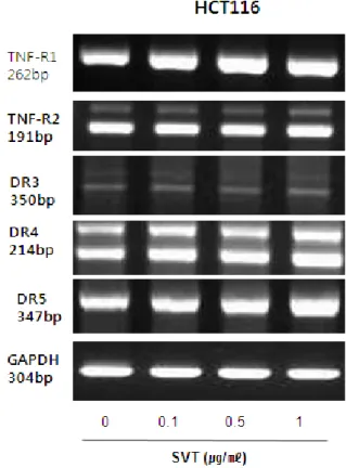
B. Expression of death receptors in HCT116 colon cancer cells by SVT
Apoptosis can be induced by stimulation of DRs
Fig. 1. Effect of SVT on cell viability in HCT116 colon cancer cells
Concentration-dependent effect of SVT on the cell viability assay in HCT116 or CCD112. After treatment of SVT (0.1, 0.5 and 1 ㎍/㎖) for 24 hr, the cells were harvested by trypsinization and stained with 0.2% trypan blue. Relative cell survival rate was determined by counting live and dead cells. The results were expressed as a percentage of viable cells.
Columns, means of three experiments, with triplicates of each experiment; bars, SD.
expression. Therefore, to investigate expression of DRs in cancer cells undergoing apoptotic cell death, we performed RT-PCR analysis. RT-PCR analysis showed that SVT treatment increased TNF-R1, DR4 and DR5 mRNA levels in a concentration dependent manner, but TNF-R2, DR3 expression levels were not changed by SVT in HCT116 cells(Fig. 2).
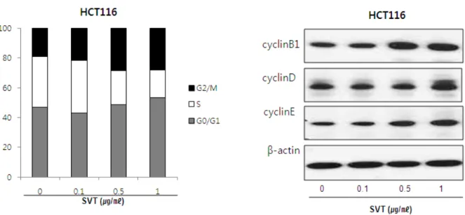
C. Cell cycle arrest at the G0/G1 and G2/M phase
To evaluate whether arrest of the cells in specific cell cycle could be related with cell death, I analyzed cell cycle after treatment of SVT. As increasing the concentration of SVT over 0.1 ㎍/㎖, the number of cells distributed in S phase was decreased significantly compared to the cells in other phases. The percentage of the cells in the S phase was about 35% in the untreated both cells, but the number was gradually decreased up to about 17% by the treatment of SVT (1 ㎍/㎖), whereas, SVT arrested the cell in both the G0/G1 phase and the G2/M phase (Fig.3). The percentage of the cells presented in the G0/G1 phase was 47.7% (control), 43.3% (0.1 ㎍/㎖), 51.4% (0.5 ㎍/㎖)
Fig. 2. Effect of SVT on DRs expression in HCT116 colon cancer cells
Cells were treated with SVT (0.1, 0.5, and 1 ㎍/㎖) for 24 hr, and total RNA were extracted and examined for expressions of TNF-R1, TNF-R2, DR-3, -4, -5 and GAPDH by RT-PCR. GAPDH was used as an internal control to show equal RNA loading.
Each band is representative for three experiments.
Fig. 3. Cell cycle analysis of HCT116 colon cancer cells treated with various doses of SVT
DNA content was analyzed by flow cytometry as described in the Materials and Methods. HCT116 colon cancer cells were treated with 0 to 1 ㎍/㎖ SVT for 24 hr. Each panel is representative of three similar experiments with triplicates.
Fig. 4. Effect of SVT on apoptotic cell death of HCT116 colon cancer cells
HCT116 were treated with SVT (0.1, 0.5 and 1 ㎍/㎖) for 24 hr, and then labeled with DAPI and TUNEL solution.
Total number of cells in a given area was determined by using DAPI nuclear staining (fluorescent microscope). The green color in the fixed cells marks TUNEL-labeled cells.
The apoptotic index was determined as the DAPI-stained TUNEL-positive cell number/total DAPI stained cell number (magnification, 200×). Columns, means of three experiments, with triplicates of each experiment; bars, SD. * : p < 0.05, significantly different from SVT-untreated control cells.
and 54.3% (1 ㎍/㎖) in colon cancer cell, respectively.
The percentage of the cells presented in the G2/M phase was 18.8%(control), 23.1%(0.1 ㎍/㎖), 29.1%(0.5 ㎍/㎖) and 29.3%(1 ㎍/㎖) in colon cancer cell, respectively.
Moreover, to investigate the influence of SVT upon cell cycle regulatory proteins such as cyclin B1, cyclin D, cyclin E, I performed western blot analysis, showing that SVT increased the cyclin B1, cyclin D and cyclin E expressions concentration dependently in HCT116 colon cancer cells (Fig. 3).
D. Apoptotic cell death by SVT
To determine the inhibition of cell growth by SVT was due to the induction of apoptotic cell death, we evaluated the changes in the chromatin morphology of cells by using DAPI staining followed by TUNEL staining assays, and then the double labeled cells were analyzed by fluorescence microscope. Conversely well with cell growth inhibition, DAPI-stained TUNEL-positive cells were significantly increased in SVT treated cells dose-dependently. The treatment of SVT over 1 ㎍/㎖
resulted in about 60-70% induction of apoptotic cell death in HCT116 colon cancer cells (Fig. 4).
E. Effect of SVT on the expression of apoptotic regulatory proteins
To figure out the relationship between the induction of apoptotic cell death and the expression of their regulatory proteins by SVT, expression of apoptotic cell death related proteins was investigated by Western blots. The expression of
Caspase3 Cle-Caspase3
Caspase3 Cle-Caspase3 Caspase9 Cle-Caspase9 Bax
Bcl2
Caspase8 →
Cle-Caspase8 →
PARP Cle-PARP
β-actin
Fig. 5. Effect of SVT on the expression of apoptosis regulatory proteins
Expression of apoptosis regulatory proteins was determined using Western blot analysis. The HCT116 colon cancer cells were treated with different concentrations of SVT (0.1, 0.5, and 1 ㎍/㎖) for 24 hr. Equal amounts of total proteins (50
㎍/lane) were subjected to 12% or 8% SDS-PAGE.
Expression of PARP, caspase-3, and -9, Bax, Bcl-2, and β -actin were detected by Western blotting using specific antibodies. β-actin protein here was used as an internal control. Each band is representative for three experiments.
anti-apoptotic protein Bcl-2 was significantly decreased; however, the expression of pro-apoptotic proteins, Bax, cleaved form of caspase-3, -8 and -9 were significantly and dose-dependently increased by treatment of SVT in HCT116 colon cancer cells (Fig. 5).
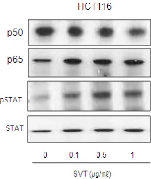
F. Effect of SVT on the activation of NF-κB signal molecules and STAT3
The Phosphorylation of IκB in a NF-κB signal pathway and STAT3 is associated with proliferation and maintenance of tumors. Thus, activation of NF-κB signal molecules and STAT3 pathway was investigated. We found that the ex- pression of NF-κB signal molecules and p- STAT3 were not changed by SVT in HCT116
Fig. 6. Effect of SVT on the activation of NF-κ B/STAT3 in HCT116 colon cancer cells
The colon cancer cells were treated with different con- centrations of SVT(0.1, 0.5 and 1 ㎍/㎖) for 24 hr. Equal amounts of total proteins(50 ㎍/lane) were subjected to 8%
SDS-PAGE. And β-actin were detected by Western blotting using specific antibodies of p50, p65, p-STAT3 and b-actin protein here was used as an internal control.
Each band is representative for three experiments.
colon cancer cells (Fig. 6).
Ⅳ. Discussion
The central and noteworthy finding in this study is the confirmation of anti-cancer efficacy of SVT against colon cancer, HCT116 cells through inducton of apoptosis via increase of DR expres- sions, cell cycle arrest, impact on cellular proteins including PARP, capases-3, -9, Bax and Bcl-2 in the classical apoptotic pathway.
Apoptosis is the process of programmed cell death that majorly involves in anti-cancer effects in cancers, and it is also considered to be a promising mechanism to overcome the acquired chemore- sistance12,13,18). According to several researches, it was found that natural agents-induced apoptotic
cell death could be selectively triggered by increase of DR expression19-23). In other words, expression of DR induces activation of caspase-8 and then make the apoptosis going through the activation of pro-apoptotic proteins including caspases-9 and -3, as well as Bax in the DR dependent apoptotic systems24,25). In the present study, Similar with the above, my data represented that SVT inhibited cell proliferation of HCT116 colon cancer cells in a concentration-dependent manner through about 60-70% induction of apoptotic cell death, and that it also increased TNF-R1, DR4 and DR5 mRNA levels dose-dependently, triggering the subsequent increase of the expression of pro-apoptotic proteins including PARP, Bax, cleaved form of caspase-3, -8, and -9, and concomitant decrease of that of anti-apoptotic protein, Bcl-2.
Meanwhile, the induction of apoptosis is also closely related with the regulation of cell cycle progression in which cyclin dependent kinases (CDK), CDK inhibitor and cyclins are involved.
CDK inhibitors are tumor suppressor proteins down-regulating the cell cycle progression by binding active CDK-cyclin complexes and thereby inhibiting their kinase activities26,27). Cyclin D is rate limiting for the progression of the cell cycle from the G1 phase to the S phase, during which DNA replication occurs28). Cyclin E, an essential cyclin that is expressed in late G1 and early S phase, regulates the phosphorylation of many proteins that are important for cell proliferation29). cyclin B1 is likely to be most important in mitotic regulation the target of multiple mitotic checkpoints, which is related with a mechanism by which the p53 tumor suppressor inhibits G2/M transition28,29).
Cell-cycle checkpoints, receptor collectives are responsible for the control of cell-cycle phase progression, which is emerging as a novel target of anti-cancer agents for increasing cytotoxicity of chemotherapy30-33). G0/G1 or G2/M arrest have also been associated with enhanced apoptosis, for defects in the G1 or G2/M checkpoint may lead a cancer cell to enhanced apoptosis through correcting abnormal proliferation of damaged cells or inducing
mitosis of them34-37).
According to Sauter et al’s report38), down- regulation of cyclin D through antisense treatment led to the apoptosis of melanoma cells and caused tumor shrinkage of xenotransplants in nude mice.
Moreover, Son et al. demonstrated that SVT- induced G0/G1 and G2/M phase arrest, accompanied by the reduction of cell distribution into the S phase, is a possible mechanism for cell growth inhibition leading to apoptotic cell death. In this study, consistent with the above, SVT caused a weak G0/G1 cell cycle arrest and a strong G2/M arrest in the colon cancer. However, expression of G1 or G2/M phase regulating protein cyclin D, E or cyclin B1 was upregulated in the colon cancer HCT116 cells, unlike the above prostate cancer cells.
In addition, activated NF-κB or STAT3 plays important roles in cell growth, proliferation, survival, differentiation, apoptosis, metastasis, and angiogenesis39-41). Alternatively, agents inactivating NF-κB or STAT3 can inhibit cancer growth through undergoing apoptosis. Park et al42). reported that bee venom inhibited prostate cancer growth through induction of apoptosis via inactivation of NF-κB, and Saydmohammed et al43). demonstrated that Curcumin suppresses constitutive activation of STAT-3 by up-regulating protein inhibitor of activated STAT-3 (PIAS-3) in ovarian and endometrial cancer cells. Unexpectedly, my data revealed that SVT didn’t affect the activity of NF- κB and STAT3 in colon cancer cells, HCT116, although it was confirmed that SVT inhibit the colon cancer growth through induction of apoptosis via enhancement of DR expression and subsequent caspase cascade events. Taken together, as the activated apoptotosis related pathway doesn’t reflect all of the characteristics of the classical apoptotic pathways from cytochrome C release to caspase cascade events relying on anti-cancer agents and cancer cell type10,11), present study revealed partially different findings including conversely increased G1 or G2/M related cyclins, unaffected NF-κB and STAT3 activity, from the previous findings.
Although further study is needed to reconfirm the results in this study and to elucidate more concrete mechanism, consequently, These present data provide that SVT could be useful candidate compounds to enhance tumor growth inhibiting ability of chemotherapeutics through overcoming the resistance via enhancement of DR expression and the related apoptosis.
Ⅴ. References
1. Jemal A, Siegel R, Ward E, Hao Y, Xu J, Thun MJ. Cancer statistics. CA Cancer J Clin. 2009 ; 59(4) : 225-49.
2. Danaei G, Van der Hoorn S, Lopez AD, Murray CJL, Ezzati M. The comparative risk assessment collaborating group(cancers), causes of cancer in the world: Comparative risk assessment of nine behavioural and environmental risk factors.
Lancet. 2005 ; 366(9499) : 1784-93.
3. Scheele J, Altendorf-Hofmann A. Resection of colorectal liver metastases. Langenbeck’s Arch Surg. 1999 ; 384(4) : 313-27.
4. Goldberg RM, Sargent DJ, Morton RF, Fuchs CS, Ramanathan RK, Williamson SK, Findlay BP, Pitot HC, Alberts SR. A randomized controlled trial of fluorouracil plus leucovorin, irinotecan and oxaliplatin combinations in patients with previously untreated metastatic colorectal cancer. J Clin Oncol. 2004 ; 22(1) : 23-30.
5. Kamb A, Wee S, Lengauer C. Why is cancer drug discovery so difficult? Nature Rev Drug Discovery. 2007 ; 6(2) : 115-20.
6. Dalerba P, Cho RW, Clarke MF. Cancer Stem Cells: Models and Concepts. Annual Rev Med.
2007 ; 58 : 267-84.
7. Strasser A, O’Connor L, Dixit VM. Apoptosis signaling. Ann Rev Biochem. 2001 ; 69 : 217-45.
8. Salvase GS, Abrams JM. Caspase activation—
stepping on the gas or releasing the brakes Lessons from humans and flies. Oncogene. 2004 ; 23(16) : 2774-84.
9. Debatin KM, Krammer PH. Death receptors in chemotherapy and cancer. Oncogene. 2004 ; 23(16) : 2950-66.
10. Balan KV, Demetzos C, Prince J, Dimas K, Cladaras M, Han Z, Wyche JH, Pantazis P.
Induction of apoptosis in human colon cancer HCT116 cells treated with an extract of the plant product, Chios mastic gum. In Vivo. 2005 ; 19(1) : 93-102.
11. Balan KV, Prince J, Han Z, Dimas K, Cladaras M, Wyche JH, Sitaras NM, Pantazis P.
Antiproliferative activity and induction of apoptosis in human colon cancer cells treated in vitro with constituents of a product derived from Pistacia lentiscus L. var. chia. Phytomedicine.
2007 ; 14(4) : 263-72.
12. Efimova EV, Liang H, Pitroda SP, Labay E, Darga TE, Levina V, Lokshin A, Roizman B, Weichselbaum RR, Khodarev NN. Radioresistance of Stat1 overexpressing tumour cells is associated with suppressed apoptotic response to cytotoxic agents and increased IL6-IL8 signalling. Int J Radiat Biol. 2009 ; 85(5) : 421-31.
13. Maduro JH, Noordhuis MG, ten Hoor KA, Pras E, Arts HJ, Eijsink JJ, Hollema H, Mom CH, de Jong S, de Vries EG, de Bock GH, van der Zee AG. The prognostic value of TRAIL and its death receptors in cervical cancer. Int J Radiat Oncol Biol Phys. 2009 ; 75(1) : 203-11.
14. Siigur E, Aaspollu A, Siigur J. Sequence diversity of Vipera lebetina SVT gland serine proteinase homologs-result of alternative- splicing or genome alteration. Gene. 2001 ; 263 : 199-203.
15. Alves RM, Antonucci GA, Paiva HH, Cintra AC, Franco JJ, Mendonça-Franqueiro EP, Dorta DJ, Giglio JR, Rosa JC, Fuly AL, Dias-Baruffi M, Soares AM, Sampaio SV. Evidence of caspase-mediated apoptosis induced by l-amino acid oxidase isolated from Bothrops atrox snake venom. Comp Biochem Physiol A Mol Integr Physiol. 2008 ; 151(4) : 542-50.
16. Park MH, Son DJ, Kwak DH, Song HS, Oh
KW, Yoo HS, Lee YM, Song MJ, Hong JT.
Snake venom toxin Inhibits Cell Growth through Induction of Apoptosis in Neuroblastoma Cells.
Arch Pharm Res. 2009 ; 32(11) : 1545-54, 17. Son DJ, Park MH, Chae SJ, Moon SO, Lee JW,
Song HS, Moon DC, Kang SS, Kwon YE, Hong JT. Inhibitory effect of SVT toxin from Vipera lebetina turanica on hormone-refractory human prostate cancer cell growth: induction of apoptosis through inactivation of nuclear factor kappaB. Mol Cancer Ther. 2007 ; 6(2) : 675-83.
18. O’Donovan TR, O’Sullivan GC, McKenna SL.
Induction of autophagy by drugresistant esophageal cancer cells promotes their survival and recovery following treatment with chemotherapeutics.
Autophagy. 2011 ; 7(5) : 509-24.
19. Kang YJ, Kim IY, Kim EH, Yoon MJ, Kim SU, Kwon TK, Choi KS. Paxilline enhances TRAIL-mediated apoptosis of glioma cells via modulation of c-FLIP, survivin and DR5. Exp Mol Med. 2011 ; 43(1) : 24-34.
20. Li J, Yu W, Tiwary R, Park SK, Xiong A, Sanders BG, Kline K. α-TEA-induced death receptor dependent apoptosis involves activation of acid sphingomyelinase and elevated ceramide- enriched cell surface membranes. Cancer Cell Int. 2010 ; 10 : 40.
21. Zhu H, Liu XW, Ding WJ, Xu DQ, Zhao YC, Lu W, He QJ, Yang B. Up-regulation of death receptor 4 and 5 by celastrol enhances the anti-cancer activity of TRAIL/Apo-2L. Cancer Lett. 2010 ; 297(2) : 155-64.
22. Kim EJ, Park SY, Lee JY, Park JH. Fucoidan present in brown algae induces apoptosis of human colon cancer cells. BMC Gastroenterol.
2010 ; 10 : 96.
23. Tang Y, Li X, Liu Z, Simoneau AR, Xie J, Zi X. Flavokawain B, a kava chalcone, induces apoptosis via up-regulation of death-receptor 5 and Bimexpression in androgen receptor negative, hormonal refractory prostate cancer cell lines and reduces tumor growth. Int J Cancer. 2010 ; 127(8) : 1758-68.
24. Sun SY. Understanding the role of the death
receptor 5/FADD/caspase-8 death signaling in cancer metastasis. Mol Cell Pharmacol. 2011 ; 3(1) : 31-4.
25. Elrod HA, Sun SY. Modulation of death receptors by cancer therapeutic agents. Cancer Biol Ther. 2008 ; 7(2) : 163-73.
26. Graña X, Reddy EP. Cell cycle control in mammalian cells: role of cyclins, cyclin depend- ent kinases(CDKs), growth suppressor genes and cyclin-dependent kinase inhibitors (CKIs).
Oncogene. 1995 ; 11(2) : 211-9.
27. Morgan DO. Effects of pulmonary gas embolism on circulation and respiration in the dog. VI.
Influence of body position on the effects of pulmonary gas embolism. Nature. 1995 ; 374 : 131-4.
28. Stewart ZA, Westfall MD, Pietenpol JA. Cell- cycle dysregulation and anticancer therapy.
Trends Pharmacol Sci. 2003 ; 24(3) : 139-45.
29. Zhao J, Dynlacht B, Imai T, Hori T, Harlow E.
Expression of NPAT, a novel substrate of cyclin E-CDK2, promotes S-phase entry. Genes Dev. 1998 ; 12(4) : 456-61.
30. Senderowicz AM, Sausville EA. Preclinical and clinical development of cyclin-dependent kinase modulators. J Natl Cancer Inst. 2000 ; 92(5) : 376-87.
31. Jackson JR, Gilmartin A, Imburgia C, Winkler JD, Marshall LA, Roshak A. An indolocarbazole inhibitor of human checkpoint kinase (Chk1) abrogates cell-cycle arrest caused by DNA damage. Cancer Res. 2000 ; 60(3) : 566-72.
32. Hirose Y, Berger MS, Pieper RO. Abrogation of the Chk1 mediated G2 checkpoint pathway potentiates Temozolomide-induced toxicity in a p53-independent manner in human glioblastoma cells. Cancer Res. 2001 ; 61(15) : 5843-9.
33. Sausville EA, Arbuck SG, Messmann R, Headlee D, Bauer KS, Lush RM, Murgo A, Figg WD, Lahusen T, Jaken S, Jing X, Roberge M, Fuse E, Kuwabara T, Senderowicz AM. Phase I trial of 72 hour continuous infusion UCN-01 in patients with refractory neoplasms. J Clin Oncol.
2001 ; 19(8) : 2319-33.
34. Carlson B, Lahusen T, Singh S, Loaiza-Perez A, Worland PJ, Pestell R, Albanese C, Sausville EA, Senderowicz AM. Down-regulation of cyclin D1 by transcriptional repression in MCF-7 human breast cancer cells induced by flavopiridol. Cancer Res. 1999 ; 59(18) : 4634-41.
35. Bible KC, Kaufmann SH. Cytotoxic synergy between flavopiridol(NSC 649890, L86-8275) and various antineoplastic agents: the importance of sequence of administration. Cancer Res. 1997 ; 57(16) : 3375-80.
36. Shapiro GI, Supko JG, Patterson A, Lynch C, Lucca J, Zacarola PF, Muzikansky A, Wright JJ, Lynch TJ Jr, Rollins BJ. A Phase II trial of the cyclin-dependent kinase inhibitor flavopiridol in patients with previously untreated stage IV non-small cell lung cancer. Clin Cancer Res.
2001 ; 7(6) : 1590-9.
37. Tyagi AK, Singh RP, Agarwal C, Chan DC, Agarwal R. Silibinin strongly synergizes human prostate cancer DU145 cells to doxorubicin- induced growth inhibition, G2-M arrest, and apoptosis. Clin Cancer Res. 2002 ; 8(11) : 3512-9.
38. Sauter ER, Yeo UC, von Stemm A, Zhu W, Litwin S, Tichansky DS, Pistritto G, Nesbit M, Pinkel D, Herlyn M, Bastian BC. Cyclin D1 is a candidate oncogene in cutaneous melanoma.
Cancer Res. 2002 ; 62(11) : 3200-6.
39. Shan XL, Zhou XY, Yang J, Wang YL, Deng YH, Zhang MX. Inhibitory effect and mechanism of cucurbitacin E on the proliferation of ovarian cancer cells and its mechanism. Chin J Cancer.
2010 ; 29(1) : 20-4.
40. Colomiere M, Ward AC, Riley C, Trenerry MK, Cameron-Smith D, Findlay J, Ackland L, Ahmed N. Cross talk of signals between EGFR and IL-6R through JAK2/STAT3 mediate epithelial-mesenchymal transition in ovarian carcinomas. Br J Cancer. 2009 ; 100(1) : 134-44.
41. Sun M, Liu C, Nadiminty N, Lou W, Zhu Y, Yang J, Evans CP, Zhou Q, Gao AC. Inhibition of Stat3 activation by sanguinarine suppresses prostate cancer cell growth and invasion.
Prostate. 2011 ; 72(1) : 82-9.
42. Park MH, Choi MS, Kwak DH, Oh KW, Yoon DY, Han SB, Song HS, Song MJ, Hong JT.
Anti-cancer effect of bee venom in prostate cancer cells through activation of caspase pathway via inactivation of NF-κB. Prostate.
2010 ; 61 : 801-12.
43. Saydmohammed M, Joseph D, Syed V. Curcumin suppresses constitutive activation of STAT-3 by up-regulating protein inhibitor of activated STAT-3(PIAS-3) in ovarian and endometrial cancer cells. J Cell Biochem. 2010 ; 110(2) : 447-56.