J. Exp. Biomed. Sci. 13 (2007) 55–60
A Quantitative Comparison of Fibroblasts, Collagen and Elastic Fiber Densities in the Young and Aged Rat Skin
In-Yong Song1, Myung-A Jeong2 and Jae-Hyoung Lee3,†
1Department of Cosmetology, Mokpo Science College, Mokpo 530-730, Korea.
2Department of Health Science, Graduate School, Chosun University, Gwangju 501-759, Korea.
3Department of Physical Therapy, Wonkwang Health Science College, Iksan 570-750, Korea
Computerized image processing and analysis system was used for quantitative assessment of skin tissue components in color histological sections. The purpose of this study was to determine that the changes in the collagen fiber density and elastic fiber density in dermis in the rat skin as aging. And also to determine that the correlation between fibroblast density and collagen fiber density and elastic fiber density in the aged rat skin. Ten weeks old (130~150 g) eight and fifty-four weeks old (300~350 g) eight female Sprague-Dawley rats were used. The full-thickness skin biopsy specimens were prepared serial sections and stained with hematoxylin and eosin, Masson's trichrome and Verhoeff-van Gieson.
The collagen fiber and the elastic fiber were identified using the image analysis processing system and then calculated the collagen fiber density rate and the elastic fiber density rate in the dermis. It also identified fibroblast and calculated fibroblast density in the dermis. By using a Student's t-test, a decrease in the collagen fiber density rate (t=-4.650, P<0.001) and the elastic fiber density rate (t=-6.494, P<0.001) of dermis can be observed in aged rats as compared with the young rats. A Student's t-test showed a significantly less fibroblast density in the aged rats than the young rats (t=-4.497, P<0.001). There were significantly positive correlation between the fibroblast density and the collagen fiber density rate (r=.69, P<0.001) and the elastic fiber density rate (r=.91, P<0.001). These results indicate that the aging may decrease the collagen fiber density and elastic fiber density due to reduced the proliferative and synthetic activity of fibroblast in the dermis.
Key Words: Image analysis, Collagen fiber, Elastic fiber, Fibroblast, Aged rat
서 론
피부는 인체에서 가장 큰 단일 기관으로 물리적, 화학적 및 생물학적 유해자극으로부터 몸을 지키는 보호기능, 외부 로부터의 자극을 수용하는 감각기능, 땀을 통한 체내 노폐물 을 배출하는 배설기능과 체온조절기능, 비타민 D 합성 기능, 내부 장기의 이상 및 감정을 표현하는 기능, 외부로부터의 물질유입을 제한하고 수분과 전해질의 외부 유출을 방지하 는 장벽기능 등 다양한 기능을 가지고 있으나 시간이 경과함 에 따라 자연적으로 세포 및 조직단위에서 기능적, 구조적, 생화학적으로 비가역적 퇴행성 변화가 유발되는 내재성 노 화현상이 나타난다 (Lavker et al., 1989). 나이가 증가하면 피
부의 두께가 얇아지고 주름이 형성되며, 건조하고 거칠어지 며, 반점 및 검버섯이 형성되는 등 외관적으로 표피에서 전 형적으로 변화를 보인다. 또한 진피의 콜라겐섬유 (collagen fiber) 및 탄력섬유 (eleastic fiber)의 수와 크기 및 배열 변화 등 진피의 구조적 변화가 초래되며 이로 인해 피부의 장력과 탄력성이 감소한다 (Lavker et al., 1987).
진피에 존재하는 방추형의 길고 납작한 섬유모세포 (fibro- blast)는 콜라겐, 탄력소 및 proteoglycan, fibronectin, lamine 등 진피의 세포외 기질을 합성하여 세포외로 분비하는 중요 한 역할을 하는 세포이다 (Smith et al., 1986). 콜라겐은 진피 세포외 기질의 대부분을 차지하고 있으며 (Uitto et al., 1989), 피부 노화현상은 섬유모세포의 콜라겐 및 탄력소 합성능의 감소로 기인한다 (Oikarinen, 1994).
Varani 등 (2006)은 젊은 사람과 노인 피부에서 분리한 피 부 섬유모세포를 배양하여 프로콜라겐 합성능을 비교한 결 과 노인 피부에서 분리한 섬유모세포의 프로콜라겐 합성능 이 유의하게 감소하였으며, Dumas 등 (1994)은 젊은 여성과 노인 여성의 피부에서 분리한 섬유모세포의 제 I형 및 제 III
*논 문 접 수: 2007년 1월 22일 수정재접수: 2007년 3월 12일
†교신저자: 이재형, (우) 570-750 전북 익산시 신용동 344-2, 원광보건대학 물리치료과
Tel: +82-63-840-1223, Fax: +82-63-840-1229 e-mail: [email protected]
형 프로콜라겐 합성능을 비교한 결과 나이가 증가함에 따라 제 I형 및 제 III형 프로콜라겐의 합성능이 비례하여 감소하 였고, Mays 등 (1995)은 흰쥐 피부 섬유모세포를 배양하여 콜라겐 합성능을 비교한 결과 세대가 증가할수록 콜라겐이 합성능이 감소하고 분해율이 증가함을 보고하는 등 사람 및 동물 섬유모세포를 배양한 실험에서 피부 섬유모세포의 활 성이 진피의 콜라겐 합성능에 영향을 주고 있음을 시사한다.
Shin 등 (2005)은 노인 생검 피부를 대상으로 한 연구에 서 노화에 따라 섬유모세포 내 프로콜라겐 합성능이 감소하 였다고 보고하였고, Mays 등 (1991)은 24개월령의 흰쥐의 피부에서 콜라겐 합성능이 현저하게 감소하였다고 보고하는 등 나이에 증가함에 따라 콜라겐 합성능이 감소하여 노화가 섬유모세포의 활성과 관련이 있음을 시사하고 있다.
진피 내 콜라겐섬유 및 탄력섬유의 구조적, 정량적 변화 에 대해서는 전자현미경 (electron microscopy) (Bouissou et al., 1988), 투과전자현미경 (transmission electron microscopy) (El- Domyati et al., 2002)을 비롯하여 최근에는 공촛점 레이저 주 사현미경 (confocal laser scanning electron microscopy) (Bernstein et al., 1996), 다광자 레이저 주사 단층사진 (multiphoton laser scanning tomography) (Koehler et al., 2006) 등을 사용한 연구 가 이루어지고 있기는 하나 피부조직 절편을 염색하여 광학 현미경 하에서 폐쇄회로 카메라로 포착한 영상을 영상분석 프로그램을 이용하여 피부의 형태를 관찰하고 콜라겐섬유 및 탄력섬유 등을 정량적으로 분석하는 방법이 빈번하게 사 용되고 있다 (Gogly et al., 1997; Moragas et al., 1998; Sanders et al., 1999; Noorlander et al., 2002; Lee et al., 2003; Lee et al., 2004).
본 연구는 내재성 노화에 따른 피부 진피 내 콜라겐섬유 밀도 및 탄력섬유 밀도 변화를 영상분석방법을 이용하여 정 량적으로 비교하고 섬유모세포와 콜라겐섬유 밀도 및 탄력 섬유 밀도와의 상관성을 규명하였다.
재료 및 방법
1. 실험 동물
실험 동물은 피부병변이 없는 Sprague-Dawley계의 생후 8주령 암컷 흰쥐 8마리와 생후 52주령의 암컷 흰쥐 8마리를 bioclean cage에 2마리씩 분리하여 2주간 사육하였다. 사육실 의 온도는 23±2℃, 습도는 50~60%로 조절하였고, 명암주기 는 자연광으로 하였으며 고형사료 (익스트루전트 E.P, 삼양 유지사료, 원주)와 물은 24시간 완전 자유 급식으로 공급하 였다. 2주 간 예비 사육 후 10주령 8마리 (130~150 g)와 54 주령 8마리 (300~350 g)를 실험에 사용하였다.
2. 조직학적 검사
흰쥐를 에테르 마취 하에 희생시킨 후 등의 피부를 약 1.5×1.5 mm의 크기로 적출하고 조직의 수축을 방지하기 위 해 카드보드에 붙인 다음 즉시 10% phosphate buffered for- malin 용액으로 고정시켰다. 고정한 조직 표본을 자동침투기 (Citadel 1000, Shandon, Life Science International Ltd., Astmoor, Rumcorn, England, WA7 1PR)에 넣어 계열 상승 알코올로 탈수하고, xylene 투명과정을 거쳐 조직이 직각이 되도록 파 라핀으로 포매한 다음 회전식 박절기 (Rotary Microtome HM 340E, Microm Laborgeräte GmbH, Robert-Bosch Strasse 49, D-6909 Walldorf, Germany)를 사용하여 피부표본 중앙부에서 수직 방향으로 4 µm의 두께로 박절하여 연속 절편을 제작하 였다. 조직 절편을 유리 슬라이드에 정위치로 부착하여 1시 간 동안 60℃의 slide warmer에 건조시킨 다음 xylene으로 탈파라핀하고 하강 계열 알코올을 거쳐 함수, 수세시킨 후 각각 hematoxylin-eosine, Masson trichrome, Verhoeff-van Gieson 염색을 하였다.
1) 섬유모세포 밀도 계측
Hematoxylin-eosin 염색한 조직 슬라이드를 광학현미경 (Olympus BX 50, Olympus Optical Co., Ltd., 2-43-2, Hatagaya, Shibuya-Ku, Tokyo, Japan)으로 관찰하여 400배로 확대한 후 CCD카메라 (IK-642K Toshiba CCD color camera, Toshiba Co., 1-1-1 Shibatori, Minato-Ku, Tokyo, Japan)로 영상을 포착하여 컴퓨터 화면에서 섬유모세포의 수를 계측한 다음 관찰한 영 상의 총면적으로 나누어 단위면적 mm2 당 섬유모세포 밀도 를 산출하였다.
2) 콜라겐섬유 밀도 비율 계측
Masson's trichrome 염색한 조직 슬라이드를 광학현미경 (Olympus BH2)으로 관찰하여 200배로 확대한 후 CCD카메 라로 영상을 포착하여 TIF (tagged image file)로 저장하였다.
모든 영상은 광학현미경 빛의 강도와 영상분석 프로그램의 영상조절 기능을 이용하여 실제 염색과 가장 유사한 영상을 얻었다. 포착한 영상을 450 MHz 펜티엄 프로세서와 영상 보드 (Image Pro, Media Cybernetics, Inc., 8484 Georgia Avenue, Silver Spring, MD 20910, U.S.A.)가 장착된 개인용 컴퓨터를 이용하여 영상분석 프로그램 (Image-Pro® Plus ver 4.01, Media Cybernetics, Inc., 8484 Georgia Avenue, Silver Spring, MD 20910, U.S.A.)으로 포착한 영상의 휘도값을 조절하여 콜라 겐섬유의 색과 배경의 색을 분리한 다음 청색으로 염색된 콜라겐섬유의 면적을 계측하여 영상의 총면적으로 나누어 단위면적 당 콜라겐섬유가 차지하는 면적의 비율 (%)을 산 출하였다.
3) 탄력섬유 밀도 비율 계측
Verhoeff-van Gieson 염색한 조직 슬라이드를 광학현미경
으로 관찰하여 200배로 확대한 후 CCD카메라로 영상을 포 착하여 TIF로 저장하였다. 모든 영상은 광학현미경 빛의 강 도와 영상분석 프로그램의 영상조절 기능을 이용하여 실제 염색과 가장 유사한 영상을 얻었다. 포착한 영상을 450 MHz 펜티엄 프로세서와 영상보드가 장착된 개인용 컴퓨터를 이 용하여 영상분석 프로그램으로 포착한 영상의 휘도값을 조 절하여 탄력섬유의 색과 배경의 색을 분리한 다음 갈색으로 염색된 탄력섬유의 면적을 계측하여 영상의 총면적으로 나 누어 단위면적 당 탄력섬유가 차지하는 면적의 비율 (%)을 산출하였다.
3. 통계분석
젊은 쥐와 노령 쥐의 진피 내 섬유모세포 밀도, 콜라겐섬 유 밀도, 탄력섬유 밀도의 차이를 규명하기 위해 10주령과 54주령 흰쥐의 진피 내 섬유모세포 밀도, 콜라겐섬유 밀도 비율, 탄력섬유 밀도 비율을 군 간 t-검정하였다. 또한 진피
내 섬유모세포 밀도, 콜라겐섬유 밀도 비율, 탄력섬유 밀도 비율의 상관관계를 규명하기 위해 Pearson 상관분석을 하였 다. 유의 수준은 0.05로 하였다. 모든 통계분석은 SPSS/WIN 10.0을 사용하였다.
결 과
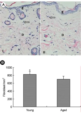
Hematoxylin-eosin 염색에서 방추형의 길고 납작한 섬유 모세포가 진피에 산재하고 있었다 (Fig. 1A). 섬유모세포의 밀도는 10주령 흰쥐가 825.37±78.78/mm2, 54주령 흰쥐가 703.35±74.66/mm2였으며 이를 t-검정한 결과 54주령 흰쥐 진피 내 섬유모세포의 밀도가 10주령 흰쥐보다 유의하게 낮 았다 (t=-4.497, P<0.001) (Fig. 1B).
Masson's trichrome 염색에서 콜라겐섬유는 청색으로 진하 게 염색되었으며, 10주령 흰쥐의 콜라겐섬유들은 비교적 치
B A
Fig. 1A. Black dot like stained fibroblast (arrow) is shown in the dermis (D) below the epidermis (E) in the young (I) and aged rat skin (II). The aged rat skin showed less fibroblasts in the dermis than in the young rat skin. Hematoxylin & Eosin, × 200.
Fig. 1B. Comparison of fibroblast density between young and aged rat skin. There was significantly higher fibroblast density in the young rat skin than in the aged rat skin as determined by group t-test (*: P<0.001). Each value represents the mean ± S.D. (n=8).
B A
Fig. 2A. Bluish-stained collagen fiber (arrow) is shown in the dermis (D) below the epidermis (E) in young (I) and aged rat skin (II). In the young rat skin, collagen fiber bundles show well org- anized in tightly packed bundles. The aged rat skin showed less collagen fibers in the dermis than in the young rat skin. Masson- trichrome stained, × 200.
Fig. 2B. Comparison of collagen density rate between young and aged rat skin. There was significantly higher collagen density rate in young rat skin than in the aged rat skin as determined by group t-test (*: P<0.001). Each value represents the mean ± S.D. (n=8).
밀하고 질서정연하게 배열되어 있으나 54주령 흰쥐는 콜라 겐섬유 다발이 엉성하고 절편으로 나누어져 무질서하게 배 열되어 있었으며 유두 진피 아래에서 부분적으로 콜라겐섬 유의 소실이 나타났고 망상 진피에서 콜라겐섬유사이의 간 격이 넓게 나타났다 (Fig. 2A). 진피 내 콜라겐섬유 밀도의 비율은 10주령에서 62.09±4.91%, 54주령에서 53.68±5.31%
였으며 이를 t-검정한 결과 54주령의 진피 내 콜라겐섬유 밀 도의 비율이 10주령 흰쥐보다 유의하게 낮았다 (t=-4.650, P<
0.001) (Fig. 2B).
Verhoeff-van Gieson 염색에서 탄력섬유는 갈색으로 염색되 었으며 10주령 흰쥐의 유두 진피의 미세한 탄력섬유가 기저 막을 향해 수직으로 배열되는 경향을 보였으며 54주령 흰쥐 의 유두 진피에서는 미세한 탄력섬유가 위축되어 있었다. 진 피의 망상층에서 분포하는 탄력섬유는 54주령 쥐의 경우 그 수가 감소하는 경향을 보였다 (Fig. 3A). 진피 내 탄력섬유
밀도의 비율은 10주령 흰쥐에서 2.61±0.22%, 54주령 흰쥐에 서 2.08±0.26%였으며, 진피 내 탄력섬유 밀도의 비율을 t- 검정한 결과 54주령 흰쥐 진피의 탄력섬유 밀도의 비율이 10 주령 흰쥐보다 유의하게 낮았다 (t=-6.494, P<0.001) (Fig. 3B).
진피 내 섬유모세포의 수와 콜라겐섬유 밀도의 비율 및 탄력섬유 밀도의 비율을 Pearson 상관분석한 결과 섬유모세 포의 수와 콜라겐섬유 밀도의 비율 간 유의한 상관관계가 있 었으며 (r=.69, P<0.001), 섬유모세포의 수와 탄력섬유 밀도의 비율 간에도 유의한 상관관계를 나타냈다 (r=.91, P<0.001).
고 찰
노화는 피부의 주요 구조요소인 콜라겐의 변화를 야기하 며 (Fisher et al., 1997), 제 I형 및 제 III형 콜라겐 합성능 감 소는 내재성 피부 노화의 특성이다. 콜라겐은 α-사슬이 삼중 나선을 형성하는 3개의 다단백질로 α-사슬에 따라 20종 이 상의 콜라겐이 있으나 사람의 피부에는 I, III, IV, V, VI, VII 형 콜라겐이 주로 분포하고 있으며 진피에는 제 I형 콜라겐 이 80~85%, 제 III형 콜라겐이 10~15%, IV, V, VI, VII형 콜 라겐이 5% 정도이며 피부 건조 중량의 75%를 차지하고 있 다 (Uitto et al., 1989). 유두 진피층과 망상 진피층에 분포하 는 콜라겐섬유는 크기 및 배열이 다른데 유두 진피층에는 직경 0.2~1 µm의 폭이 좁고 길이가 짧은 제 III형 콜라겐이 기질에 비교적 성기게 묻혀 있으며 혈관분포가 많고 망상 진 피층에서는 직경 2~15 µm의 굵고 긴 제 III형 콜라겐이 다 발을 이루며 피부 표면에 평행하게 배열하여 기질에 묻혀있 으며 피부의 골격 구조를 유지하고 피부 장력을 제공한다 (Lavker et al., 1987). 그러나 노화가 진행함에 따라 콜라겐섬 유와 다발이 매년 1% 정도씩 감소하며 (Shuster et al., 1975), 콜라겐섬유 다발의 배열이 엉성하고 무질서해지며 개개의 콜라겐섬유가 두꺼워 지고 (Kligman et al., 2000), 이에 따라 피부의 유연성이 상실된다 (Bailey et al., 1998). Moragas 등 (1998)은 3.5개월부터 86세 나이의 97명의 배 피부 부검 표 본을 Picro-Sirius 염색하여 영상분석장치를 사용하여 콜라겐 섬유를 분석한 결과 진피 내 콜라겐섬유 다발의 밀도가 어린 이부터 중년까지 서서히 증가하다가 60대 이후 급속하게 감 소하고 콜라겐섬유 다발의 배열이 단순해지고 수평배열 섬 유가 우세해지며 이질성이 증가한다고 하였으며, Gogly 등 (1997)도 영상분석장치를 사용하여 10~75세의 피부의 콜라 겐섬유를 비교한 결과 나이가 증가함에 따라 섬유의 굵기가 유의하게 두꺼워졌으며 진피 내 점유면적이 유의하게 감소 하였다고 보고하였다. 본 연구에서도 Masson's trichrome 염 색에서 10주령 흰쥐의 콜라겐섬유가 더 진하게 염색되었고 콜라겐섬유들은 비교적 치밀하고 질서정연하게 배열되어 있 으나 54주령 흰쥐는 콜라겐섬유 다발이 절편으로 나누어져 A
B
Fig. 3A. Brownish-stained elastin fiber (arrow) is shown in the dermis (D) below the epidermis (E) in the young rat skin (I) and aged rat skin (II). The aged rat skin showed less elastic fibers in the dermis than in the young rat skin. Verhoef van Gieson stained,
× 200.
Fig. 3B. Comparison of elastic fiber density rate between young and aged rat skin. There was significantly higher elastic fiber den- sity rate in young rat skin than in the aged rat skin as determined by group t-test (*: P<0.001). Each value represents the mean ± S.D. (n=8).
엉성하고 무질서하게 배열되어 있었으며, 진피 내 콜라겐섬 유의 면적을 영상분석기로 측정하여 비교한 결과 54주령 흰 쥐의 진피 내 콜라겐섬유 밀도가 10주령 흰쥐보다 유의하게 낮게 나타나 노화에 따라 콜라겐섬유가 감소함을 보여 주 었다.
진피에 소량 존재하는 탄력섬유는 무정형 단백인 탄력 소와 미세섬유단백으로 구성되어 있으며 변형된 피부가 원 래의 모습으로 돌아오도록 탄력을 제공하는 역할을 한다 (Kanitakis, 2002). 나이 증가에 따라 탄력섬유가 점진적으로 퇴행과정을 거치며 진피에서 탄력섬유의 감소와 구조 변화 에 의해 피부의 탄력성이 감소하고 주름이 형성되고 처지는 피부 노화현상이 일어난다 (Braverman and Fonferko, 1982).
노화에 따라 유두 진피에서 탄력섬유의 수가 감소하고 위축 이 유발되며 망상 진피에서도 탄력섬유에 작은 구멍이 생기 고 엉성해지며 (Bouissou et al., 1988), 탄력섬유가 납작해지 고 표면이 거칠어지고 섬유면적의 감소, 미세원섬유 감소 등 구조 변화가 유발된다 (Tsuji and Hamada, 1981). Gogly 등 (1997)이 영상분석장치를 사용하여 10~75세의 피부의 탄 력섬유를 비교한 결과 나이가 증가함에 따라 진피 내 점 유면적이 유의하게 감소하였다고 보고하였다. 본 연구에서 Verhoeff-van Gieson 염색에서 진피 내 탄력섬유를 관찰한 결 과 54주령 흰쥐의 탄력섬유가 위축되어 있었고 탄력섬유의 밀도를 측정하여 비교한 결과 54주령 흰쥐의 유두 진피 내 탄력섬유 밀도가 10주령 흰쥐보다 유의하게 낮아 노화가 진 행됨에 따라 탄력섬유의 크기와 수가 감소됨을 확인하였다.
진피에 존재하는 섬유모세포는 콜라겐, 탄력소 및 무정형 기질을 합성하여 세포외로 분비하는 중요한 역할을 하는 세 포이다 (Smith et al., 1986). 피부 노화현상은 섬유모세포의 콜 라겐 및 탄력소 합성능의 감소로 기인한다 (Oikarinen, 1994).
Mays 등 (1995)은 노령 흰쥐 피부 섬유모세포의 콜라겐 합 성능이 5배 감소하고 콜라겐 분해율이 33~45% 증가함을 보 고하였고, Varani 등 (2006)은 18~29세의 젊은 사람과 80세 이상의 노인 피부에서 분리한 피부 섬유모세포의 프로콜라 겐 합성능을 비교한 결과 노인 피부에서 분리한 섬유모세포 의 프로콜라겐 합성능이 유의하게 감소하였으며, 생검 피부 를 대상으로 한 조직학적 검사 결과 젊은 사람의 섬유모세포 는 세포 표면의 78%가 콜라겐섬유와 부착되어 있는 반면 노 인의 섬유모세포 표면은 58%가 부착되어 있으며 이는 노화 에 따라 기계적 자극의 수준이 낮아지기 때문이라 하였다.
또한 Dumas 등 (1994)은 19세부터 68세 여성의 피부에서 분 리한 섬유모세포의 제 I형 및 제 III형 프로콜라겐 합성능을 비교한 결과 나이가 증가함에 따라 선상으로 감소하고, Shin 등 (2005)은 평균 22세와 71세의 생검 피부를 대상으로 한 연구에서 노화에 따라 섬유모세포 내 프로콜라겐 mRNA 발 현이 감소하였다고 보고하는 등 섬유모세포의 활성이 내재
성 노화에 관련이 있음을 시사하고 있다.
본 연구에서 hematoxylin-eosin 염색에서 진피 내 섬유모 세포 밀도를 계측하여 비교한 결과 54주령 흰쥐의 진피 내 섬유모세포 밀도가 10주령 흰쥐보다 유의하게 낮아 노화가 진행됨에 따라 섬유모세포의 밀도가 감소되었고, 섬유모세포 의 밀도와 콜라겐섬유 밀도 및 탄력섬유 밀도가 유의한 상관 관계를 보여 섬유모세포의 수가 감소함에 따라 콜라겐섬유 와 탄력섬유의 밀도가 감소하여 섬유모세포가 단백합성능에 관여하고 있음을 시사하였다. 본 연구 결과 피부가 노화됨에 따라 콜라겐섬유 밀도 및 탄력섬유 밀도가 유의하게 감소하 였으며 이는 섬유모세포의 증식 및 합성 활성의 감소에 기인 한 것으로 사료된다.
REFERENCES
Bailey AJ, Paul RG, Knott L. Mechanisms of maturation and ageing of collagen. Mech Ageing Dev. 1998. 106: 1-56.
Bernstein EF, Chen YQ, Kopp JB, Fisher L, Brown DB, Hahn PJ, Robey FA, Lakkakorpi J, Uitto J. Long-term sun exposure alters the collagen of the papillary dermis. Comparison of sun-protected and photoaged skin by northern analysis, immunohistochemical staining, and confocal laser scanning microscopy. J Am Acad Dermatol. 1996. 34: 209-218.
Bouissou H, Pieraggi MT, Julian M, Savit T. The elastic tissue of the skin. A comparison of spontaneous and actinic (solar) aging. Int J Dermatol. 1988. 27: 327-335.
Braverman IM, Fonferko E. Studies in cutaneous aging: I. The elastic fiber network. J Invest Dermatol. 1982. 78: 434-443.
Dumas M, Chaudagne C, Bonte F, Meybeck A. In vitro biosyn- thesis of type I and III collagens by human dermal fibroblasts from donors of increasing age. Mech Ageing Dev. 1994. 73:
179-187.
El-Domyati M, Attia S, Saleh F, Brown D, Birk DE, Gasparro F, Ahmad H, Uitto J. Intrinsic aging vs. photoaging: a com- parative histopathological, immunohistochemical, and ultra- structural study of skin. Exp Dermatol. 2002. 11: 398-405.
Fisher GJ, Wang ZQ, Datta SC, Varani J, Kang S, Voorhees JJ.
Pathophysiology of premature skin aging induced by ultra- violet light. N Engl J Med. 1997. 337: 1419-1428.
Gogly B, Godeau G, Gilbert S, Legrand JM, Kut C, Pellat B, Goldberg M. Morphometric analysis of collagen and elastic fibers in normal skin and gingiva in relation to age. Clin Oral Investig. 1997. 1: 147-152.
Kanitakis J. Anatomy, histology and immunohistochemistry of normal human skin. Eur J Dermatol. 2002. 12: 390-401.
Koehler MJ, Konig K, Elsner P, Buckle R, Kaatz M. In vivo assessment of human skin aging by multiphoton laser scan- ning tomography. Opt Lett. 2006. 31: 2879-2881.
Kligman LH, Schwartz E, Sapadin AN, Kligman AM. Collagen loss in photoaged human skin is overestimated by histoche- mistry. Photodermatol Photoimmunol Photomed. 2000. 16:
224-228.
Lavker RM, Zheng PS, Dong G. Morphology of aged skin. Clin Geriatr Med. 1989. 5: 53-67.
Lavker RM, Zheng PS, Dong G. Aged skin: a study by light, transmission electron, and scanning electron microscopy. J Invest Dermatol. 1987. 88: 44-51.
Lee JH, Song IY, Kim JG. Acceleration of wound healing and collagen deposition in rat skin by high voltage pulsed current stimulation. J Korean Soci Phys Ther. 2003. 15: 745-754.
Lee JH, Lee JS, Kil EY. Keratinocyte proliferation in aged rat skin by high voltage pulsed current stimulation. J Exp Biomed Sci.
2004. 10: 361-366.
Mays PK, McAnulty RJ, Campa JS, Laurent GJ. Age-related changes in collagen synthesis and degradation in rat tissues.
Importance of degradation of newly synthesized collagen in regulating collagen production. Biochem J. 1991 276: 307 -313.
Mays PK, McAnulty RJ, Campa JS, Laurent GJ. Age-related alterations in collagen and total protein metabolism deter- mined in cultured rat dermal fibroblasts: age-related trends parallel those observed in rat skin in vivo. Int J Biochem Cell Biol. 1995. 27: 937-945.
Moragas A, Garcia-Bonafe M, Sans M, Toran N, Huguet P, Martin- Plata C. Image analysis of dermal collagen changes during
skin aging. Anal Quant Cytol Histol. 1998. 20: 493-499.
Noorlander ML, Melis P, Jonker A, Van Noorden CJ. A quan- titative method to determine the orientation of collagen fibers in the dermis. J Histochem Cytochem. 2002. 50: 1469-1474.
Oikarinen A. Aging of the skin connective tissue: how to measure the biochemical and mechanical properties of aging dermis.
Photodermatol Photoimmunol Photomed. 1994. 10: 47-52.
Sanders JE, Goldstein BS, Leotta DF, Richards KA. Image pro- cessing techniques for quantitative analysis of skin structures.
Comput Methods Programs Biomed. 1999. 59: 167-180.
Shin MH, Rhie GE, Park CH, Kim KH, Cho KH, Eun HC, Chung JH. Modulation of collagen metabolism by the topical appli- cation of dehydroepiandrosterone to human skin. J Invest Dermatol. 2005. 124: 315-323.
Shuster S, Black MM, McVitie E. The influence of age and sex on skin thickness, skin collagen and density. Br J Dermatol.
1975. 93: 639-643.
Smith LT, Holbrook KA, Madri JA. Collagen types I, III, and V in human embryonic and fetal skin. Am J Anat. 1986. 175: 507 -521.
Tsuji T, Hamada T. Age-related changes in human dermal elastic fibres. Br J Dermatol. 1981. 105: 57-63.
Uitto J, Olsen DR, Fazio MJ. Extracellular matrix of the skin: 50 years of progress. J Invest Dermatol. 1989. 92: 61S-77S.
Varani J, Dame MK, Rittie L, Fligiel SE, Kang S, Fisher GJ, Voorhees JJ. Decreased collagen production in chronologi- cally aged skin: roles of age-dependent alteration in fibroblast function and defective mechanical stimulation. Am J Pathol.
2006. 168: 1861-1868.