한국전문물리치료학회지 제 10 권 제 4호
KAUTPT Vo l. 10 No. 4 2003.
Ini tiation and Tennination of Electromyographic Activity in the Early Hemiparetic Wrist
Chung Yi-jung , M.Sc. , P.T.
Dep t. of Rehabilitation Therapy , Th e Graduate School , Yonsei University Cho Sang-hyun , Ph.D. , M.D.
Dep t. of Physical Th erapy , College of Health Science , Y onsei University Institute of Health Science , Y onsei University
Kwon Oh-yun , Ph.D. , P.T.
Dep t. of Physical Th erapy , College of Health Science , Yonsei University
Dep t. of Ergotherapy , Th e Gr aduate School of Health and Environment , Yonsei
U피versityInstitute of Health Science , Y onsei University Lee Young-hee , Ph.D. , M.D.
Dep t. of Rehabilitation Medicine , Wonju College of Medicine , Yonsei University
국문요약초기 편마비 환자의 손목에서 근수축 개시 및 종료의 지연
정이정
연세대학교 대학원 재활학과
조상현
연세대학교 보건과학대학 물리치료학과 및 보건과학연구소
권오윤
연세대학교 보건과학대학 물리치료학과, 보건환경대학원 인간공학치료학과, 보건과학연구소
이영희
연세대학교 원주의과대학 재활의학교실
본 연구는 초기 편마비 환자의 손목에서 표면근전도 분석을 통해 근수축 개시 및 종 료의 특성들을 알아보고, 임상적인 치료방법의 기초를 제안하고자 실시하였다. 연구대상 자는 원주기독병원에 뇌졸중으로 입원한 환자 중 발병 후 3 개월 미만인 13 명과 원주시 에 거주하는 대조군 7 명이었다. 근수축 개시 및 종료의 지연은 표면근전도를 이용하여 손목굽힘근과 손목펴짐근에서 손목관절의 굽힘과 펌동작 시 3초의 근전도 신호음에 따 라 가장 빠르고 강하게 최대 등척성 수축과 이완을 하여 신호를 수집하였다. 그 결과 편 마비 환자의 환측은 건측과 대조군에 비해 손목관절 굽힘과 펌동작에서 근수축 개시및 종료가 유의하게 지연되었으며, 개시보다 종료가 더 유의하게 지연되었다. 따라서 초기 뇌졸중 환자의 근약화는 근육의 개시 및 종료의 반응시간 지연에 영향을 준다고
Corresponding author: Chung Yi-jung !!obilitv 3 004 (âJJz otma i/. com
nerve physiology.
Th e purpose of this study is going recognize the agility of wrist muscle con- traction in hemiplegia caused by cerebrovascular accident (CV A). This may
and an offset of
with of
by and ea r1 y problems
limbs after brain injury
to the physiologic change of the nervous system by muscle weakness
agility than spasticity regarding
studies , it was recognized that there were efficiency and relationship
recruitrnent (De Luca , 1993; Kupa et al , 1995; Toffola et al , 2001). However , be-
of the existing studies the injury
discriminate whether
atrophy that is peripheral nervous system by a progress of time or neurological damage of an initial central nervous sys-
(N ewham and Hsiao , 2001). 80 the functional evaluations were not able
a nation in the affected sides (Hammond et
upper related
recent
to
to u ·m
was the
the cause of muscle weakness is due to
민 것
of and loss
delayed contractíon
of of motor
이「 하금
x •
야
LL 벼
는 구 있 연
wrist of the
understanding more
becomes are
a
劉
m
ιu
nr
onset
볼 수 있다. 앞으로 운동조절과 연관되어 기능적인 회복을 유도할 수 첩성 훈련과 큰섬유 동원의 효율성을 증진시키기 위한 치료방법들이 이다
slnce stroke , it is hard to
basic
contractíon
weakness of al , 1988).
Th e functional
핵섬단어: 근약화; 뇌졸중; 손목굽힘근; 손목펴짐근; 표면근전도.
an classified
decrease the
that most
provide
assume
muscle muscle cause
tem not
the recovery of their proximal upper
1i mb than the distal upper 1i mb relatively.
Agility and coordination are required in the initiation and termination of repetitive activities of grasp
of upper limbs , there is a limitation , and this syndrome can see what is limited on
phasic
da i1 y living. Researchers
lyze a muscle contraction delay of an on- and offset in a wrist joint
hemiparesis who used an electromyogram in order to evaluate the agility of muscle contraction until now (Angel , 1981; Chae et al , 2002; Hammond et al , 1988). Wh en a wrist joint moves , cocontraction of ago-
and antagonist related to spasticity occurred in the chronic hemiplegic patient who used an electromyogram and delyed muscle contraction of initiation and termi-
After a cerebral injury , which may fol- or head trauma , many pa- of physical collectively as the "upper motomeuron" syndrome (Gempe r1 ine et al , 1995). These signs include spastic hyper- tonia , muscular weakness , and impaired
ana- release
the of to and
studied
한국전문물리치료학회지 제 10권 제 4호
KAUTPT Vo l. 10 No. 4 2003.
muscle contraction in constellation In troduction
movement coordination.
Wh en the functional a
a stroke
known suffer
motor tients slgns low
mst
set
한국전문물리치료학회지 제 10권 제 4호
KAUTPT Vo l. 10 No. 4 2003.
hemiplegia. Therefore , this study observes muscular response time regarding motor control during the rehabilitation stage of early stroke patients. The agile and con- centrated treatment strategy related the muscular reaction time will be used as good information.
Metbods
Subjects
Thirteen stable patients with a history of unilateral CV A 9 to 87 days pre- viously , and seven healthy control sub-
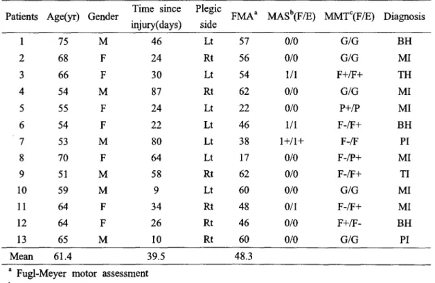
Table 1. Clinical characteristics of patients
jects with a similar balance of age and sex were tested. Inclusion criteria included 1) an interval of at least 3 months from stroke onset; 2) unilateral lesion; 3) man- ual muscle testing that is poor or above in wrist flexor and extensor; 4) ability to follow three second maximal isometric contraction command. Control groups were excluded if they reported previous injury or current
0πhopedicproblems in their body. Characteristics of the subjects are summarized in Table 1.
(N =13)
Ti me since Plegic
~".a ".~bPatients Age(yr) Gender FMA a MASD(FIE) MMTC(FIE) Diagnosis injury(days) side
75 M 46 Lt 57 0/0 G/G BH
2 68 F 24 Rt 56 0/0 G/G MI
3 66 F 30 Lt 54 111 F+IF+ TH
4 54 M 87 Rt 62 0/0 G/G MI
5 55 F 24 Lt 22 0/0 P+ /P MI
6 54 F 22 Lt 46 111 F-IF+ BH
7 53 M 80 Lt 38 1+11+ F-IF PI
8 70 F 64 Lt 17 0/0 F- /P+ MI
9 51 M 58 Rt 62 0/0 F-IF+ TI
10 59 M 9 Lt 60 0/0 G/G MI
11 64 F 34 Rt 48 0/1 F-IF+ MI
12 64 F 26 Rt 46 0/0 F+IF- BH
13 65 M 10 Rt 60 0/0 G/G PI
Mean 6 1. 4 39.5 48 .3
a Fugl-Meyer motor assessment
b Modifted Ashworth scale C Manual muscle testing
E: extensor , F: flexor , BH: basal ganglia
hemoπhage,MI: middle cerebral
앙tery infar따ionPI: pontine infarction , TH: thalamic hemorrhage , TI: thalamic infarction
한국전문물리치료학회지 제 10권 저14호
KAUTPT Vo 1. 10 No. 4 2003.
FC ’‘
ECR
Fïgure 1. The ’ forearm based skateboard' and the placement of the electrodes on the flexor carpi radialis and extensor car- pi radialis muscles
Instruments
The surface electromyography (sEMG) signal was detected with an active paral- lel-bar electrode (b ar size: 1 mm by 10 mm , located 10 mm apart differential electrode (DelSys)l). The electrodes were placed on the flexor carpi radialis and ex- tensor carpi radialis muscle belly (Cram et al , 1998) (Figure 1). The EMG signals were digitally band-pass filtered at 20-450 Hz and notch filtered at 60 Hz to reduce noise. A sampling frequency of 1000 Hz was used. After collection , the data were transferred to a personal com- puter for data reduction.
The Acqknowledge 3.72 program 2) was employed to set up the required parame- ters and to store the EMG signal as com- puter files.
1) Delsys Inc. Boston , MA. u. S. A.
2) Biopac systems Inc. C A. U.S .A.
Procedures
Su에ects
were instructed to contract the wrist flexor or extensor as forcefully and quickly as possible against the confine- ment of the apparatus in response to an audible beep , and to relax the muscle as quickly as possible as soon as the beep terminated. For wrist flexion , all subjects were asked to respond to audible beeps consisting of three
πialsof 3-s contractions. The trials were presented in a balanced random order in order to min- imize subject anticipation , The procedure was repeated for wrist extension. Delay in recruitment of the EMG signal was de- fined as the time ínterval between the on- set of the audible beep and the onset of the EMG signal (F igure 2).
Delay in termination of the EMG signal
was defmed as the time ínterval between
the offset of the audible beep and the
offset of the EMG signal. The onset of
the EMG activity was defined by com-
한국전문물리치료학회지 제10권 저14호
KAUTPT Vo l. 10 No. 4 2003
Oft. t.\ jπ....
:...‘’
)j ... - ~“표월;않Jft앙회브생 g보l않i흐E훌효l힘j침
Bt、Cp Cl'
na、,\>
dat “
3 ‘
l'('Fi gure 2. EMG raw data during wrist isometric contraction of the patient #11
puter-based onset and offset detennination techniques (ICCs=.932/.977). This exhibits
si맹ficantly
higher reliab i1i ty (Chung et al , 2003). The parameters were evaluated by low pass software filter (50 Hz) and the other parameters evaluated were the number of samples assessed in the sliding window (25 ms) and the magnitude of the deviation from the baseline required to indicate the threshold (3 SD).
Processing was done using MatLab ’ signal processing toolk :it3).
Staûsûcal analysis
An independent t-test was used to ex- amine the differences between sides (flexion and extension , onset time and offset time) and paired t-test of with in the sides. All statistical analyses were perfonned using SPSS 10.0 for windows.
3) Math Works Inc. MA. U.S .A.
A P value level of <.05 was used as the level of significance.
Res띠ts
The results of study were as follows.
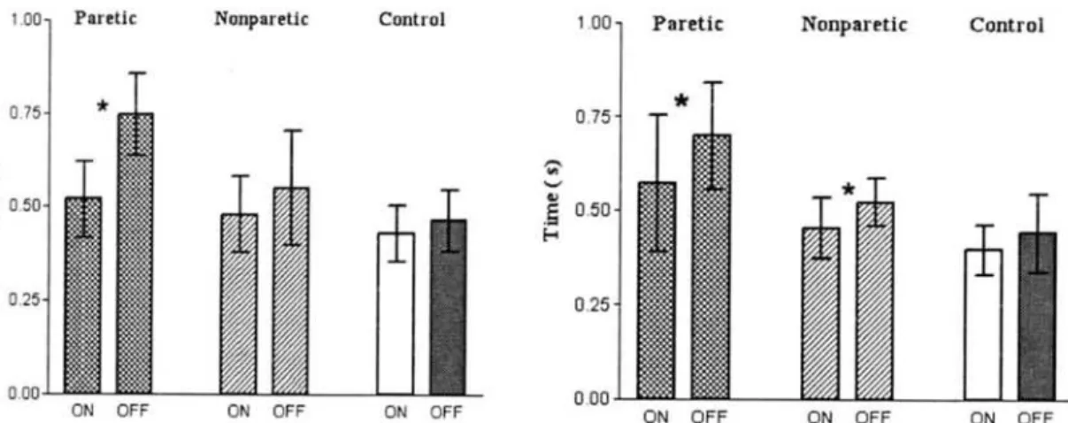
The onset and offset of muscle con- traction were significantly delayed on the more paretic than nonparetic and control sides (Table 2). Offset was significaotly more delayed than the onset 00 the pa- retic sides (Figure 3).
Discussion
Slower initiation of movements has been shown in perfonnance of simple re- action time tasks by persons with unilat- eral cerebral damage (Benton and Joynt , 1959; DeRenzi and Faglioni , 1965; Howes and Boller , 1979).
S따face el없omyographicstudies have documented slower muscle re-
한국전문물리치료학회지 제10 권 제4호
KAUTPT Vo l. 10 No. 4 2003 .
Table 2. Comparison of delay in contraction onset and offset of the wrist flexor and extensor muscles between side
Offset delay .44( .1 0) .52(.06)*
.70 (.1 4)"f Wrist extension
째-액 뼈
-m
m 씨 뻐
하
-mn
ν π
m -4
4 5 Onset delay Offset delay 0
.4 3(.08) .4 6(.08) .4 8(.10) .55( .1 6) .52(. 1O )"f .75(.11) All values in the cells are mean (SD): second
.Si맹ificantly
different from control sides with independent t-test (p <.05) t Significantly different from nonparetic sides with paired t-test (p <.05)
Wrist flexion Sides
Control
·κ
빠 ·κ 뼈‘ 않
N
Control
Non따lp찌aretic
Paretic
0.75
~、
‘'" ‘”
@틀 0.50
•
0.25
Control NonpareUc
(‘)
‘ζa·R
。 FF
Fi gure 3. Comparison of delay in contraction onset and offset of the wrist flexor and extensor muscles with in the sides
。N
。 FF
。 FF 。N
。N
0.00
。 FF
O FF
ON。FF 。N
ON
Values of the bar are
mean土SD.ON: onset delay , OFF: offset delay
.Si빼ficantdifference between onset delay and offset delay (p<.05).
of eight hemiparetic subjects.
and associates (1 978) found that the onset of EMG activity was longer for the hemi- paretic biceps during both elbow
and forearm supination.
Norton (1 977) noted delayed recruitments of agonist contraction in the hemiparetic upper limb.
The c1assical explanation for the paresis Nakamura
ml- sponsiveness (An gel , 1981; Nakamura et al ,
1978).
We found delay m
of musc1e
빠
’
m
. m
뼈
m
a
‘ a
flexion and Sahrmann con-
σaction of the hemiparetic wrist compared to nonparetic and control wrist (Table 2).
An gel (1 981) showed that the latency from a visual signal to onset of EMG ac- tivity in arm musc1es was longer in four
and
tlat lO n
한국전문물리치료학회지 제 10권 제 4호
KAUTPT Vo l. 10 No. 4 2003.
advanced by Hughlings Jackson is that there is a loss of descending excitation from major descending pathways to the spinal cord (Lassek , 1970). Th e final mo- tor output among stroke survivors can be modulated by changes in descending and propriospinal excitatory and inhibitory in- puts into the spinal intemeurons and al- pha motoneurons as well as neuroplastic changes consequent to brain injury (D obkin , 1996; Nudo et al , 2001).
Co-contraction of antagonist mus c1 es is a widely recognized c1 inical problem and was expected in paretic forearms (Hammond et al , 1988; Sahrmann and Norton , 1977). Wh ereas spasticity hardly appeared in initial hemiparetic patients in this study , it seems that primary cerebral cortex dysfunction causes specific impair- ments in processing. This presented to be contributed to neurological deficit that is the reorganization of the corticospinal tract , change of characteristic motor unit and activation of spinal intemeuron (Adams et al , 1990; Bohannon and Walsh , 1992; Colebatch et al , 1986;
Newham and Hsiao , 2001).
We found a significant delay in termi- nation times compared to initiation times in paretic agonist mus c1 es. Subject pre- sented longer than in control wrist flexor and extensor mus c1 es (Figure 3).
Consistent with prior studies , we found a significant delay in termination of mus c1 e contraction of the hemiparetic upper limb compared to the nonparetic upper limb (B eneck et al , 1983; Dewald et al , 1995;
Sahrmann and Norton , 1977). Th is is at- tributed to loss of supraspinal inhibitory influence on the normal intemeuronal pool (Ghez , 1991) , spasticity , abnormal co-con- traction of antagonist and agonist mus c1 es , and abnormal co-activation of synergistic mus c1 es (Chae et al , 2002). The main ef- fect is on the lateral reticulospinal tract that is the most nearly corticospinal tract that the loss of inhibitory delivery (Delwaide and Young , 1985). So a reduc- tion in corticospinal input may also result in increased dependence on undamaged vestibulospinal , reticulospinal , and tectospi- nal pathways not a spasticity and abnor- mal co-contraction (D elwaide and Y oung ,
1985).
Conclusion
Th is study also suggests that since mus c1 e weakness of early stroke patients affects the functional delay of mus c1 e contraction in upper limbs , further studies must be focused on treatment to improve mus c1 e ag i1i ty and mus c1 e fiber recruit- ment efficiency that can induce the func- tional recovery correlated to motor contro 1.
References
Adams RW , Gandevia SC , Skuse NF.
Th e distribution of mus c1 e weakness in upper motoneuron lesions affecting the lower limb. Brain. 1990;113(5):
1459-1476.
한국전문물리치료학회지 제 10권 제 4호
KAUTPT Vo l. 10 No. 4 2003
An gel RW. Electromyographic pattems during ballistic movements in normals and hemiplegic patients. Prog Clin Neurophysiology. 1981;9:347-357.
Beneckee , Conrad , Meinck , et a 1.
Elelctromyographic analysis of bicy- cling on an ergoneter for evaluation of spasticity of lower limbs in man.
Adv Neuro 1. 1983;39:1045-1046.
Benton , Joyn t. Reaction time in unilatera1 cerebral disease. Confm Neuro 1. 1959;
19:247-256.
Bohannon RW , Walsh S. Nature , reli- abi1ity , and predictive value of mus- cle performance measures in patients with hemiparesis following stroke.
Ar ch Phys Med Rehabi 1. 1992;73(8):
721-725.
Chae J , Yang G , Park BK , et a 1. Delay in initiation and termination of mus- cle contraction , motor impairment , and physical disability in upper limb hemiparesis. Muscle Nerve. 2002;
25(4):568-575.
Chung YJ , Cho SH , Lee JH , et a l.
Reliability of the onset time determÍ- nation during maximal isometric con- traction in surface EMG. J Kor Acad Univ Tra Phys The r. 2003; 1 :51-62.
Colebatch JG , Gandevia SC , Spira PJ.
Voluntary muscle strength in hemi- paresis: Distribution of weakness at the elbow. J Neurol Neurosurg Psychiatry. 1986;49(9): 1019-1024.
Cram JR, Kasman GS , Holtz J.
lntroduction to Surface Electro- myography. Gaithersburg , Aspen Pub. ,
1998.
De Luca CJ. Use of the surface EMG signal for performance evaluation of back muscles. Muscle Nerve. 1993;
16(2):210-216.
Delwaide PJ , Young RR. Clinical neuro- physi010gy in spasticity. New York , E1sevier Science , 1985.
DeRenzi , Faglion i. Comparative efficiency of intelligence and vigilance tests in detecting hemispheric cerebral damage.
Cortex. 1965;1:410-433.
Dewald , Pope , Given , et
떠.Abnorma1 muscle coactivation pattems during isometric torque generation at the el- bow and shou1der in hemiparetic subjects. Brain. 1995;118:495-510.
Dobkin BH. Neur010gic Rehabilitation:
Prob1ems of Medical Managemen t.
Vo 1. 47. Philade1phia , F. A. Davis , 1996.
Gemperline JJ , Allen S , Walk D , et a 1.
Characteristics of motor unit dis- charge in subjects with hemiparesis.
Muscle Nerve , 1995;18:1101-1114.
Ghez C. Voluntary movemen t. In: Kandal ER , Schwartz JH , Jessell TM.
Principles of Neura1 Science , Norwalk , CT , App1eton & Lange , 199 1.
Hammond MC , Fitts SS , Kra ft G H, et a 1.
Co-contraction in the
h얹nipareticfore-
따m:
Quna titative EMG evaluation. Ar ch Phys Med Rehabi1. 1988;69:348-35 1.
Hammond MC , Kr aft GH , Fitts SS.
Recruitment and termination of elec-
tromyographic activity in the hemi-
한국전문물리치료학회지 저]10권 제 4호