The degenerative spinal cord disease cervical spondylotic myelopathy (CSM), and cervical myelopathy caused by trauma, can result in debilitating symptoms affecting quality of life. This study used acupotomy and other Korean medicine treatments (acupuncture, herbal medicine, and physical therapy) to improve the symptoms of CSM and cervical myelopathy. The visual analog scale, the modified Japanese Orthopaedic Association scale (mJOA scale), the Nurick grading system, and the American Spinal Injury Association impairment scale were used as the evaluation criteria to determine the effectiveness of treatment. The functional status of both patients improved from mild to moderate, with improved gait, local sensation, and level of pain. The degree of spinal cord injury remained the same. The findings of this study suggest that combined Korean medicine treatments including acupotomy may be helpful in the treatment of CSM and cervical myelopathy.
©2021 Korean Acupuncture & Moxibustion Medicine Society. This is an open access article under the CC BY- NC-ND license (http://creativecommons.org/licenses/by-nc-nd/4.0/).
Article history:
Submitted: November 28, 2020 Revised: January 19, 2021 Accepted: January 26, 2021 Keywords:
acupotomy, cervical spondylosis, conservative treatment, Korean traditional medicine, myelopathy
https://doi.org/10.13045/jar.2020.00465 pISSN 2586-288X eISSN 2586-2898
Case Report
Treatment of Cervical Myelopathy with Acupotomy Combined with Korean Medicine Treatments: Two Clinical Cases
Yu-Kyeong Park, Sangha Woo, Jae Hoon Kim, Jung Hee Lee, Yun-Kyu Lee, Hyun-Jong Lee, Jae Soo Kim*
Department of Acupuncture and Moxibustion, College of Korean Medicine, Daegu Haany University, Daegu, Korea
ABSTRACT
Journal of Acupuncture Research
Journal homepage: http://www.e-jar.org
Introduction
Cervical spondylotic myelopathy (CSM) is a common, age related degenerative spinal cord disease which causes a combination of changes such as mechanical spinal cord compression due to degenerative changes in the cervical spine, and herniation of the intervertebral disc, ossification of the posterior ligament, ischemic damage due to blood circulation disorders in the spinal cord, and trauma [1]. CSM is a chronic disease that progresses slowly and is without an effective cure. In addition, CSM is becoming a public health concern as the morbidity rate increases with age and the population is aging [2]. Causes of cervical myelopathy include spinal trauma (for example, a road traffic accident), stenosis, infection, and cancer. Treatment goals are to manage and relieve symptoms of CMS and cervical myelopathy. Chief complaints include gait disturbance, and hand movement and sensory disorders, which cause discomfort in daily life. In severe cases, permanent sequelae may remain, thus an accurate diagnosis and treatment are required. Based on the degree of injury, CSM
and cervical myelopathy can be treated either conservatively or surgically. If symptoms are severe, surgical treatments such as central-separated laminectomy, interbody fusion, and osteotomy, are considered [3,4]. Conservative treatments include drug therapy, injection therapy, and physical therapy, amongst others [5].
Conservative treatment is more effective than surgical treatment in patients with mild symptoms [3,4].
Recently, Korean medicine treatments including acupuncture, moxibustion, and acupotomy have been studied for the treatment of various spinal diseases [2,5-7]. Acupotomy has been used to restore soft tissue damage, and the original dynamic state of abnormal lesions of the body by removing adhesions, nodules, and scars [5]. However, reviews report that there are only a few studies using combined Korean medicine treatment including acupotomy for the cervical region [6,7]. Therefore, this report describes the results for 2 patients 1 with CSM and 1 with cervical myelopathy, who were treated with combined Korean medicine including acupotomy.
* Corresponding author. Jae Soo Kim
Department of Acupuncture and Moxibustion medicine, Daegu Oriental hospital of Daegu Haany University, 136, Sincheondong-ro, Suseong-gu, Daegu, 706-828, Korea E-mail: [email protected]
ORCID: Yu-Kyeong Park https://orcid.org/ 0000-0003-0087-953X, Sangha Woo https://orcid.org/ 0000-0002-0446-5644, Jae Hoon Kim https://orcid.org/ 0000-0002-1116- 8277, Jung Hee Lee https://orcid.org/ 0000-0002-2771-659X, Yun-Kyu Lee https://orcid.org/ 0000-0001-8806-9501, Hyun-Jong Lee https://orcid.org/ 0000-0003-1260-0778, Jae Soo Kim https://orcid.org/ 0000-0003-4101-8058
©2021 Korean Acupuncture & Moxibustion Medicine Society. This is an open access article under the CC BY-NC-ND license (http://creativecommons.org/licenses/by-nc-
nd/4.0/).
Case Report Case 1 Patient
48-year-old female Chief complaints
Neck pain
Bilateral upper extremity numbness
Reduced upper and lower extremity muscle strength Onset date
Early October 2019 Present illness
In early October 2019, the patient experienced sudden neck pain, bilateral upper extremity numbness, and bilateral upper and lower extremity weakness. On October 15
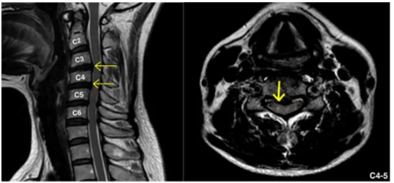
th, she was diagnosed with CSM at a hospital in Seoul. A cervical and lumbar spine magnetic resonance image (MRI) showed extrusion at C4-5, and protrusion at C3-4 (Fig. 1). Surgery was recommended however, the patient refused surgery and opted for Korean medicine treatment at the Department of Acupuncture at Daegu Korean Medicine Hospital of Daegu Haany University on October 31
st, 2019.
Medical history Arrhythmia
Lumbar spinal stenosis MRI Refer to Fig. 1.
Case 2 Patient
60-year-old male Chief complaints
Neck pain Lower back pain
Distal arm, hand, and left lower extremity numbness Onset date
December 25
th, 2019 Present illness
On December 25
th, 2019, the patient had a traffic accident.
Consequently, he experienced neck and lower back pain, and right upper and left lower extremity numbness. He was admitted to the Department of Acupuncture at Daegu Korean Medicine Hospital of Daegu Haany University on December 26
th, 2019. On January 3
rd, 2020, he was diagnosed with cervical myelopathy at C5-6 and a c-spine MRI showed protrusion at C3-4 and C5-6 at the Department of Radiology in Daegu (Fig. 2).
Medical history
Lumbar stenosis at L4-5 Bulging at L3-4 and L5-S1 Protrusion at C5-6 Bulging at C3-5 MRI Refer to Fig. 2.
Methods
The 2 patients presented at the Department of Acupuncture at Daegu Korean Medicine Hospital of Daegu Haany University between October 31
st, 2019, and May 2
nd, 2020, who were diagnosed with myelopathy, among patients who received outpatient treatment and inpatient treatment. The patients gave informed consent (i.e., the treatment was explained and the patient agreed to be involved in the study). The Institutional Review Board of the hospital approved this study (IRB no.: DHUMC-D-20016- AMD-01).
Methods of treatment
① Acupotomy treatment
1. Treatment duration and frequency
In Case 1, acupotomy treatment was administered between October 31
st, 2019, and February 12
th, 2020. She received treatment once during hospitalization (November 30
th, 2019, to December 6
th, 2019), and 9 times during outpatient treatment (October 31
st, 2019, to February 12
th, 2020).
Fig. 1. C-spine MRI of Case 1.
A T2-weighted image of a C-spine MRI scan performed on October 15
th, 2019. The image on the left is the sagittal view and the discs of cervical vertebrae are herniated in the C3-4 and C4-5 areas, compressing the spinal cord (a yellow arrow indicates the herniation of the discs, and a red dot border indicates a high signal). The image on the right is the axial view of the C4-5 area, and the spinal cord was marked using high- shaded features.
Red dot border, spinal cord; high shade, yellow arrow.
MRI, magnetic resonance image.
Figure 1. C-spine MRI of Case 1
This is a T2-weighted image of a C-spine MRI scanned at October 15, 2019. The image on the left is the s agittal view and the discs of cervical vertebrae are herniated in the C3-4 and C4-5 areas, compressing the s pinal cord.(A yellow arrow indicates the herniation of the discs, and a red dot border indicates a high signa l.) The image on the right is the axial view of the C4-5 area, and the spinal cord was marked with high-sha ded features.(The red dot border is the spinal cord, and the high shade is indicated by a yellow arrow.)
Fig. 2. C-spine MRI of Case 2
A T2-weighted image of a C-spine MRI scan performed on January 3
rd, 2020. The image on the left, the sagittal view, shows the discs of cervical vertebrae herniated in the C3-4 and C5-6 areas, compressing the spinal cord, and shows high signal findings on the spinal cord at the height of C5-6. A yellow arrow indicates the herniation of the discs, and a red dot border indicates a high signal. The image on the right is the axial view of the C5-6 area. The spinal cord shows high signal findings.
Red dot border, spinal cord; high shade, yellow arrow.
MRI, magnetic resonance image.
Figure 2. C-spine MRI of Case 2
This is a T2-weighted image of a C-spine MRI scanned at January 3, 2020. The image on the left the sagit
tal view. the discs of cervical vertebrae are herniated in the C3-4 and C4-5 areas, compressing the spinal c
ord, and shows high signal findings on the spinal cord at the height of C5-6.A yellow arrow indicates the h
erniation of the discs, and a red dot border indicates a high signal.) The image on the right is the axial vie
w of the C5-6 area. the spinal cord shows high signal findings.(The red dot border is the spinal cord, and t
he high shade is indicated by a yellow arrow.)
In Case 2, the patient received 10 acupotomy treatments between January 3
rd, 2020 to May 2
nd, 2020. He received treatment once during hospitalization (December 26
th, 2019, to January 16
th, 2020), and 9 times during outpatient treatment (January 17
th, 2020 to May 2
nd, 2020).
The treatment was performed for each patient every 1 or 2 weeks, 10 times each.
2. Treatment tools
A 7-cm sterilized needle made of stainless steel (Dongbang Medical, Korea) was used. The stagnation is cylindrical and 1 mm in diameter, with a flat blade attached to the tip of the needle. The tip of the blade used was either 0.5 mm or 0.8 mm.
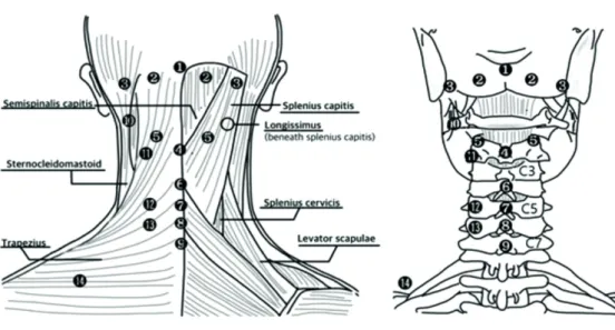
3. Treatment sites [6]
There were 9 primary treatment sites (❶-❾) selected (Fig. 3) which followed the acupotomy points of the cervical region [6].
Depending on the patients’ condition and site of pain (with a tenderness score of 5) additional treatment sites were used ( - ) (Fig. 3).
4. Operation method [5,6]
After sterilizing the treatment site with povidone-iodine and alcohol, the acupotomy needle was inserted towards the facet joint.
The blade tip of the needle was held parallel to the articular surface of the facet joint and handled up/down to incise the adhesions surrounding the facet joint capsule.
② Other Korean medicine treatments 1. Acupuncture
Acupuncture treatments were performed on the days the patient did not receive acupotomy during hospitalization, and for the duration of outpatient care. For acupuncture treatment 0.20 × 30 mm disposable sterilized needle made of stainless steel (Dongbang Medical, Korea) were used, and left in place for 15 minutes.
During hospitalization, acupuncture was performed twice a day (morning and afternoon) and once a day for outpatient treatment.
Acupuncture was performed by choosing the acupoints between BL10ㆍBL11ㆍSI9ㆍSI11ㆍSI12ㆍSI13ㆍSI14ㆍSI15ㆍGB20 and GB21, including sites of pain with tenderness, and the aim was to trigger local muscle reactions at a depth of 1.0-3.0 mm.
2. Herbal medicine
During the hospitalization period, herbal medicine was administrated orally using 3 packs per day (approximately 120 cc per pack) after meals. The herbal medicine included Mangeumtang gamibang (mainly consist of Lindera aggregate 16 g, Angelica sinensis 12 g, Cinnamomum 12 g, Atractylodes lancea 12 g, Bombyx mori L. 8 g, Aralia continentalis 6 g per day) for Case 1.
Case 2 received Oyaksoonkisan gamibang(mainly consist of Lindera aggregate 12 g, Angelica sinensis 12 g, Angelica dahurica 6 g, Poncirus trifoliata 6 g, Aralia continentalis 6 g per day).
3. Physical therapy
Interferential current therapy, microwave therapy, dry cupping treatment, and percutaneous transdermal muscle thermotherapy (hot pack) were applied to the points of pain in the lumbar or cervical region.
Evaluation criteria
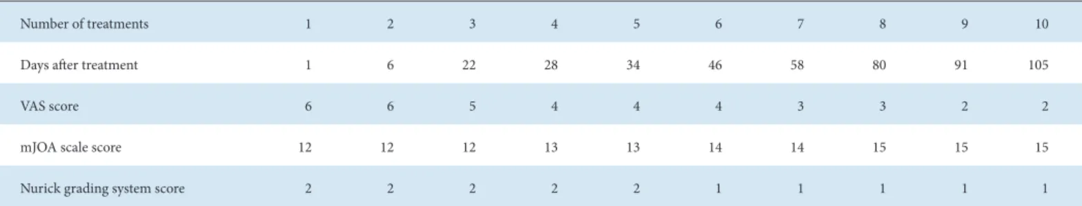
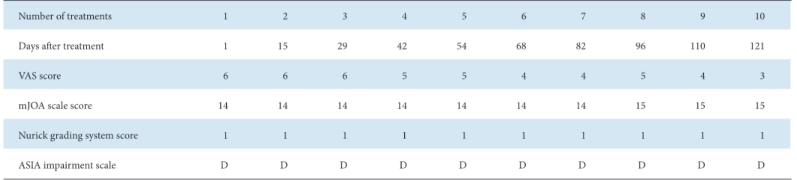
The visual analog scale (VAS) [8], the modified Japanese Orthopaedic Association scale (mJOA) [9], and the Nurick grading system [10] were used to evaluate the patients’ symptoms and treatment results. The American Spinal Injury Association (ASIA) impairment scale [11] was also used in Case 2 where a road traffic accident had occurred. All evaluation criteria were checked after every treatment.
① VAS
The questionnaire using the VAS [8] was performed before and after the treatment, with 0 as the painless state and 10 as the most severe pain, and the patient indicated their level of pain accordingly.
Fig. 3. Acupotomy sites. ❶ Upper site of GV16 ❷ 2-2.5 cm side down of external occipital protuberance (GB20) ❸ 4.5-5 cm side down of external occipital protuberance (GB12) ❹ Origin of trapezius muscle, insertion of nuchal ligament (GV16) ❺ Facet joint, side of C2 spinous process (BL10) ❻ C4 spinous process ❼ C5 spinous process ❽ C6 spinous process ❾ C7 spinous process (GV14) ❿ C1(the atlas) transverse process C2(the axis) transverse process 2cm side to the C5 spinous process 2cm side to the C6 spinous process the middle of the upper trapezius.
11 1214
13