Multidisciplinary approach for treatment of primary hepatic choriocarcinoma in adult male patient
Yongchel Ahn1, Ji-Hoon Kim2, Cheon-Soo Park2, Tae-Eun Kim3, Shin-Hwang4, and Sung-Gyu Lee4
Departments of 1Hematology-Oncology, 2Surgery, and 3Pathology, Gangneung Asan Hospital, University of Ulsan College of Medicine, Gangneung, 4Department of Surgery, Division of Hepatobiliary Surgery and Liver
Transplantation, Asan Medical Center, University of Ulsan College of Medicine, Seoul, Korea
Choriocarcinoma is a rare malignant germ cell tumor and it usually occurs in the gonads (ovary or testis) and uterus.
Primary hepatic choriocarcinoma (PHC) is a variant of choriocarcinoma featuring sole liver presentation without any evidence of gonodal involvements. Adult male patients with PHC carry dismal prognosis and their median survival period was less than 5 months. We herein present a first Korean case of a 54-year-old male patient with adult PHC, who was treated by surgical resection and chemotherapy through a multidisciplinary approach. (Ann Hepatobiliary Pancreat Surg 2018;22:164-168)
Key Words: Liver; Choriocarcinoma; Surgery; Chemotherapy
Received: November 9, 2017; Revised: December 20, 2017; Accepted: December 30, 2017 Corresponding author: Cheon-Soo Park
Department of Surgery, Gangneung Asan Hospital, University of Ulsan College of Medicine, 38 Bangdong-gil, Sacheon-myeon, Gangneung 25440, Korea
Tel: +82-33-610-3226, Fax: +82-33-641-8120, E-mail: [email protected]
Copyright Ⓒ 2018 by The Korean Association of Hepato-Biliary-Pancreatic Surgery
This is an Open Access article distributed under the terms of the Creative Commons Attribution Non-Commercial License (http://creativecommons.org/
licenses/by-nc/4.0) which permits unrestricted non-commercial use, distribution, and reproduction in any medium, provided the original work is properly cited.
Annals of Hepato-Biliary-Pancreatic Surgery ∙ pISSN: 2508-5778ㆍeISSN: 2508-5859
INTRODUCTION
Choriocarcinoma is one of the germ cell tumors com- posed exclusively of syncytioblastic and cytotrophoblastic cells.1 Primary hepatic choriocarcinoma (PHC) is charac- terized by liver involvement without a detectable primary lesion in gonads, retroperitoneum, mediastinum, bladder or prostate.2 It is extremely rare but highly aggressive germ cell tumor subtype that tends to disseminate even in an early stage.3 To date, there exist only 5 case reports involving 9 adult male patients and most of the patients were diagnosed only after tumor rupture or distant meta- stasis (Table 1). Herein, we report the first case of an adult male patient with PHC in Korea who was managed by palliative resection and subsequent systemic chemotherapy.
CASE
A 54-year-old man, a known hepatitis B carrier, and heavy alcoholic, was referred to our institution with a liv-
er mass on routine abdominal ultrasonography. We per- formed physical examination, laboratory tests, and imag- ing workup. He had no abdominal pain on palpation and the initial tumor markers were in normal level: -fetopro- tein (AFP) 3.4 IU/ml (normal range: 0-5 IU/ml), proteins induced by vitamin K deficiency or antagonists-II (PIVKA-II) 17 mAU/ml (normal range: 0-40 mAU/ml), Carbohydrate antigen (CA) 19-9 7.4 U/ml (normal range:
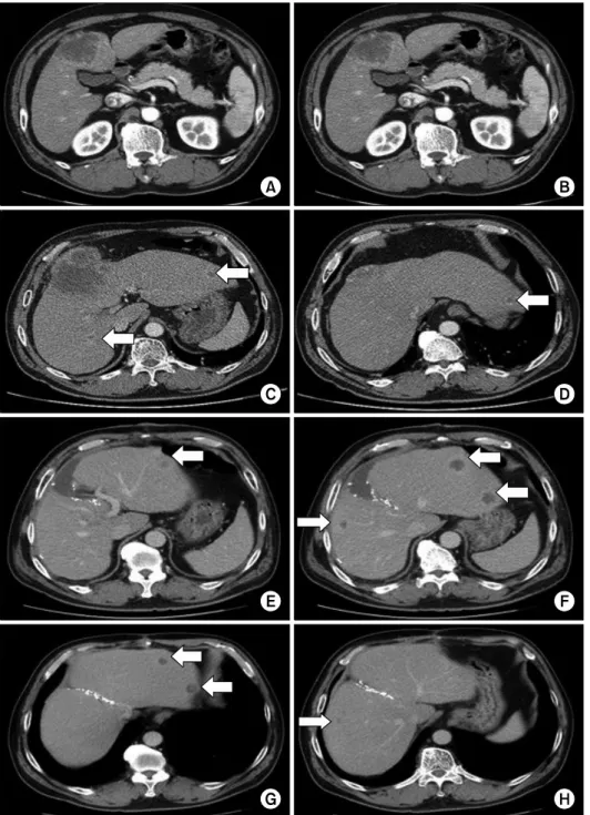
0-37 U/ml), and chorioembryonic antigen (CEA) 2.6 ng/ml (normal range: 0-6 ng/ml). Dynamic abdomen com- puted tomography (CT) scan (Fig. 1A) and magnetic reso- nance imaging of the liver showed the presence of a 6.3-cm sized exophytic mass in the segment IV of the liver. The imaging revealed peripheral wall enhancement and internal hemorrhage.
We suspected that it could be an atypical hepatocellular carcinoma or intrahepatic cholangiocarcinoma, thus ultra- sonography-guided liver biopsy was conducted. The pathologic report revealed poorly differentiated carcinoma in a massive necrotic background. The results of im- munohistochemical staining were CK 19(+), CK 7(+), CK
Table 1. Review of adult male patients with primary hepatic choriocarcinoma Authors Year Country Age
(years) Initial serum -hCG Tumor location Treatment Diagnosis Survival outcome Fernández
Alonso et al.1
1992 Spain 62 41,500 U/ml Liver, LM, KM,MM, LNM, BM
Chemotherapy Autopsy Died after 12 months
Arai et al.2 2001 Japan 65 820 ng/ml Liver Chemotherapy Autopsy Died after 1.5 months Shi et al.8 2010 China 39 560-16,500 mIU/ml Liver Resection and
chemotherpy
Biopsy Died after 6 months 2010 China 45 560-16,500 mIU/ml Liver, LM, BM Chemotherapy Autopsy Died after 2 months 2010 China 48 560-16,500 mIU/ml Liver, AM Chemotherapy Autopsy Died after
3 months 2010 China 36 560-16,500 mIU/ml Liver, LM Chemotherapy Autopsy Died after
5 months 2010 China 40 560-16,500 mIU/ml Liver Resection and
chemotherpy
Biopsy Died after 8 months Bakhshi et
al.7
2012 India 40 Undescribed Liver Resection Biopsy Died after
10 days Sekine et
al.3
2013 Japan 49 53,000 IU/ml Liver, LM, LNM,AM, KM, SM
None Autopsy Died after
2 months Present
case
2015 Korea 54 54,075 mIU/ml Liver Resection and
chemotherapy
Biopsy Died after 18 months hCG, human chorionic gonadotropin; LM, lung metastasis; KM, kidney metastasis; MM, mediastinum metastasis; LNM, lymph node metastasis; BM, bone metastasis; AM, adrenal metastasis; SM, spleen metastasis
20(+), hepatocyte(–) and glypican 3(+), but it was difficult to reach a final diagnosis because the viable tissue compo- nent was very small (less than 5%). A multidisciplinary discussion was held with surgeons, physicians and pathol- ogists, and the patient was recommended to primarily re- ceive hepatic tumor resection.
Three weeks after liver biopsy, he was admitted for surgery. The indocyanine green retention rate at 15 mi- nutes (ICG R15) was 23.1%. Preoperative chest CT scan (Fig. 1B-D) showed the absence of distant metastasis, but increase in size of the primary hepatic lesion (11 cm) and another four small (1-1.5 cm) masses in both the hepatic lobes. Considering high ICG R15 value and large left hep- atic volume (43% of the total liver volume) on cirrhotic background liver, a palliative resection with left medial sectionectomy was performed to confirm the diagnosis as well as to reduce the tumor burden. Liver contour was in early cirrhotic phase with nodularity and the main mass was firmly attached to the greater omentum in the oper- ative findings. Palliative left medial sectionectomy was performed, but we could not resect the other nodules be- cause they were neither localizable nor palpable. The op-
eration was carried out for 6 hours and the total bleeding amount was 1,235 ml. Since his vital signs were stable, no further transfusions were given.
After one week, choriocarcinoma was pathologically confirmed, so we checked -human chorionic gonado- tropin (hCG) subunit level (54,075 mIU/ml, normal range:
0-10 mIU/ml) and bilateral scrotal ultrasonography showed no abnormal findings. One week later, remnant tumors increased from 1-1.5 cm to 2-2.5 cm on post- operative CT scan (Fig. 1E, F). The patient was recom- mended to receive systemic chemotherapy after multi- disciplinary tumor board discussion. Immunohistochemical stains showed CK 19(+), CK 7(+), MOC-31(+), hep- atocyte(–), glypican 3(+), cluster of differentiation (CD) 31(+), AFP(–), and -hCG(+, diffuse). Gross appearance of the tumor was a multi-lobulated hemorrhagic mass, measuring 11.1×8.5×8.5 cm in size. It extended to the ex- tracapsular portion with adhesion to the omentum.
Microscopically, it consisted of a biphasic proliferation of mononuclear cytotrophoblasts and multinucleated syncy- tiotrophoblasts (Fig. 2). Cellular pleomorphism, brisk mi- totic activity, extensive necrosis, and hemorrhage were
Fig. 1. Pre- and postoperative computed tomography (CT) scan findings. Preoperative CT scan showed that the initial tu- mor diameter was 6.3 cm (A).
The size of the main tumor in- creased to 11 cm after 3 weeks (B), and new nodules (arrow) were found on the right and left lobes in the same CT scan (C and D); CT scan taken 1 week after surgery showed increase in the sizes of the remnant tu- mors (arrows) to up to 2.5 cm (E and F); Follow-up CT after chemotherapy showed a de- crease in the size of the rem- nant multiple tumors (arrows) to achieve partial response by RECIST (G and H).
observed.
Three weeks after surgery, the medical oncologists ini- tiated the standard chemotherapy regimen of EMA-CO (Etoposide on day 1-2, Methotrexate on day 1, Dactinomycin on day 1-2, Cyclophosphamide on day 8, and Vincristine on day 8). Four cycles of chemotherapy were given without significant adverse events and the pa- tient achieved partial response (PR) based on Response Evaluation Criteria in Solid Tumors (RECIST, version 1.1)4 on imaging follow-up (Fig. 1G, H). Serial -hCG
levels were checked at the beginning of each cycle. The level rapidly went down to 1,003 mIU/ml at the fourth cycle but did not decrease further afterward.
Subsequently, we planned to perform curative resection if the remnant lesions exhibited responsiveness, after team discussions.
But the next follow up CT after the seventh cycle re- vealed progressive disease (PD) by RECIST. At this time, we decided to switch to another palliative regimen called EMA-EP (Etoposide on day 1 and 8, Methotrexate on day
Fig. 2. Pathological findings. Gross appearance showed the multilobulated hemorrhagic mass with extracapsular mass extension (arrows) (A); Biphasic pleomorphic cells of cytotrophoblasts and syncytiotrophoblasts (arrow), and hemorrhage and necrosis (arrowhead) were observed (H&E stain, ×200) (B); Diffuse expression of -hCG was identified in immunohistochemistry staining (×200) (C).
1, Dactinomycin on day 1-2, and Cisplatin on D8). After two cycles, the patient achieved stable disease (SD) by RECIST and the -hCG level dropped down to 269 mIU/ml. The patient was alive for 10 months after initial surgery and was on the fourth EMA-EP cycle in a good performance state. Despite nine cycles of chemotherapy, hepatic metastases progressed rapidly. Finally, he died due to hepatic failure subsequent to only 18 months after ini- tial surgery.
DISCUSSION
Choriocarcinoma comprises only 1% of all germ cell neoplasms and is more common in women.5,6 It is a
-hCG secreting tumor which is pertinent to uterus and pregnancy.7 PHC is defined as a choriocarcinoma in the liver, which excludes metastasis from another primary ori- gin, especially from the testis in men.8 The majority of PHC is an infantile subtype, which represents metastasis from occult placental choriocarcinoma. Pathogenesis of adult PHC is not clearly understood, but the tumor is as- sumed to arise from abnormal migration of germ cells during embryogenesis or from different histogenetic origin.9
So far, only 9 adult male patients with PHC have been presented in the last 20 years in English literature (Table 1),1-3,7,8 and a majority of them (8 patients) were Asian.
Their mean age at the time of diagnosis was 47.6 years (ranging from 36 to 65 years), and initial serum -hCG level was elevated except in an undescribed case. Primary hepatic involvement was observed in half of the patients
but the other half had disseminated disease at diagnosis or on autopsy. Since PHC grows rapidly, 6 patients were diagnosed by postmortem inspection, usually in ruptured or metastasized state. Only 4 patients were able to under- go curative surgical resection.
The overall prognosis of PHC in adult male patients had been consistently poor with a median survival period of 5 months (ranging from 10 days to 12 months). And owing to its paucity of incidence, there have been no opti- mal diagnostic and therapeutic strategies to improve pa- tient outcomes. In our case, we could not anticipate PHC at first and our liver biopsy yielded no diagnostic clues.
Only after surgical resection, we reached a final diagnosis and checked serum -hCG level. In men in their 40’s or in 50’s with atypical and quickly growing hepatic lesions, it can be difficult to distinguish hepatocellular carcinoma with sarcomatous change or cholangiocarcinoma from PHC. Since cytotrophoblasts are histologically similar to poorly-differentiated carcinoma, diagnostic accuracy of liver biopsy can be unsatisfactory. However, Sekine et al.3 emphasized that liver biopsy should be performed without hesitation in order to enable earlier treatment of such an aggressive tumor. We presume that liver biopsy for earlier histological confirmation is still important and it can be also helpful to investigate the changes in the levels of se- rum -hCG as well as other tumor markers such as AFP, CA 19-9, and CEA, especially when the tumor is rapidly progressive in middle-aged men. Employment of im- munohistochemical battery of antibodies against CK,
-hCG, HPL and Ki-67 when reviewing pathologic slides is also of great importance.8
The role of chemotherapy in the treatment of PHC is unclear due to the tumor’s rapid clinical deterioration, but some authors reported a pathologic complete response and 2-year disease-free survival with etoposide and cisplatin in gastric choriocarcinoma.10 Cisplatin and 5-fluorouracil combination has been challenged as adjuvant or palliative chemotherapy but the clinical outcome was not sat- isfactory as per another published report.8 We applied EMA-CO and EMA-EP regimens to our patient. These regimens are widely used in female gestational tropho- blastic tumor patients and have demonstrated clinical effi- cacy with moderate toxicities.
PHC in adult male patients is known to be a very ag- gressive subset of germ cell tumors and is commonly found in the Asian population. Because of its highly ma- lignant potential, tumor proliferates and disseminates rapidly. In middle-aged men with atypically and rapidly deteriorating hepatic lesions, we should suspect PHC as a differential diagnosis of hepatic tumors. When the pathologic finding of liver biopsy is uncertain, checking additional serum -hCG level can be of importance since delayed diagnosis of PHC can be fatal.
REFERENCES
1. Fernández Alonso J, Sáez C, Pérez P, Montaño A, Japón MA.
Primary pure choriocarcinoma of the liver. Pathol Res Pract 1992;188:375-377; discussion 378-379.
2. Arai M, Oka K, Nihei T, Hirota K, Kawano H, Kawasaki T, et al. Primary hepatic choriocarcinoma: a case report.
Hepatogastroenterology 2001;48:424-426.
3. Sekine R, Hyodo M, Kojima M, Meguro Y, Suzuki A, Yokoyama T, et al. Primary hepatic choriocarcinoma in a 49-year-old man: report of a case. World J Gastroenterol 2013;
19:9485-9489.
4. Eisenhauer EA, Therasse P, Bogaerts J, Schwartz LH, Sargent D, Ford R, et al. New response evaluation criteria in solid tu- mours: revised RECIST guideline (version 1.1). Eur J Cancer 2009;45:228-247.
5. Scholz M, Zehender M, Thalmann GN, Borner M, Thoni H, Studer UE. Extragonadal retroperitoneal germ cell tumor: evi- dence of origin in the testis. Ann Oncol 2002;13:121-124.
6. Fine G, Smith RW Jr, Pachter MR. Primary extragenital chorio- carcinoma in the male subject. Case report and review of the literature. Am J Med 1962;32:776-794.
7. Bakhshi GD, Borisa AD, Bhandarwar AH, Tayade MB, Yadav RB, Jadhav YR. Primary hepatic choriocarcinoma: a rare cause of spontaneous haemoperitoneum in an adult. Clin Pract 2012;2:e73.
8. Shi H, Cao D, Wei L, Sun L, Guo A. Primary choriocarcinoma of the liver: a clinicopathological study of five cases in males.
Virchows Arch 2010;456:65-70.
9. Garcia RL, Ghali VS. Gastric choriocarcinoma and yolk sac tu- mor in a man: observations about its possible origin. Hum Pathol 1985;16:955-958.
10. Waseda Y, Komai Y, Yano A, Fujii Y, Noguchi N, Kihara K.
Pathological complete response and two-year disease-free surviv- al in a primary gastric choriocarcinoma patient with advanced liver metastases treated with germ cell tumor-based chemo- therapy: a case report. Jpn J Clin Oncol 2012;42:1197-1201.