Ann Hepatobiliary Pancreat Surg 2018;22:274-281
https://doi.org/10.14701/ahbps.2018.22.3.274
Case Report
Long-term complete response after transcatheter arterial chemoembolization and stereotactic body radiation therapy in
a patient with hepatocellular carcinoma at the caudate lobe
Yong-Kyu Chung1, Shin Hwang1, Gi-Young Ko2, and Sang Min Yoon3
Departments of 1Surgery, 2Diagnostic Radiology, and 3Radiation Oncology, Asan Medical Center, University of Ulsan College of Medicine, Seoul, Korea
It is expected that a combination of transcatheter arterial chemoembolization (TACE) with stereotactic body radiation therapy (SBRT) may induce synergistic therapeutic effects in hepatocellular carcinoma (HCC), which would result in a high rate of complete therapeutic response. In this study, we present the 5-year clinical course of a patient who had HCC at the caudate lobe, which was treated with TACE and SBRT. A 53-year-old male was diagnosed with an 8 cm-sized HCC at the caudate lobe with compression of the inferior vena cava (IVC). For fear of pulmonary meta- stasis, we decided to perform sequential TACE-radiotherapy instead of upfront hepatectomy, although the tumor ap- peared resectable. The first session of TACE, SBRT with 12 fractions, and the second session of TACE were sequen- tially performed. The patient was administered metformin for chemoprevention. Over the course of a 5-year follow-up, there was no evidence of HCC recurrence. We reported the clinical sequence of a patient showing complete therapeutic response of HCC at the caudate lobe after a combination of TACE and radiotherapy. This type of combined locore- gional treatment can be a therapeutic option for HCC at the caudate lobe with marginal resectability. (Ann Hepatobiliary Pancreat Surg 2018;22:274-281)
Key Words: Treatment response; Recurrence; Radiotherapy
Received: May 31, 2018; Revised: June 1, 2018; Accepted: June 29, 2018 Corresponding author: Shin Hwang
Department of Surgery, Asan Medical Center, University of Ulsan College of Medicine, 88 Olympic-ro 43-gil, Songpa-gu, Seoul 05505, Korea Tel: +82-2-3010-3930, Fax: +82-2-3010-6701, E-mail: [email protected]
Copyright Ⓒ 2018 by The Korean Association of Hepato-Biliary-Pancreatic Surgery
This is an Open Access article distributed under the terms of the Creative Commons Attribution Non-Commercial License (http://creativecommons.org/
licenses/by-nc/4.0) which permits unrestricted non-commercial use, distribution, and reproduction in any medium, provided the original work is properly cited.
Annals of Hepato-Biliary-Pancreatic Surgery ∙ pISSN: 2508-5778ㆍeISSN: 2508-5859
INTRODUCTION
Hepatocellular carcinoma (HCC) is the fifth most com- mon malignancy worldwide and is one of the leading causes of cancer-related deaths.1,2 Hepatic resection (HR) is considered to be the preferred treatment method for HCC but is also considered to be a challenging surgical procedure in the presence of liver cirrhosis. HR resection for HCC encircling the retrohepatic inferior vena cava (IVC) increases the risk of an intraoperative tumor spread during the handling of the hypervascular mass.3-6
Transcatheter arterial chemoembolization (TACE) rep- resents one of the locoregional therapies for HCC. TACE often improves outcomes in patients with unresectable HCCs, but it has not been considered a curative treatment because of high recurrence rates. We previously reported that preoperative TACE for resectable HCC may ad-
versely affect post-resection prognosis, irrespective of pathological responses. Thus, we suggested that pre- operative TACE should be avoided for patients with re- sectable small HCCs.7
With recent advances in the radiotherapy techniques, stereotactic body radiation therapy (SBRT) has been con- sidered an alternative treatment option for a small HCC not suitable for hepatic resection and other locoregional treatments.8-10 More recently, volumetric-modulated arc therapy, one of the most sophisticated linear accelerator- based treatment modalities, has become available even with gated delivery. This method allows dose rate-chang- ing intensity modulation with gantry rotation and may provide more conformal dose distribution while reducing treatment delivery time and monitor units.
It is expected that a combination of TACE with SBRT may induce synergistic therapeutic effects, which would
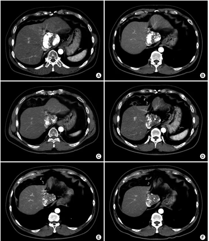
Fig. 2. CT findings of the liver mass at the caudate lobe at admission with pre-enhancement phase (A), arterial phase (B), portal phase (C) and delayed phase (D).
Fig. 1. Computed tomography (CT) findings of the liver mass (A) at the caudate lobe taken 3 years before admission. This mass compresses the retrohepatic inferior vena cava (B).
276 Ann Hepatobiliary Pancreat Surg Vol. 22, No. 3, August 2018
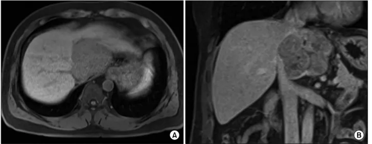
Fig. 3. Magnetic resonance imaging showing a hypervascular mass (A) compressing the inferior vena cava (B).
Fig. 4. Positron emission tomography showed a vague and heterogeneously hypermetabolic mass.
result in a high rate of complete therapeutic response. In the present study, we report the 5-year clinical course of a patient who had HCC at the caudate lobe and was treat- ed with TACE and SBRT.
CASE
A 53-year-old male was admitted for examination of a liver mass at the caudate lobe. The mass was first detected during a routine health screening 3 years before. It was 7 cm in size at that time with no elevation of tumor mark- ers (Fig. 1). During a follow-up with liver ultrasono- graphy, its size had increased very slowly. He had a past history of hepatitis B virus (HBV) infection. During the precedent follow-up, HBV surface antigen (HBsAg) be- came seronegative with the appearance of anti-HBs. At admission, HBV DNA was not detectable on polymerase chain reaction assays; Serum alpha-fetoprotein (AFP) was 3.2 ng/ml and prothrombin-induced by vitamin K absence or antagonist-II (PIVKA-II) was 988 mAU/ml.
Computed tomography (CT) revealed an 8 cm-sized hy- pervascular mass with calcification at the caudate lobe of the liver, which was abutted with the IVC (Fig. 2). Magnetic resonance imaging showed a hypervascular mass com- pressing the IVC (Fig. 3). These two imaging studies, with tumor marker findings, strongly suggested diagnosis of HCC. Positron emission tomography showed an 8 cm-sized heterogeneously hypermetabolic mass (maximal standardized uptake value [SUV]=4.0) (Fig. 4), which was not regarded as being a hypermetabolic uptake.
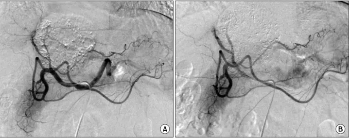
Since this mass was directly attached to the IVC, thus there was a high risk of tumor cell spread through the short hepatic veins, which can induce early pulmonary metastasis. For fear of such a catastrophic pulmonary metastasis, we decided to perform sequential TACE-radio- therapy instead of upfront hepatectomy, although the tu- mor appeared resectable. The tumor was primarily fed by the left hepatic artery and conventional TACE was per- formed using chemoinfusion of cisplatin and embolization with Lipiodol and Gelfoam (Fig. 5).
Four days later, a liver CT scan was taken, in which some suspected viable tumor portion was found in the lip- iodolized HCC (Fig. 6). We decided to carry out the pre-
Fig. 6. Liver CT scan of pre-enhancement (A) and arterial phase (B) taken 4 days after transcatheter arterial chemoembolization (TACE) shows a lipiodolized mass.
Fig. 5. Hepatic arteriography shows the feeding arteries from the left hepatic artery (A) and post-embolization status (B).
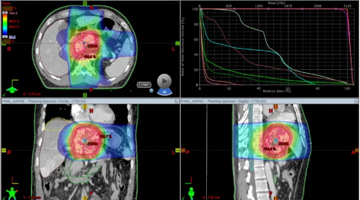
planned sequential TACE-radiotherapy. A CT simulation was taken 3 weeks after the first session of TACE (Fig.
7). SBRT of 30 Gy with 12 fractions for 2 week was per- formed in order to avoid irradiation-associated bleeding from the gastric antrum.
Six weeks later, after SBRT, a follow-up CT scan showed a slight decrease in the size of the lipiodol-uptake HCC, in which we could not exclude the possibility of a viable tumor (Fig. 8). At this time, the serum PIVKA-II level was normalized. Thus, the second session of TACE was performed, in which suspicious tumor staining was found in both lobes, thus chemoinfusion and lip- iodol/gelfoam embolization was performed via the right and left hepatic arteries (Fig. 9).
Follow-up CT scans taken every 3 months for 5 years showed no changes of the partial lipiodolized lesion at the caudate lobe without definite arterial enhancement (Fig.
10). A Chest CT scan was taken concurrently, twice per year, showing no evidence of pulmonary metastasis. After finishing follow-up of the first 5 years with no evidence of HCC recurrence, the follow-up interval at the out- patient clinic was extended to 4 months.
The patient showed normal blood glucose levels, but metformin 500 mg has been prescribed twice per day since the second session of TACE for the purpose of chemoprevention.
278 Ann Hepatobiliary Pancreat Surg Vol. 22, No. 3, August 2018
Fig. 7. CT simulation for stereotactic beam radiotherapy with avoidance of exposure to the gastric antrum.
Fig. 8. Liver CT scan of arterial phase (A, B) shows suspected viable tumor portion in the lipiodolized mass.
DISCUSSION
HCC located at the caudate lobe with encircling in- volvement of the retrohepatic IVC has often been treated with HR through various surgical approach methods.3-6 The transhepatic approach, dividing the hepatic paren- chyma to expose the paracaval portion, is a feasible meth- od used to reduce the risk of tumor handling-associated tumor spread. Downstaging with preoperative TACE had
been attempted, but its therapeutic effect was worse than expected.7,11 HR, used only a few months after TACE, should be avoided because the integrity of the HCC cells is loosened, by which the risk of intraoperative tumor spread increases. The preoperative waiting period after TACE is usually recommended to be at least 3-6 months.
So far, it is not recommended to perform any preoperative locoregional treatment of resectable or marginally resect- able HCCs located at the caudate lobe.
Fig. 9. Second session of transcatheter arterial chemoembolization shows suspicious tumor staining in both lobes (A) and post-em- bolization status.
Considering that both HR and TACE for use with HCC at the caudate lobe often resulted in inferior outcomes compared with those at other liver portions, as well as ra- diofrequency ablation (RFA), is usually not applicable for HCCs at the caudate lobe; it is not simple to choose the treatment modality for patients with HCC at the caudate lobe. HCC with portal vein invasion has been attempted with a combination of TACE and radiotherapy, which re- sulted in improved outcomes.12,13 This therapeutic ap- proach encouraged us to perform a combination of TACE and radiotherapy for HCC at the caudate lobe with a cura- tive intent.
Our protocol for a combination of TACE and radio- therapy with a curative intent in patients with marginally resectable HCC, including HCC at the caudate lobe, in- cludes conventional TACE once or twice in 1-month in- tervals and then subsequent SBRT within 1 month after the last TACE. Follow-up protocol includes monthly dy- namic liver CT scans twice, bimonthly CT scans twice and then routine CT scan follow-up every 3 months. A blood tumor marker study is performed during every visit to the outpatient clinic. A chest CT scan is performed concurrently with every other liver CT scan. During fol- low-up, if tumor recurrence is detected, we perform repeat TACE, RFA, cryoablation, or even HR as indicated. We think that only hypervascular HCC is indicated for a com- bination of TACE and radiotherapy. If vascularity of the HCC is not definitely increased, we prefer performing up- front HR without performing any locoregional treatment
in the case of HCC with marginal resectability.
Recently, the therapeutic role of radiotherapy for HCC has been emphasized more than before. The local control rate of SBRT is reported to be similar to that of RFA,8,9 which suggests the possibility of SBRT as an ablative therapy for small HCC. Specifically, respiratory-gated volumetric-modulated arc therapy can shorten the treat- ment delivery time when compared with SBRT using stat- ic beams. Although the clinical impact of a prolonged treatment delivery time on SBRT at a high dose per frac- tion is not yet clear, from a biological perspective, a pro- longed delivery time has shown a detrimental effect on tumor control by reducing cell killing in cell lines or in xenograft models of tumors with a low alpha/beta ratio.14-16
The clinical sequence of our present case appears to be unique. The mass at the caudate lobe grows very slowly, indicating malignant transformation within the dysplastic nodule during follow-up over several years. At the time of admission, this lesion was diagnosed of overt HCC.
During 5-year follow-up after TACE-radiotherapy, it re- gressed slowly but probably the non-tumorous portion remained. These sequences are not typical, thus not being presented in detail in literature.
In our present case, metformin has been administered for chemoprevention although he was not diabetic.
Metformin is a biguanide agent used to treat type 2 dia- betes mellitus. It regulates the blood sugar by improving insulin sensitivity and reducing hepatic glucose output through inhibition of gluconeogenesis and glycogenolysis.
280 Ann Hepatobiliary Pancreat Surg Vol. 22, No. 3, August 2018
Fig. 10. Follow-up CT scans showing no changes of the partial lipiodolized lesion at the caudate lobe taken 1 month (A), 1 year (B), 2 years (C), 3 years (D), 4 years (E) and 5 years (F) after the second session of TACE.
Recently, metformin has proven capable of inhibiting can- cer cell growth by inducing cell cycle arrest and enhanc- ing apoptosis.17 A considerable number of studies have found that metformin plays a chemopreventive role in other
cancers and is associated with reduced risk for HCC.18,19 We recently presented that the administration of metfor- min showed a tendency to reduce the tumor recurrence rate and helped induce significant improvement in overall
survival in patients who underwent HR for HCC.20 We suggest administering metformin in HCC patients with glucose intolerance or overt diabetes mellitus.
In conclusion, we presented the clinical sequence of a patient showing complete response of HCC at the caudate lobe after a combination of TACE and radiotherapy. This type of locoregional treatment can be a therapeutic option for HCC at the caudate lobe with marginal resectability.
REFERENCES
1. Forner A, Llovet JM, Bruix J. Hepatocellular carcinoma. Lancet 2012;379:1245-1255.
2. El-Serag HB. Hepatocellular carcinoma. N Engl J Med 2011;
365:1118-1127.
3. Wang ZG, Lau W, Fu SY, Liu H, Pan ZY, Yang Y, et al.
Anterior hepatic parenchymal transection for complete caudate lobectomy to treat liver cancer situated in or involving the para- caval portion of the caudate lobe. J Gastrointest Surg 2015;19:
880-886.
4. Ahanatha Pillai S, Sathyanesan J, Perumal S, Ulagendra Perumal S, Lakshmanan A, Ramaswami S, et al. Isolated caudate lobe re- section: technical challenges. Ann Gastroenterol 2013;26:150-155.
5. Liu P, Qiu BA, Bai G, Bai HW, Xia NX, Yang YX, et al.
Choice of approach for hepatectomy for hepatocellular carcino- ma located in the caudate lobe: isolated or combined lobectomy?
World J Gastroenterol 2012;18:3904-3909.
6. Chaib E, Ribeiro MA Jr, Souza YE, D'Albuquerque LA. Anterior hepatic transection for caudate lobectomy. Clinics (Sao Paulo) 2009;64:1121-1125.
7. Ha TY, Hwang S, Lee YJ, Kim KH, Ko GY, Ii Gwon D, et al.
Absence of benefit of transcatheter arterial chemoembolization (TACE) in patients with resectable solitary hepatocellular carci- noma. World J Surg 2016;40:1200-1210.
8. Jeong Y, Jung J, Cho B, Kwak J, Jeong C, Kim JH, et al.
Stereotactic body radiation therapy using a respiratory-gated volumetric-modulated arc therapy technique for small hep- atocellular carcinoma. BMC Cancer 2018;18:416.
9. Ohri N, Dawson LA, Krishnan S, Seong J, Cheng JC, Sarin SK, et al. Radiotherapy for hepatocellular carcinoma: new indications and directions for future study. J Natl Cancer Inst 2016;108.
djw133.
10. Rim CH, Seong J. Application of radiotherapy for hepatocellular carcinoma in current clinical practice guidelines. Radiat Oncol J 2016;34:160-167.
11. Kang WH, Hwang S, Song GW, Lee YJ, Kim KH, Ahn CS, et al. Prognostic effect of transarterial chemoembolization-induced complete pathological response in patients undergoing liver re- section and transplantation for hepatocellular carcinoma. Liver Transpl 2017;23:781-790.
12. Yoon SM, Ryoo BY, Lee SJ, Kim JH, Shin JH, An JH, et al.
Efficacy and safety of transarterial chemoembolization plus ex- ternal beam radiotherapy vs sorafenib in hepatocellular carcino- ma with macroscopic vascular invasion: a randomized clinical trial. JAMA Oncol 2018. doi: 10.1001/jamaoncol.2017.5847. [in press]
13. Im JH, Yoon SM, Park HC, Kim JH, Yu JI, Kim TH, et al.
Radiotherapeutic strategies for hepatocellular carcinoma with portal vein tumour thrombosis in a hepatitis B endemic area.
Liver Int 2017;37:90-100.
14. Kwon JH, Bae SH, Kim JY, Choi BO, Jang HS, Jang JW, et al. Long-term effect of stereotactic body radiation therapy for primary hepatocellular carcinoma ineligible for local ablation therapy or surgical resection. stereotactic radiotherapy for liver cancer. BMC Cancer 2010;10:475.
15. Wang JZ, Li XA, D'Souza WD, Stewart RD. Impact of pro- longed fraction delivery times on tumor control: a note of cau- tion for intensity-modulated radiation therapy (IMRT). Int J Radiat Oncol Biol Phys 2003;57:543-552.
16. Zheng XK, Chen LH, Yan X, Wang HM. Impact of prolonged fraction dose-delivery time modeling intensity-modulated radia- tion therapy on hepatocellular carcinoma cell killing. World J Gastroenterol 2005;11:1452-1456.
17. Ben Sahra I, Regazzetti C, Robert G, Laurent K, Le Marchand-Brustel Y, Auberger P, et al. Metformin, independent of AMPK, induces mTOR inhibition and cell-cycle arrest through REDD1. Cancer Res 2011;71:4366-4372.
18. Chen HP, Shieh JJ, Chang CC, Chen TT, Lin JT, Wu MS, et al. Metformin decreases hepatocellular carcinoma risk in a dose-dependent manner: population-based and in vitro studies.
Gut 2013;62:606-615.
19. Donadon V, Balbi M, Mas MD, Casarin P, Zanette G.
Metformin and reduced risk of hepatocellular carcinoma in dia- betic patients with chronic liver disease. Liver Int 2010;30:750- 758.
20. Kang WH, Tak E, Hwang S, Song GW, Jwa E, Lee YJ, et al.
Metformin-associated chemopreventive effects on recurrence af- ter hepatic resection of hepatocellular carcinoma: from in vitro to a clinical study. Anticancer Res 2018;38:2399-2407.