ISSN 2234-3806 • eISSN 2234-3814
510 www.annlabmed.org https://doi.org/10.3343/alm.2021.41.5.510 Ann Lab Med 2021;41:510-513
https://doi.org/10.3343/alm.2021.41.5.510
Letter to the Editor
Clinical Microbiology
Serological Evidence of Coxiella burnetii and SARS- CoV-2 Co-infection: A Case Report
Hee Sue Park , M.D., Ph.D.1, Pan Kee Bae , Ph.D.2, Hye Won Jeong , M.D., Ph.D.3, Bo Ra Son , M.D., Ph.D.4, and Kyeong Seob Shin , M.D., Ph.D.4
1Department of Laboratory Medicine, Chungbuk National University Hospital, Cheongju, Korea; 2BioNano Health Guard Research Center (H-GUARD), Daejeon, Korea; 3Department of Internal Medicine, Chungbuk National University College of Medicine, Cheongju, Korea; 4Department of Laboratory Medicine, Chungbuk National University College of Medicine, Cheongju, Korea
Dear Editor,
The most common symptoms of coronavirus disease 2019 (CO- VID-19) are respiratory symptoms that are not easily distinguish- able from those of other acute respiratory infections [1]. As bac- terial/fungal co-infections are reported in 8% of COVID-19 pa- tients, diagnosing them is critical for appropriate treatment [2].
Pathogens that cause co-infection with severe acute respiratory syndrome coronavirus 2 (SARS-CoV-2) include influenza A/B vi- ruses, Mycoplasma pneumoniae, Acinetobacter baumannii, Candida albicans, and Legionella pneumophila [2]. Q fever is asymptomatic in approximately 60% cases, but a flu-like illness with high fever, myalgia, headache, and cough, which lasts for one to three weeks, may occur in acute infection and then re- solve spontaneously [3]. We report the case of a 37-year-old man who was diagnosed as having Coxiella burnetii and SARS- CoV-2 co-infection. To the best of our knowledge, this is the first such report in Korea. The study was approved by the Institu- tional Review Board of the Chungbuk National University Hospi- tal, Cheong ju, Korea (IRB number: 2020-03-025).
A 37-year-old man presented to the Chungbuk National Uni- versity Hospital in May 2020 with fever, cough, and sputum de- velopment, which had started three days before hospital pre- sentation. The patient was a farmer, with no epidemiologic link
to COVID-19-confirmed cases. Physical examination revealed a temperature of 38.8°C, blood pressure of 126/64 mm Hg, pulse rate of 95/minute, and respiratory rate of 18/minute. A complete blood count revealed a hemoglobin level of 151 g/L, white blood cell count of 2.32×10⁹ cells/L (absolute neutrophil count 1.37×
109 cells/L, absolute lymphocyte count 0.64×10⁹ cells/L), and platelet count of 116×109/L. Other blood tests revealed elevated levels of C-reactive protein (50.5 mg/L) and lactate dehydroge- nase (12.07 µkat/L) and slightly elevated levels of D-dimer (12.05 nmol/L), AST (1.12 µkat/L), and ALT (0.83 µkat/L). The patient’s prothrombin time and activated partial-thromboplastin time tests were within the reference ranges (Table 1). Chest X-ray showed no active lung lesion. A real-time reverse transcription (real-time RT-PCR) (Allplex 2019-nCoV Assay, Seegen, Seoul, Korea) test for SARS CoV 2 was performed on admission day. The result was negative in a naso/oropharyngeal swab and positive in spu- tum. Multiplex RT-PCR results for M. pneumoniae, Chlamydia pneumoniae, L. pneumophila, Bordetella pertussis, Streptococ- cus pneumoniae, Haemophilus influenzae (Allplex Pneumo- Bacter Assay, Seegen) and influenza A/B virus (Sofia fluores- cence immunoassay, Quidel, San Diego, CA, USA) were all neg- ative. Considering the patient’s occupation, serological tests for C. burnetii, Leptospira interrogans, and Orientia tsutsugamushi
Received: September 23, 2020 Revision received: November 18, 2020 Accepted: March 15, 2021
Corresponding author: Kyeong Seob Shin, M.D., Ph.D.
Department of Laboratory Medicine, Chungbuk National University College of Medicine, 1 Chungdae-ro, Seowon-gu, Cheongju 28644, Korea Tel: +81-43-269-6240
Fax: +81-43-271-5243 E-mail: [email protected]
© Korean Society for Laboratory Medicine
This is an Open Access article distributed under the terms of the Creative Commons Attribution Non-Commercial License (https://creativecommons.org/licenses/by-nc/4.0) which permits unrestricted non-commercial use, distribution, and reproduction in any medium, provided the original work is properly cited.
1 / 1 CROSSMARK_logo_3_Test
2017-03-16 https://crossmark-cdn.crossref.org/widget/v2.0/logos/CROSSMARK_Color_square.svg
Park HS, et al.
C. burnetii and SARS-CoV-2 co-infection
https://doi.org/10.3343/alm.2021.41.5.510 www.annlabmed.org 511
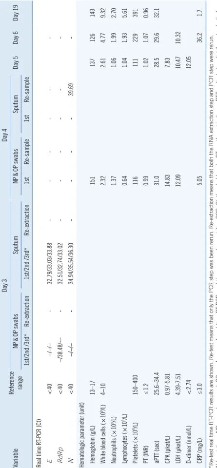
Table 1. Laboratory results after symptom onset VariableReference range
Day 3Day 4 Day 5Day 6Day 19NP&OP swabsSputumNP&OP swabsSputum 1st/2nd /3rd*Re-extraction1st/2nd /3rd*Re-extraction 1stRe-sample1stRe-sample Real time RT-PCR (Ct) E <40–/–/–-32.79/33.03/33.88--- RdRp <40–/38.48/-–-32.51/32.74/33.02--- N <40–/–/–-34.94/35.54/36.30----39.69- Hematologic parameter (unit) Hemoglobin (g/L)13–17151137126143 White blood cells (×109/L)4–102.322.614.779.32 Neutrophils (×109/L)1.371.061.992.70 Lymphocytes (×109/L)0.641.041.935.61 Platelets (×109/L)150–400116111229391 PT (INR)≤1.20.991.021.070.96 aPTT (sec)25.6–34.431.028.529.632.1 CPK (µkat/L)0.97-5.8114.837.83 LDH (µkat/L)4.39-7.5112.0910.4710.32 D-dimer (nmol/L)<2.7412.05 CRP (mg/L)≤3.05.0536.21.7 *Re-test real time RT-PCR results are shown. Re-test means that only the PCR step was been rerun. Re-extraction means that both the RNA extraction step and PCR step were rerun. Abbreviations: NP, nasopharyngeal; OP, oropharyngeal; real-time RT-PCR, real-time reverse transcription PCR; Ct, threshold cycle; PT, prothrombin time; aPTT, activated partial-thromboplastin time; CPK, creatinine phosphokinase; LDH, lactate dehydrogenase; CRP, C-reactive protein.
Park HS, et al.
C. burnetii and SARS-CoV-2 co-infection
512 www.annlabmed.org https://doi.org/10.3343/alm.2021.41.5.510 were performed on admission day and were all negative.
The next day, SARS-CoV-2 RT-PCR test results were negative.
To rule out a false-positive result in the first RT-PCR test, we re- extracted and re-tested the original samples. After re-extraction, the sputum tested negative. In addition, serological antibody tests for SARS-CoV-2 infection were performed to rule out a false-positive RT-PCR result. A sandwich ELISA targeting the SARS-CoV-2 receptor binding domain (RBD) of the spike pro- tein (SP) was conducted the next day. Briefly, the SARS-CoV-2 RBD antigen was attached to a 96-well plate and diluted serum was applied. After washing to remove unbound substance, the detection antibody (horseradish peroxidase-conjugated anti-hu- man IgG or IgM) was added. After washing away excess detec- tion antibody, the optical density at 450 nm in each well was measured using a microplate reader. The anti-RBD IgG anti- body test was positive on the day after admission (Fig. 1). Based on the positive SARS-CoV-2 RT-PCR result in sputum and posi- tive sandwich ELISA result, the patient was diagnosed as having SARS-CoV-2 infection. Follow-up serological tests showed pa- tient seroconversion indicating C. burnetii infection. Therefore, co-infection with C. burnetii and SARS-CoV-2 was confirmed in the follow-up period.
Although the severity of COVID-19 varies from mild to life- threatening, bacterial or fungal co-infection in COVID-19 pa- tients increases the risk of mortality [4]. Therefore, clinicians should consider the variable clinical severity of COVID-19 and the possibility of co-infection, which may cause the same symp- toms as COVID-19 but can aggravate the patient’s condition and require additional laboratory testing for diagnosis.
Real-Time RT-PCR is a standard method for diagnosing
SARS-CoV-2 infection, as it gives minimal false-positive results [5]. Considering that negative conversion of real-time RT-PCR test results takes more than two weeks for SARS-CoV-2 infection [6], the patient might have had SARS-CoV-2 infection in the past. On days 15 to 29 of COVID-19, the sensitivity of real-time RT-PCR is 70.7%, whereas that of ELISA is 100% [7]. In sero- logical tests for SARS-CoV-2, various target proteins, such as RBD, nucleocapsid protein, and SP, can be used, and, when these tests are used in combination with molecular tests, the sensitivity and specificity of COVID-19 diagnosis are increased [7]. In our case, a false-positive real-time RT-PCR result could not be ruled out, but SARS-CoV-2 infection was assumed, con- sidering the results of additional serological tests. We believe that the negative conversion of the real-time RT-PCR result was due to a low viral load or virus remnant. Anti-SARS-CoV-2 IgM is less sensitive than IgG [8], and a negative IgM result on days 3 and 4 is considered false. It is necessary to further evaluate the diagnostic performance of serological tests for COVID-19.
In Q fever, serological tests have been used to diagnose acute infection, and seroconversion from negative to positive occurs one to three weeks after symptom onset [3]. Although viral loads do not differ between asymptomatic and symptomatic COVID-19 patients, our patient’s symptoms at the time of hospi- tal presentation are more likely to have been due to Q fever [9].
Other laboratory results were nonspecific, but lymphopenia was notable on admission. Lymphopenia is rarely observed in Q fe- ver but is common in COVID-19, for which it is a prognostic in- dicator [10].
To the best of our knowledge, this is the first report on C. bur- netii and SARS-CoV-2 co-infection, and serologic testing played
Fig. 1. Serological diagnosis of co-infection with Coxiella burnetii and SARS-CoV-2. Serological tests for SARS-CoV-2 IgM/IgG anti-RBD and C. burnetii were positive, indicating co-infection.
Abbreviations: SARS-CoV-2, severe acute respiratory syndrome coronavirus 2; RBD, SARS-CoV-2 receptor binding domain.
0 1 2 3 4 5 6 7 8 9 10 11 12 13 14 15 16 17 18 19 20 Symptoms
SARS-CoV-2 RNA
Undetectable level
Days
Scrology (SARS-CoV-2) 1.95 1.57 2.35
1.95
1:64 1:1,024
1:1,028 0.76 0.76
<1:16
Hospitalization
<1:16 <1:16
anti-RBD lgM Ab (■) anti-RBD lgM Ab (▲)
anti-phase I (■)/II (□) lgM Ab anti-phase I (▲)/II (△) lgM Ab
Positive/Negative Cut off (OD 1.1) Serology (Q fever)
Park HS, et al.
C. burnetii and SARS-CoV-2 co-infection
https://doi.org/10.3343/alm.2021.41.5.510 www.annlabmed.org 513
an important role in the diagnosis. For accurate COVID-19 diag- nosis, clinicians should consider a multidisciplinary approach and utilize accurate and rapid diagnostic tools.
ACKNOWLEDGEMENTS
We thank all clinicians and laboratory technologists in Chung- buk National University Hospital and BioNano Health Guard Research Center.
AUTHOR CONTRIBUTIONS
Park HS designed the study and wrote the manuscript; Bae PK carried out the experiment and analyzed the data; Son BR col- lected the data; Jeong HW provided clinical information and discussed the manuscript; Shin KS designed the study and ed- ited the manuscript. All authors have read the approved the fi- nal manuscript.
CONFLICTS OF INTEREST
The authors declare no conflict of interest.
RESEARCH FUNDING
Not applicable.
ORCID
Hee Sue Park https://orcid.org/0000-0002-8378-6066
Pan Kee Bae https://orcid.org/0000-0003-4488-5261 Hye Won Jeong https://orcid.org/0000-0002-1063-8476 Bo Ra Son https://orcid.org/0000-0001-9020-4303 Kyeong Seob Shin https://orcid.org/0000-0002-1680-1510
REFERENCES
1. Zha L, Shen J, Tefsen B, Wang Y, Lu W, Xu Q. Clinical features and out- comes of adult COVID-19 patients co-infected with Mycoplasma pneu- moniae. J Infect 2020;81:e12-5.
2. Rawson TM, Moore LS, Zhu N, Ranganathan N, Skolimowska K, Gil- christ M, et al. Bacterial and fungal co-infection in individuals with coro- navirus: a rapid review to support COVID-19 antimicrobial prescribing.
Clin Infect Dis 2020;71:2459-68.
3. Gikas A, Kokkini S, Tsioutis C. Q fever: clinical manifestations and treat- ment. Expert Rev Anti Infect Ther 2010;8:529-39.
4. Zhou F, Yu T, Du R, Fan G, Liu Y, Liu Z, et al. Clinical course and risk factors for mortality of adult inpatients with COVID-19 in Wuhan, China:
a retrospective cohort study. Lancet 2020;395:1054-62.
5. Hong KH, Lee SW, Kim TS, Huh HJ, Lee J, Kim SY et al. Guidelines for laboratory diagnosis of Coronavirus Disease 2019 (COVID-19) in Korea.
Ann Lab Med 2020;40:351-60
6. Carmo A, Pereira-Vaz J, Mota V, Mendes A, Morais C, da Silva AC, et al.
Clearance and persistence of SARS-CoV-2 RNA in patients with COV- ID-19. J Med Virol 2020;92: 2227-31.
7. Lou B, Li T, Zheng S, Su Y, Li Z, Liu W, et al. Serology characteristics of SARS-CoV-2 infection since exposure and post symptom onset. Eur Respir J 2020;56:2000763.
8. Zhang Z, Hou Y, Li D, Li F. Diagnostic efficacy of anti‐SARS‐CoV‐2 IgG/
IgM test for COVID‐19: a meta‐analysis. J Med Virol 2020; 93:366-74 9. Zou L, Ruan F, Huang M, Liang L, Huang H, Hong Z, et al. SARS-CoV-2
viral load in upper respiratory specimens of infected patients. N Engl J Med 2020;382:1177-9.
10. Terpos E, Ntanasis-Stathopoulos I, Elalamy I, Kastritis E, Sergentanis TN, Politou M, et al. Hematological findings and complications of COV- ID-19. Am J Hematol 2020; 95:834-47.