J Korean Surg Soc 2012;83:403-407 http://dx.doi.org/10.4174/jkss.2012.83.6.403
CASE REPORT
Journal of the Korean Surgical Society
JKSS
pISSN 2233-7903ㆍeISSN 2093-0488
Received May 29, 2012, Revised October 24, 2012, Accepted October 29, 2012 Correspondence to: Hyung-Il Seo
Department of Surgery, Pusan National University Hospital, Pusan National University School of Medicine, 179 Gudeok-ro, Seo-gu, Busan 602-739, Korea
Tel: +82-51-240-7238, Fax: +82-51-247-1365, E-mail: [email protected]
cc Journal of the Korean Surgical Society is an Open Access Journal. All articles are distributed under the terms of the Creative Commons Attribution Non-Commercial License (http://creativecommons.org/licenses/by-nc/3.0/) which permits unrestricted non-commercial use, distribution, and reproduction in any medium, provided the original work is properly cited.
Primary leiomyosarcoma of gallbladder
Eun Young Park, Hyung-Il Seo, Sung Pil Yun, Suk Kim
1, Joo Yeun Kim
2, Koon Taek Han
Departments of Surgery, 1Radiology, and 2Pathology, School of Medicine, Pusan National University, Biomedical Research Institute, Pusan National University Hospital, Busan, Korea
Malignant mesenchymalneoplasms of the gallbladder are extremely rare with only 105 cases of primary gallbladder sarcoma having been described. It has a very aggressive behavior and is usually diagnosed at advanced stages. Therefore, curative surgical management may not be possible. We performed a radical cholecystectomy (S4b + S5 segmentectomy), omentec- tomy and small bowel resection in a 54-year-old patient with locally invasive leiomyosarcoma of the gallbladder. Further studies are needed to confirm the benefit of aggressive treatment for patients with leiomyosarcoma of the gallbladder.
Key Words: Leiomyosarcoma, Gallbladder neoplasms
INTRODUCTION
Adenomatous hyperplasia of gallbladder is the most common benign mesenchymal proliferation, accounting for more than 40% of tumor-like lesions of the organ [1,2].
The malignant degeneration of adenomatous hyperplasia is rare. Malignant mesenchymal neoplasms of the gall- bladder are extremely rare and only 105 isolated cases of primary gallbladder sarcoma have been reported. A varie- ty of tumor types have been reported (such as leiomayo- sarcoma, rhadomyosarcom, angiosarcoma, Kaposi’s sar- coma, malignant fibrous histiocytoma, and synovial sar- coma). Leiomyosarcoma is the most common type of pri- mary gallbladder sarcoma. Leiomyosarcomas are usually diagnosed at an advanced stage therefore surgical man- agement is not a therapeutic option. Consensus about the management of leiomyosarcomas is limited due to limited
experience with to this type of tumor. We present a case of leiomyosarcoma of the gallbladder treated by radical chol- ecystectomy and small bowel resection.
CASE REPORT
A 54-year-old male patient presented to the outpatient clinic with complaints of a palpable mass in the right up- per quadrant of the abdomen, nausea and some weight loss. On physical examination, a firm and fixed mass was found in the abdomen. The levels of hepatic and biliary en- zymes were normal. The tumor markers (carcinoembyonic antigen, carbohydrate antigen 19-9, alpha-fetoprotein) were also normal. Endoscopic ultrasound examination re- vealed a gallbladder mass without direct invasion of liver parenchyma and duodenum (Fig. 1). Abdominal com-
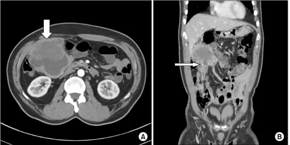
Fig. 2. Computed tomography (CT) image with intravenous contrast shows 9 cm-sized exophytic gallbladder mass that invaded abdominal wall (A, thick arrow) and small bowel (B, thin arrow).
Fig. 3. Coronal T2WI shows 9 cm-sized well-defined and hete- rogenousexophytic mass lesion in gallbladderaccompanied by invasion into surrounding tissues.
Fig. 1. Endoscopic ultrasound shows 6.7 × 7.1 cm-sized huge mass on gallbladder body extended to omentum but not invaded into duodenum.
puted tomography (CT) and magnetic resonance chol- angiopancreatography (MRCP) demonstrated a 9-cm mass lesion that was attached to the small bowel and ab- dominal wall, but had not invaded the liver parenchyma (Figs. 2, 3). The stage of tumor using the standard tu- mor-node-metastasis staging system was T4N0M0. An anomalous union of the pancreatobiliary duct was not demonstrated on MRCP. The 18F-fluorodeoxyglucose
positron emission tomography/CT that was performed to check for distal metastasis revealed a hypermetabolic le- sion (maximum standardized uptake value [SUVmax]
14.5) in the entire gallbladder (Fig. 4).
We performed a simultaneous radical cholecystectomy (S4b + S5 segmentectomy), omentectomyand small bowel segmental resection. We also did dissection of lymph no- des around the hepatoduodenal ligaments and the celiac
Fig. 4. A hypermetabolic lesion (maximum standardized uptake value [SUVmax] 14.5) in entire gallbladder was shown on 18F- fluorodeoxyglucose positron emission tomography/computed tomography.
Fig. 6. (A) Tumor cells showed positive immunoreactivity for smooth muscle actin (A, ×200) and vimentin (B, ×400).
Fig. 5. Higher magnification of spindle cell component displaying marked nuclear atypia and mitosis (62/10 high-power fields [HPFs]; H&E, ×400).
trunk. The size of the tumor was 7.0 × 5.5 cm. Histopatho- logy showed a malignant spindle cell tumor consisting of leiomyosarcoma with marked nuclear atypia and mitosis (62/10 high-power fields [HPFs]) (Fig. 5). The neoplastic cells infiltrated the muscularispropia layers of the small bowel and the soft tissue of the abdominal wall. The tumor cells showed positive immunoreactivity for smooth mus- cle actin (SMA) (Fig. 6A) and vimentin (Fig. 6B). There was negative immunoreativities for the following markers;
caldesmon, cytokeratin (CK) 7, CK19, CK20, CD31, CD34, c-kit, calponin, desmin, myoglobin, HMB45, high molec- ular weight cytokeratin (HMWCK), PanCK, and S100.
Necrosis was observed in 5% of the tumor. Of the 22 re- sected lymph nodes, a metastatic lymph node was not contained.
The patient developed a high fever and tarchycardia on the 3rd postoperative day. A pulmonary thromboemolism in the upper left anterior lobe’s segmental branch was re- vealed in a chest CT therefore, the patients received throm- bolysis followed by heparin anticoagulation therapy. The patient was discharged from the hospital on the 12th post- operative day.
Multiple liver metastasis and seeding metasitasis on the peritoneum were found in an abdominal CT 1 month postoperatively. The patient subsequently underwent ad- juvant chemotherapy composed of mesna, adriamycin,
ifosfamide and dacarbazine (MAID) for 3 months.
DISCUSSION
Sarcomas of the gallbladder are rare and represent about 1.5% of all malignant gallbladder diseases. A variety of tumor types have been described including leiomyo- sarcoma, rhabdomyosarcoma, angiomyosarcoma, Kapo- si’s sarcoma, malignant fibrous histiyocytoma, and syno- vial sarcoma [2]. Leiomyosarcoma of the gallbladder is an especially rare malignant tumor. By 1984, 105 cases of pri- mary sarcomas of the gallbladder had been reported, with primary leiomyosarcomas accounting for 7% of them [3].
The diagnoses were established in accordance with the new World Health Organization classifications for soft tis- sue tumors and the most recent soft tissue criteria published. Leiomyosarcoma is defined as a malignant tu- mor composed of cells showing distinct smooth muscle features. In the macroscopy, leiomyosarcoma typically forms a fleshy mass, with colors varying from grey to white to tan. Large examples often display hemorrhage, necrosis or a cystic change. The typical histopathologic pattern of leiomyosarcoma is that of intersecting, sharply marginated groups of spindle cells. In the immunopheno- type, desmin, h-caldesmon, and SMA were positive in a great majority of leiomyosarcomas. None of these are ab- solutely specific to smooth muscle and positivity for two of these markers were more supportive of leiomyosarco- ma than positivity for one alone. Immunostains may be fo- cally positive on CD34, epithelial membrane antigen (EMA), keratin and S100. A diagnosis should be made on the appropriate morphologic features, not only on the im- munostains. In this case, immunopositive staining was strong for SMA and vimentin but the typical histopatho- logic pattern of leiomyosarcoma had appeared in the hem- atoxylin and eosin stain. Therefore other types of the sar- coma were excluded from the diagnosis.
Leiomyosarcoma is more frequent in women between the ages of 50 and 75 years and usually has a poor prognosis. The presence of gallstones are invariable and the symptoms presented are those of chronic cholecystitis [4]. Histopathologically, the majority of these tumors are-
high grade and display an epithelioid morphology; but cases with features of well-differentiated leiomyosarcoma have been described.
According to the National Comprehensive Cancer Network clinical practice guideline in oncology ver. I.
2011, patients with a resectable intraabdominal sarcoma should undergo immediate surgical treatment with a grossly negative margin and and possible interoperative radiation therapy. The postoperative margin status was the most important factor contributing to long-term dis- ease free survival [5]. Postoperative treatment options were dependent on the surgical outcomes and clinical, or pathological finding following surgery. Postoperative ra- diation therapy should be considered in patients with pathological findings of high grade disease following a negative margin resection (R0 resection) or for micro- scopic positive margins (R1 resection). For patients with unresectable or disseminated recurrences, preoperative RT and/or chemotherapy should be considered after a biopsy. Combination regimens with activity in soft tissue sarcoma include AD (doxorubicin, dacarbazine), AIM (doxorubicin, ifosfamide, mesna), MAID, and so on [6,7].
The single agents include dacarbazine, doxorubicin, epi- rubicin, gemcitabine, ifosfamide, liposomal doxorubicin and temozolomide [8,9].
The prognosis of sarcoma and leiomyosarcomas of the gallbladder is dismal, the five year survival rate being less than 5%. This is due to the fact that at the time of the diag- nosis or surgery. Almost 75% of cases involve the liver [10].
Our patient was also diagnosed at an advanced stage, but he had no distant organ metastasis. Because R0 resection is expected in this case, an aggressive surgical approach was attempted. However, soon after, multiple liver metastasis and peritoneal seeding metastasis were detected in post- operative evaluations. Therefore additional aggressive multimodality treatments such as surgery with chemo- therapy are the only way to increase the survival rate.
In conclusion, the five year survival rate of leiomyo- sarcoas of the gallbladder is less than 5%. However for young and healthy patients with leiomyosarcomas of the gallbladder, aggressive surgical treatment followed by ad- juvant chemotherapy should increase the survival rate de- spite high mortality and morbidity. Because of limited ex-
perience with this disease, there is no consensus about management. Further studies are needed to confirm the benefit of aggressive treatment for patients with leiomyo- sarcoma of the gallbladder. Also surgeons will have to tread very carefully in selection of candidates for surgical treatments.
CONFLICTS OF INTEREST
No potential conflict of interest relevant to this article was reported.
ACKNOWLEDGEMENTS
This work was supported by a 2-year Research Grant of Pusan National University.
REFERENCES
1. Albores-Saavera J, Henson DE, Klimstra DS. Atlas of tu- mor pathology. 3rd series, fascicle 27. Tumors of the gall- bladder, and extrahepatic bile duct and ampulla of water.
Washington DC: Armed Forces Institute of Pathology;
2000.
2. Husain EA, Prescott RJ, Haider SA, Al-Mahmoud RW, Zelger BG, Zelger B, et al. Gallbladder sarcoma: a clin- icopathological study of seven cases from the UK and Austria with emphasis on morphological subtypes. Dig Dis Sci 2009;54:395-400.
3. Newmark H 3rd, Kliewer K, Curtis A, DenBesten L, Enenstein W. Primary leiomyosarcoma of gallbladder seen on computed tomography and ultrasound. Am J Gastroen- terol 1986;81:202-4.
4. Sawan AS, Salama SI. Leiomyosarcoma of the gallbladder.
J King Abdulaziz Univ Med Sci 2010;17:80-8.
5. Anaya DA, Lev DC, Pollock RE. The role of surgical mar- gin status in retroperitoneal sarcoma. J Surg Oncol 2008;98:607-10.
6. Zalupski M, Metch B, Balcerzak S, Fletcher WS, Chapman R, Bonnet JD, et al. Phase III comparison of doxorubicin and dacarbazine given by bolus versus infusion in patients with soft-tissue sarcomas: a Southwest Oncology Group study. J Natl Cancer Inst 1991;83:926-32.
7. Grobmyer SR, Maki RG, Demetri GD, Mazumdar M, Riedel E, Brennan MF, et al. Neo-adjuvant chemotherapy for primary high-grade extremity soft tissue sarcoma. Ann Oncol 2004;15:1667-72.
8. Adjuvant chemotherapy for localised resectable soft-tissue sarcoma of adults: meta-analysis of individual data. Sarco- ma Meta-analysis Collaboration. Lancet 1997;350:1647-54.
9. Antman KH, Elias A. Dana-Farber Cancer Institute studies in advanced sarcoma. Semin Oncol 1990;17(1 Suppl 2):
7-15.
10. Fotiadis C, Gugulakis A, Nakopoulou L, Sechas M.
Primary leiomyosarcoma of the gallbladder. Case report and review of the literature. HPB Surg 1990;2:211-4.
![Fig. 5. Higher magnification of spindle cell component displaying marked nuclear atypia and mitosis (62/10 high-power fields [HPFs]; H&E, ×400).](https://thumb-ap.123doks.com/thumbv2/123dokinfo/5201582.117631/3.918.164.747.883.1108/higher-magnification-spindle-component-displaying-nuclear-atypia-mitosis.webp)
