INTRODUCTION
Thyroid core needle biopsy (CNB) has been proposed as an additional diagnostic method to ultrasound (US)-guided fine needle aspiration (FNA), mainly to overcome the limitations of nondiagnostic or inconclusive cytologic diagnosis,
Ultrasound-Guided Core Needle Biopsy Techniques for Intermediate or Low Suspicion Thyroid Nodules: Which Method is Effective for Diagnosis?
Soo Yeon Hahn, MD
1, Jung Hee Shin, MD
1, Young Lyun Oh, MD
2, Ko Woon Park, MD
11Department of Radiology and Center for Imaging Science, Thyroid Center, Samsung Medical Center, Sungkyunkwan University School of Medicine, Seoul, Korea; 2Department of Pathology, Thyroid Center, Samsung Medical Center, Sungkyunkwan University School of Medicine, Seoul, Korea
Objective: To retrospectively compare the diagnostic performances of two different ultrasound (US)-guided core needle biopsy (CNB) techniques for intermediate or low suspicion thyroid nodules.
Materials and Methods: Between August 2015 and December 2016, two different biopsy techniques were alternatively applied for 248 consecutive thyroid nodules, of which, 140 intermediate or low suspicion thyroid nodules were included in this study. In the first technique, two specimens included nodular tissue, nodular margin, and surrounding normal parenchyma (i.e., marginal target). In the second technique, two specimens were obtained from two different target areas, one for the marginal target and another for the intranodular target. The diagnostic performances of the two techniques to predict neoplasm and malignancy were compared.
Results: CNB was performed on 80 intermediate or low suspicion nodules (57.1%) using the first technique and on 60 (42.9%) using the second technique. The accuracy of the first technique for predicting neoplasm or malignancy was significantly higher than that of the second technique (100% vs. 93.3%, p = 0.032 for predicting neoplasm; 88.8% vs. 75.0%, p = 0.033 for predicting malignancy). The negative predictive value of the first technique for predicting malignancy was also significantly higher than that of the second technique (87.5% vs. 72.7%, p = 0.035).
Conclusion: For intermediate or low suspicion thyroid nodules, US-guided CNB to obtain two specimens with marginal targets is more effective for diagnosing neoplasm or malignancy than is CNB for respective marginal and intranodular targets.
Keywords: Thyroid; Ultrasound; Core needle biopsy; Technique; Thyroid cancer; Thyroid neoplasm
Received November 30, 2018; accepted after revision June 18, 2019.
Corresponding author: Jung Hee Shin, MD, Department of Radiology and Center for Imaging Science, Thyroid Center, Samsung Medical Center, Sungkyunkwan University School of Medicine, 81 Irwon-ro, Gangnam-gu, Seoul 06351, Korea.
• Tel: (822) 3410-2518 • Fax: (822) 3410-0049
• E-mail: [email protected]
This is an Open Access article distributed under the terms of the Creative Commons Attribution Non-Commercial License (https://creativecommons.org/licenses/by-nc/4.0) which permits unrestricted non-commercial use, distribution, and reproduction in any medium, provided the original work is properly cited.
which can lead to repeat FNAs or unnecessary surgery (1, 2). In addition, thyroid CNB has been recently applied to various indications, including the differentiation of rapidly growing thyroid tumors, differentiation of follicular lesions, medullary thyroid carcinoma, calcified thyroid nodules, and degenerating thyroid nodules (3-5). Furthermore, several studies have even suggested the value of CNB as a first-line diagnostic tool for the thyroid (6-9).
In 2016, the Korean Society of Thyroid Radiology revised the consensus statement and recommendations for CNB of thyroid nodules (5). Although these guidelines include 11 recommendations regarding the indications, device, procedure, clinical outcomes, and complications, an obvious CNB protocol has not been clearly established yet. Previously, we suggested the modified CNB technique which targets nodular tissue, the nodular margin, and the surrounding parenchyma simultaneously (i.e., marginal Korean J Radiol 2019;20(10):1454-1461
https://doi.org/10.3348/kjr.2018.0841
target) (10). In our later study that used this modified technique, the CNB protocol for cytologically inconclusive thyroid nodules required at least two specimens with both intranodular and marginal targets (11). Although this protocol was not based on the US findings, recent studies by Ahn et al. (12) and Kim et al. (13) suggest that a modified CNB technique may be useful in the diagnosis of thyroid nodules with specific US features.
In the present study, we attempted to modify our previously proposed CNB protocol to improve its value in clinical application. Therefore, the purpose of this study was to retrospectively compare the diagnostic performances of two different biopsy techniques for obtaining two core specimens, either by marginal only or marginal with intranodular targets, for thyroid nodules with low or intermediate suspicion US patterns.
MATERIALS AND METHODS
The Institutional Review Board of Samsung Medical Center, Sungkyunkwan University School of Medicine, approved this retrospective study and waived the informed consent requirement. However, all US-guided biopsies were conducted after obtaining informed consent from the patients.
Patients
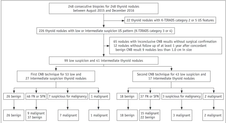
From August 2015 to December 2016, a total of 248 consecutive CNBs for 248 thyroid nodules were performed at our institution. During the period, CNB was used as a first- line biopsy or as a repeat biopsy for nodules with previous inconclusive FNA results. We retrospectively included Korean Thyroid Imaging Reporting and Data System (K-TIRADS) category 4 or 3 nodules with sizes of 1 cm or larger and final diagnoses in this analysis. A final diagnosis of malignancy was made based on surgical pathology. A final diagnosis of benign nodule was made when one of the following conditions was met: surgical diagnosis, concordant benign results after biopsy on at least two occasions, or an initial result of benign biopsy and decreased or stable size at follow-up US more than 1 year later. Then, we excluded 77 nodules without final diagnoses (inconclusive CNB results without surgical confirmation [n = 65] or lack of follow-up of at least 1 year after a concordant benign CNB result [n = 12]); 22 nodules of K-TIRADS categories 2 or 5; and 9 nodules smaller than 1.0 cm in size. Finally, the study included 135 patients (98 women and 37 men; mean age, 48.0 ± 12.4 years) with 41 intermediate and 99 low suspicion thyroid nodules (i.e., K-TIRADS category 4 or 3) (Fig. 1).
248 consecutive biopsies for 248 thyroid nodules between August 2015 and December 2016
65 nodules with inconclusive CNB results without surgical confirmation 12 nodules without follow up of at least 1 year after concordant benign CNB result 9 nodules less than 1.0 cm in size
First CNB technique for 53 low and 27 intermediate suspicion thyroid nodules
26 benign
26 benign 9 malignant 7 malignant 1 malignant 18 benign 3 malignant 2 malignant
37 benign 15 malignant
22 benign 18 benign
46 FN or SFN 7 suspicious for malignancy 1 malignant 37 FN or SFN 3 suspicious for malignancy 2 malignant Second CNB technique for 43 low suspicion and
17 intermediate thyroid nodules 226 thyroid nodules with low or intermediate suspicion US pattern (K-TIRADS category 3 or 4)
99 low suspicion and 41 intermediate thyroid nodules
22 thyroid nodules with K-TIRADS category 2 or 5 US features
Fig. 1. Study flow and outcomes of study population. Numbers represent number of thyroid nodules. CNB = core needle biopsy, FN or SFN = follicular neoplasm or suspicious for follicular neoplasm, K-TIRADS = Korean Thyroid Imaging Reporting and Data System, US = ultrasound
Ultrasonography and Ultrasonography-Guided Core Needle Biopsy Procedures
All thyroid nodules were examined using a 7–12 MHz linear transducer (iU22, Philips Medical Systems, Bothell, WA, USA). The US images were retrospectively reviewed in consensus according to the K-TIRADS by two board- certified radiologists who specialize in thyroid imaging with more than 8 years of experience. The K-TIRADS category was decided as high suspicion (5), intermediate suspicion (4), low suspicion (3), or benign (2) nodule based on the malignancy risk stratified by US patterns composed of integrated solidity, echogenicity, presence of microcalcification, orientation, and margin (14). According to the K-TIRADS, category 3 represents a low suspicion nodule (partially cystic or isohyperechoic nodule without any of the three suspicious US features, with a 3–15%
malignancy risk) and category 4, an intermediate suspicion nodule (solid hypoechoic nodule without any of the three
suspicious US features or partially cystic or isohyperechoic nodule with any of the three suspicious US features, with a 15–50% malignancy risk).
US-guided CNB was performed by one of the two board- certified radiologists who specialize in thyroid imaging with more than 8 years of experience, using a disposable 18-gauge, double-action, spring-activated needle (11 mm penetration with a 7 mm sample notch) (TSK Ace-cut, Create Medic, Yokohama, Japan). With reference to our previous study (11), we routinely obtained two specimens for thyroid core biopsies since August 2015. In particular, throughout this study period, two different biopsy techniques were alternatively applied to the consecutive thyroid nodules.
During the study period, the first and second biopsy techniques were initially performed for each of the 124 cases. Based on our exclusion criteria, 44 cases with the first technique and 64 cases with the second technique were excluded from the final population (Fig. 2). In the first
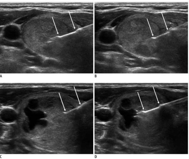
Fig. 2. Two CNB techniques.
In first biopsy technique (A, B), two specimens included nodular tissue, nodular margin, and surrounding normal parenchyma (i.e., margin target). In second technique (C, D), two specimens were obtained from two different target areas, one from marginal target (C) and another from intranodular target (D). Arrows indicate specimen notch of biopsy needle.
A
C
B
D
technique, the two specimens were targeted to include the nodular tissue, nodular margin, and the surrounding normal parenchyma simultaneously (i.e., marginal target) (10). In the second technique, the two specimens were obtained from two different target areas, one from the marginal target and another from the intranodular target. With the second technique, each biopsy specimen was placed in a separate container which contained formaldehyde solution and was labeled for identification of the different biopsy sites. All specimens were submitted for routine pathologic examination.
Histologic Analysis
All biopsy specimens were mounted on separate slides and retrospectively reviewed by one experienced pathologist who was blinded to the original pathology report.
The CNB results were assigned to 1 of 6 categories, according to the research of Jung et al. (15). According to this reporting system, category I corresponds to
nondiagnostic or unsatisfactory; category II, benign lesion;
category III, indeterminate lesion (indeterminate follicular lesion with nuclear atypia/architectural atypia); category IV, follicular neoplasm or suspicious for follicular neoplasm;
category V, suspicious for malignancy; and category VI, malignancy. Then, all thyroid nodules were divided into two subgroups: non-neoplasm (CNB categories I, II, and III) versus neoplasm (CNB categories IV, V, and VI), and malignancy (CNB categories V and VI) versus benign nodules (CNB categories I, II, III, and IV).
Statistical Analysis
Statistical analysis was performed using SPSS, version 23 (IBM Corp., Armonk, NY, USA). For diagnosing thyroid
neoplasm and malignancy, we calculated the sensitivity, specificity, positive predictive value (PPV), negative predictive value (NPV), and the accuracy of CNB. Chi-square and Fisher’s exact tests were used to compare the CNB and final diagnoses between the two different biopsy technique groups and to compare the diagnostic performances of the two different biopsy techniques to predict thyroid neoplasm and malignancy. A statistically significant difference was defined as p < 0.05.
RESULTS
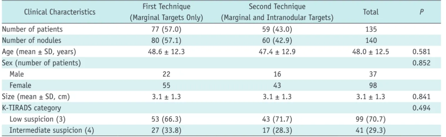
During the study period, CNB was performed for 80 intermediate or low suspicion thyroid nodules (57.1%) using the first technique (marginal targets only), and 60 nodules (42.9%) using the second technique (marginal and intranodular targets) (Table 1). Between the two groups with different biopsy techniques, there were no significant differences in the age and sex of patients, as well as the size and K-TIRADS category of the thyroid nodules.
Table 2 presents the CNB data and final diagnoses according to the biopsy technique. Between the two biopsy techniques, there were no significant differences in the CNB diagnoses (p = 0.686). According to the final diagnosis, the malignancy rate was 21.2% (17/80) in the first technique group and 33.3% (20/60) in the second technique group (p
= 0.177). There were no CNB category I or III diagnoses in this population. For predicting thyroid malignancy, neither technique demonstrated false-positive results in the CNB category V and VI nodules. In addition, neither technique produced a false-negative result in the CNB category II nodules. Among the CNB category IV nodules, false-negative diagnosis was found in nine nodules (five follicular variant
Table 1. Demographic Data of Patients with 140 Thyroid Nodules Clinical Characteristics First Technique
(Marginal Targets Only)
Second Technique
(Marginal and Intranodular Targets) Total P
Number of patients 77 (57.0) 59 (43.0) 135
Number of nodules 80 (57.1) 60 (42.9) 140
Age (mean ± SD, years) 48.6 ± 12.3 47.4 ± 12.9 48.0 ± 12.5 0.581
Sex (number of patients) 0.852
Male 22 16 37
Female 55 43 98
Size (mean ± SD, cm) 3.1 ± 1.3 3.1 ± 1.3 3.1 ± 1.3 0.841
K-TIRADS category 0.494
Low suspicion (3) 53 (66.3) 43 (71.7) 99 (70.7)
Intermediate suspicion (4) 27 (33.8) 17 (28.3) 41 (29.3)
Numbers in parentheses are percentages (%). K-TIRADS = Korean Thyroid Imaging Reporting and Data System, SD = standard deviation
of papillary thyroid carcinoma [FVPTC], three follicular thyroid carcinoma [FTC], and one classic PTC) using the first technique and 15 nodules (nine FTC, five FVPTC, and one classic PTC) using the second technique. For predicting thyroid neoplasm, the first biopsy technique produced neither false-positive nor false-negative diagnoses, while there were three false-negatives (all follicular adenomas) among the CNB category II nodules and one false-positive (benign nodule) CNB category IV nodule.
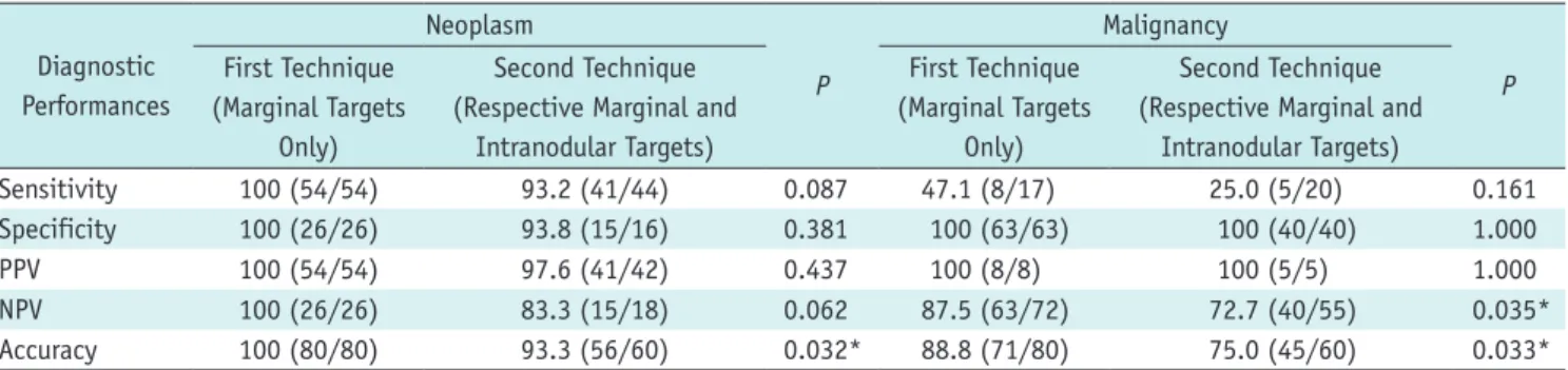
Table 3 summarizes the diagnostic performance of the two different CNB techniques for predicting thyroid neoplasm and malignancy. The accuracy of the first biopsy technique was significantly higher than that of the second biopsy technique (100% vs. 93.3%, p = 0.032 for predicting thyroid neoplasm; 88.8% vs. 75.0%, p = 0.033 for predicting thyroid malignancy). The NPV of the first biopsy technique for predicting thyroid malignancy was significantly higher than that of the second biopsy technique (87.5% vs.
72.7%, p = 0.035). The sensitivity, specificity, and PPV were comparable between the two different biopsy techniques.
In our study, one patient had subsequent perinodular hemorrhage after the first technique and another had minimal intra-parenchymal hemorrhage after the second technique, both of which were resolved by manual compression (1.4%).
DISCUSSION
We previously published results describing the ideal core number for US-guided thyroid biopsy of cytologically inconclusive nodules (11). Our results indicated that US- guided CNB for cytologically inconclusive nodules should obtain at least two cores with intranodular and additional marginal targets. Since completing that study, we routinely obtained two core specimens during thyroid biopsy at our institution. During the study period, the first and second biopsy techniques were alternatively performed for each of the 124 cases by one of the two experienced radiologists.
As described above, two core specimens were obtained targeting the margin only (i.e., the first technique) or Table 2. Data of CNB and Final Diagnoses according to Biopsy Technique
CNB Technique CNB Diagnosis Malignant Rate (%) Final Histology
First technique (marginal targets only)
II, benign follicular lesion (26) 0 (0) Benign nodule (26) IV, follicular neoplasm or suspicious
for FN (46) 9 (19.6) FA (37), FVPTC (5), FTC (3),
classic PTC (1) V, suspicious for malignancy (7) 7 (100) FVPTC (6), PD (1)
VI, malignant (1) 1 (100) Classic PTC (1)
Second technique (respective marginal and intranodular targets)
II, benign follicular lesion (18) 0 (0) Benign nodule (15), FA (3) IV, follicular neoplasm or suspicious
for FN (37) 15 (40.5) FA (21), benign nodule (1), FTC (9), FVPTC (5), classic PTC (1)
V, suspicious for malignancy (3) 3 (100) FVPTC (3)
VI, malignant (2) 2 (100) FVPTC (1), classic PTC (1)
p value 0.686 0.177
Numbers in parentheses indicate number of lesions. CNB = core needle biopsy, FA = follicular adenoma, FN = follicular neoplasm, FTC = follicular thyroid carcinoma, FVPTC = follicular variant of papillary thyroid carcinoma, PD = poorly differentiated thyroid carcinoma, PTC = papillary thyroid carcinoma
Table 3. Diagnostic Performances of Two CNB Techniques for Thyroid Neoplasm and Malignancy
Diagnostic Performances
Neoplasm
P
Malignancy First Technique P
(Marginal Targets Only)
Second Technique (Respective Marginal and
Intranodular Targets)
First Technique (Marginal Targets
Only)
Second Technique (Respective Marginal and
Intranodular Targets)
Sensitivity 100 (54/54) 93.2 (41/44) 0.087 47.1 (8/17) 25.0 (5/20) 0.161
Specificity 100 (26/26) 93.8 (15/16) 0.381 100 (63/63) 100 (40/40) 1.000
PPV 100 (54/54) 97.6 (41/42) 0.437 100 (8/8) 100 (5/5) 1.000
NPV 100 (26/26) 83.3 (15/18) 0.062 87.5 (63/72) 72.7 (40/55) 0.035*
Accuracy 100 (80/80) 93.3 (56/60) 0.032* 88.8 (71/80) 75.0 (45/60) 0.033*
Data are percentages (%). *p < 0.05 was regarded as statistically significant. NPV = negative predictive value, PPV = positive predictive value
targeting the intranodular and marginal tissue (i.e., the second technique, the same protocol proposed in the previous study) (11). The current results indicate that the first technique is better than the second technique for predicting thyroid neoplasm and malignancy. Therefore, thyroid core biopsy should obtain at least two cores with marginal targets, particularly for intermediate or low suspicion thyroid nodules.
K-TIRADS category 5, high suspicion nodules, are usually considered classic PTC (14, 16). Because classic PTC is diagnosed based on their nuclear features (17), most of them can be easily diagnosed by FNA. Therefore, even in the case of performing CNB, classic PTC is usually easily diagnosed from a single biopsy specimen if it is well targeted (intranodular target) (11). However, intranodular tissue retrieved by CNB is sometimes insufficient for differential diagnosis of follicular proliferative lesions because the presence of the capsule around the nodule is a major difference between nodular hyperplasia and follicular neoplasm (17-19). Therefore, tissue retrieval with a marginal target that includes the nodular tissue, nodular margins, and surrounding normal parenchyma is important for differentiating a follicular neoplasm from a non-neoplastic lesion (10). We introduced this theory-based technique as a “modified CNB technique” in our previous study (10).
In the present study, we demonstrated that CNB using marginal targets only (i.e., modified CNB technique only) was more effective for diagnosing cytologically inconclusive thyroid nodules with low or intermediate suspicion US patterns, compared to CNB with intranodular and additional marginal targets. On US, a large proportion of the follicular proliferative lesions, even follicular carcinomas, show low or intermediate suspicion US patterns and are finally categorized as K-TIRADS category 3 or 4 (20-24).
Low or intermediate suspicion US patterns corresponding to K-TIRADS category 3 or 4 are predominant imaging features of FVPTCs and follicular proliferative lesions, such as nodular hyperplasia, follicular adenoma, and follicular carcinoma (20-24). In this study, we found 61 follicular adenomas (43.6%), 42 benign nodules (30.0%), 20 FVPTCs (14.3%), 12 follicular carcinomas (8.6%), four PTCs (2.9%), and one poorly differentiated thyroid carcinoma (0.7%).
For the four PTCs and one poorly differentiated thyroid carcinoma, the K-TIRADS category was “intermediate suspicion, 4.”
Although CNB performed by experienced radiologists is safe and well-tolerated, there are still safety concerns (25-27). The
complication rate in our study was 1.4%, similar to that in other studies, ranging from 0% to 1.5% (28, 29). To minimize the potential for complications, we routinely used color Doppler US when we decided the appropriate biopsy route and immediately applied manual compression on the biopsy site for at least 20 minutes after each biopsy procedure.
Our study has several limitations. First, this study was not completely randomized, although we tried to apply the two different biopsy techniques alternatively to the consecutive thyroid nodules during the study period. However, in our study, no statistical differences were found in the age and sex of patients, the size and K-TIRADS category of thyroid nodules, and the CNB diagnosis between the two different biopsy techniques. Second, the accuracy of CNB depends on several factors, such as the operators’ skills and experience. However, inherent nodule characteristics can be more important factors in inducing unsatisfactory sampling under US guidance, compared to operator factors (30, 31). In this study, we included 140 thyroid nodules with relatively similar characteristics of K-TIRADS category 3 or 4. We believe that this inclusion criterion made it possible to include patients with relatively uniform inherent nodule characteristics. Third, in this study, we used an 11- mm excursion core device which retrieves a core specimen of approximately 0.7 cm in length. If we used a 16- or 22- mm excursion core device, one core specimen including nodular tissue, nodular margin, and surrounding normal parenchyma, could be enough for diagnosis.
In conclusion, for intermediate or low suspicion thyroid nodules, US-guided core biopsy to obtain two specimens with marginal targets only is more effective for diagnosing thyroid neoplasm or malignancy than CNB with respective marginal and intranodular targets.
Conflicts of Interest
The authors have no potential conflicts of interest to disclose.
ORCID iDs Jung Hee Shin
https://orcid.org/0000-0001-6435-7357 Soo Yeon Hahn
https://orcid.org/0000-0002-4099-1617 Young Lyun OH
https://orcid.org/0000-0002-9127-4642 Ko Woon Park
https://orcid.org/0000-0001-9386-5772
REFERENCES
1. Yeon JS, Baek JH, Lim HK, Ha EJ, Kim JK, Song DE, et al.
Thyroid nodules with initially nondiagnostic cytologic results:
the role of core-needle biopsy. Radiology 2013;268:274-280 2. Choi YJ, Baek JH, Suh CH, Shim WH, Jeong B, Kim JK, et al.
Core-needle biopsy versus repeat fine-needle aspiration for thyroid nodules initially read as atypia/follicular lesion of undetermined significance. Head Neck 2017;39:361-369 3. Yoon RG, Baek JH, Lee JH, Choi YJ, Hong MJ, Song DE, et
al. Diagnosis of thyroid follicular neoplasm: fine-needle aspiration versus core-needle biopsy. Thyroid 2014;24:1612- 1617
4. Lee HY, Baek JH, Ha EJ, Park JW, Lee JH, Song DE, et al.
Malignant-looking thyroid nodules with size reduction: core needle biopsy results. Ultrasonography 2016;35:327-334 5. Na DG, Baek JH, Jung SL, Kim JH, Sung JY, Kim KS, et al.;
Korean Society of Thyroid Radiology (KSThR) and Korean Society of Radiology. Core needle biopsy of the thyroid: 2016 consensus statement and recommendations from Korean Society of Thyroid Radiology. Korean J Radiol 2017;18:217- 237
6. Suh CH, Baek JH, Lee JH, Choi YJ, Kim JK, Sung TY, et al. The role of core-needle biopsy as a first-line diagnostic tool for initially detected thyroid nodules. Thyroid 2016;26:395-403 7. Kim HC, Kim YJ, Han HY, Yi JM, Baek JH, Park SY, et al. First-
line use of core needle biopsy for high-yield preliminary diagnosis of thyroid nodules. AJNR Am J Neuroradiol 2017;38:357-363
8. Suh CH, Baek JH, Choi YJ, Kim TY, Sung TY, Song DE, et al.
Efficacy and safety of core-needle biopsy in initially detected thyroid nodules via propensity score analysis. Sci Rep 2017;7:8242
9. Chung SR, Suh CH, Baek JH, Choi YJ, Lee JH. The role of core needle biopsy in the diagnosis of initially detected thyroid nodules: a systematic review and meta-analysis. Eur Radiol 2018;28:4909-4918
10. Han S, Shin JH, Hahn SY, Oh YL. Modified core biopsy technique to increase diagnostic yields for well-circumscribed indeterminate thyroid nodules: a retrospective analysis. AJNR Am J Neuroradiol 2016;37:1155-1159
11. Hahn SY, Shin JH, Oh YL. What is the ideal core number for ultrasonography-guided thyroid biopsy of cytologically inconclusive nodules? AJNR Am J Neuroradiol 2017;38:777- 781
12. Ahn S, Jung S, Kim JY, Shin JH, Hahn SY, Oh YL. Evaluation of modified core-needle biopsy in the diagnosis of thyroid nodules. Korean J Radiol 2018;19:656-664
13. Kim JH, Na DG, Lee H. Ultrasonographic echogenicity and histopathologic correlation of thyroid nodules in core needle biopsy specimens. Korean J Radiol 2018;19:673-681
14. Shin JH, Baek JH, Chung J, Ha EJ, Kim JH, Lee YH, et al.;
Korean Society of Thyroid Radiology (KSThR) and Korean Society of Radiology. Ultrasonography diagnosis and imaging-
based management of thyroid nodules: revised Korean Society of Thyroid Radiology consensus statement and recommendations. Korean J Radiol 2016;17:370-395 15. Jung CK, Min HS, Park HJ, Song DE, Kim JH, Park SY, et al.
Pathology reporting of thyroidcore needle biopsy: a proposal of the Korean Endocrine Pathology Thyroid Core Needle Biopsy Study Group. J Pathol Transl Med 2015;49:288-299
16. American Thyroid Association (ATA) Guidelines Taskforce on Thyroid Nodules and Differentiated Thyroid Cancer, Cooper DS, Doherty GM, Haugen BR, Kloos RT, Lee SL, Mandel SJ, et al.
Revised American Thyroid Association management guidelines for patients with thyroid nodules and differentiated thyroid cancer. Thyroid 2009;19:1167-1214
17. Cibas ES, Ali SZ; NCI Thyroid FNA State of the Science Conference. The Bethesda System For Reporting Thyroid Cytopathology. Am J Clin Pathol 2009;132:658-665 18. Baloch ZW, Livolsi VA. Follicular-patterned lesions of the
thyroid: the bane of the pathologist. Am J Clin Pathol 2002;117:143-150.
19. Schreiner AM, Yang GC. Adenomatoid nodules are the main cause for discrepant histology in 234 thyroid fine-needle aspirates reported as follicular neoplasm. Diagn Cytopathol 2012;40:375-379
20. Hong MJ, Na DG, Baek JH, Sung JY, Kim JH. Cytology- ultrasonography risk-stratification scoring system based on fine-needle aspiration cytology and the Korean-Thyroid Imaging Reporting and Data System. Thyroid 2017;27:953- 959
21. Rago T, Di Coscio G, Basolo F, Scutari M, Elisei R, Berti P, et al. Combined clinical, thyroid ultrasound and cytological features help to predict thyroid malignancy in follicular and Hupsilonrthle cell thyroid lesions: results from a series of 505 consecutive patients. Clin Endocrinol (Oxf) 2007;66:13-20 22. Jeh SK, Jung SL, Kim BS, Lee YS. Evaluating the degree of
conformity of papillary carcinoma and follicular carcinoma to the reported ultrasonographic findings of malignant thyroid tumor. Korean J Radiol 2007;8:192-197
23. Sillery JC, Reading CC, Charboneau JW, Henrichsen TL, Hay ID, Mandrekar JN. Thyroid follicular carcinoma: sonographic features of 50 cases. AJR Am J Roentgenol 2010;194:44-54 24. Hahn SY, Shin JH, Oh YL, Kim TH, Lim Y, Choi JS. Role of
ultrasound in predicting tumor invasiveness in follicular variant of papillary thyroid carcinoma. Thyroid 2017;27:1177- 1184
25. Park KT, Ahn SH, Mo JH, Park YJ, Park DJ, Choi SI, et al.
Role of core needle biopsy and ultrasonographic finding in management of indeterminate thyroid nodules. Head Neck 2011;33:160-165
26. Renshaw AA, Pinnar N. Comparison of thyroid fine- needle aspiration and core needle biopsy. Am J Clin Pathol 2007;128:370-374
27. Screaton NJ, Berman LH, Grant JW. US-guided core-needle biopsy of the thyroid gland. Radiology 2003;226:827-832 28. Ha EJ, Baek JH, Lee JH, Kim JK, Choi YJ, Sung TY, et al.
Complications following US-guided core-needle biopsy for thyroid lesions: a retrospective study of 6,169 consecutive patients with 6,687 thyroid nodules. Eur Radiol 2017;27:1186- 1194
29. Ha EJ, Suh CH, Baek JH. Complications following ultrasound- guided core needle biopsy of thyroid nodules: a systematic review and meta-analysis. Eur Radiol 2018;28:3848-3860 30. Nyquist GG, Tom WD, Mui S. Automatic core needle biopsy: a
diagnostic option for head and neck masses. Arch Otolaryngol Head Neck Surg 2008;134:184-189
31. Na DG, Kim JH, Sung JY, Baek JH, Jung KC, Lee H, et al.
Core-needle biopsy is more useful than repeat fine-needle aspiration in thyroid nodules read as nondiagnostic or atypia of undetermined significance by the Bethesda system for reporting thyroid cytopathology. Thyroid 2012;22:468-475