Increased Serum Cathepsin K in Patients with Coronary Artery Disease
Xiang Li,
1Yuzi Li,
1Jiyong Jin,
1Dehao Jin,
2Lan Cui,
1Xiangshan Li,
3Yanna Rei,
1,4Haiying Jiang,
5Guangxian Zhao,
1Guang Yang,
1Enbo Zhu,
1Yongshan Nan,
4and Xianwu Cheng
1,6,71Department of Cardiology, 2Intervention Laboratory, 3Central Laboratory, and 4Anesthesiology, Yanbian University Hospital, Yanji, Jilin P.R., China;
5Department of Physiology and Pathophysiology, Yanbian University Medical College, Yanji, Jilin P.R., China;
6Department of Cardiology, Nagoya University Graduate School of Medicine, Nagoya, Japan;
7Department of Internal Medicine, Kyung Hee University Hospital, Seoul, Korea.
Received: September 13, 2013 Revised: October 5, 2013 Accepted: October 11, 2013
Co-corresponding authors: Dr. Lan Cui, Department of Cardiology,
Yanbian University Hospital, Yanjishi 133000, Jilin Province, China.
Tel: 86-433-2660063, Fax: 86-433-2513510 E-mail: lancui@ybu.edu.cn and Dr. Xianwu Cheng,
Department of Cardiology,
Nagoya University Graduate School of Medicine, 65 Tsuruma-cho, Showa-ku, Nagoya 466-8550, Japan.
Tel: 81-52-744-2364, Fax: 81-52-744-2371 E-mail: chengxw0908@163.com
∙ The authors have no financial conflicts of interest.
© Copyright:
Yonsei University College of Medicine 2014 This is an Open Access article distributed under the terms of the Creative Commons Attribution Non- Commercial License (http://creativecommons.org/
licenses/by-nc/3.0) which permits unrestricted non- commercial use, distribution, and reproduction in any medium, provided the original work is properly cited.
Purpose: Cathepsin K is a potent collagenase implicated in human and animal atherosclerosis-based vascular remodeling. This study examined the hypothesis that serum CatK is associated with the prevalence of coronary artery disease (CAD). Materials and Methods: Between January 2011 and December 2012, 256 consecutive subjects were enrolled from among patients who underwent coronary angiography and percutaneous coronary intervention treatment. A total of 129 age- matched subjects served as controls. Results: The subjects’ serum cathepsin K and high sensitive C-reactive protein (hs-CRP) and high-density lipoprotein cholester- ol were measured. The patients with CAD had significantly higher serum cathep- sin K levels compared to the controls (130.8±25.5 ng/mL vs. 86.9±25.5 ng/mL, p<0.001), and the patients with acute coronary syndrome had significantly higher serum cathepsin K levels compared to those with stable angina pectoris (137.1±
26.9 ng/mL vs. 102.6±12.9 ng/mL, p<0.001). A linear regression analysis showed that overall, the cathepsin K levels were inversely correlated with the high-density lipoprotein levels (r=-0.29, p<0.01) and positively with hs-CRP levels (r=0.32, p<0.01). Multiple logistic regression analyses shows that cathepsin K levels were independent predictors of CAD (odds ratio, 1.76; 95% confidence interval, 1.12 to 1.56; p<0.01). Conclusion: These data indicated that elevated levels of cathepsin K are closely associated with the presence of CAD and that circulating cathepsin K serves a useful biomarker for CAD.
Key Words: Cathepsin K, coronary artery disease, biomarker, high sensitive C- reactive protein, myocardial ischemia
INTRODUCTION
Cathepsins are a group of lysosomal cysteine proteases belonging to the papain fam- ily.1 Recent evidence has implicated the role of cathepsins in the pathogenesis of ath- erosclerosis-based cardiovascular disease.2-4 The protein levels of cathepsins S and K were increased in atherosclerotic plaques and injury-related neointimal lesions in an-
branch block) and a history of prolonged chest pain.24 UAP was diagnosed by typical chest pain at rest in the 24 h prior to the subject’s arrival at the hospital, depressed ST
≥0.1 mV, and/or T-wave inversion on an electrocardiogram but a normal creatine kinase-MB level.
SAP was diagnosed as an invariable character of exer- tional chest pain for 3 months before the subject went to the hospital (with “invariable” meaning the same degree of ex- ertion and excitation provocation and the same location, quality, and 3- to 5-min duration), which was relieved by rest or nitroglycerin.
A total of 129 subjects who showed no evidence of cardio- vascular disease-defined as no typical chest pain on exertion, no myocardial infarction (MI) by history or electrocardio- gram, negative exercise test, and no significant luminal nar- rowing of the coronary arteries were recruited as non-CAD controls. Additionally, hypertension was defined as a systolic blood pressure >140 mm Hg, a diastolic blood pressure >90 mm Hg, and/or having received treatment for hypertension.
Diabetes mellitus was confirmed when the subject had hemo- globin A1c (HbA1c) levels ≥6.5%, a fasting plasma glucose concentration >126 mg/dL, and/or a history of any anti-hyper- glycemic medication or a previous diagnosis of diabetes.
Patients were excluded if they had prior evidence of con- genital heart disease, end-stage renal disease with mainte- nance hemodialysis, primary valvular disease, cardiomyop- athy, or secondary cardiac muscle disease caused by any known systemic condition. The study protocol was approved by the ethics committee of Yanbian University Hospital, and written informed consent was obtained from all patients and control subjects.
A blood sample was isolated prior to PCI, and HbA1c, high sensitive C-reactive protein (hs-CRP), and various lipids were measured. The gender, age, body mass index (BMI), systolic and diastolic blood pressures, medication history, and smoking history were recorded for each subject.
Laboratory examination
Human serum cathepsin K levels were determined by using ELISA kits (Biomedica Gruppe, Biomedica Medizinproduk- te, Vienna, Austria) in duplicate. Serum levels of creatinine, low-density lipoprotein (LDL), high-density lipoprotein (HDL), hs-CRP, and HbA1c were measured at the clinical laboratory of Yanbian University Hospital (Clinical Labora- tory, Yanji, China). Serum cathepsin K values are expressed as ng/mL, and the inter-assay and intra-assay coefficients of variation were <8%.
imals and humans.5-8 Among the same lines of evidence, the genetic and pharmacological inhibition of cathepsins S and K prevented atherosclerotic plaque growth and cardiovascular repair in several animal models.9-12 Thus, cathepsins repre- sent a viable target to alleviate vascular dysfunction and re- modeling in response to multiple pathogenic stresses.
Among the cathepsin family members, cathepsin K has been shown to be one of the most potent mammalian colla- genases in vivo and in vitro.11,13-15 Data from our group and others have shown that cathepsin K abounds in vascular cells (including smooth muscle cells and endothelial cells) and infiltrated macrophages of human and animal athero- genic lesions.3,16-18 The ablation of cathepsin K was shown to ameliorate obesity- and pressure overload-related cardiac dysfunction and remodeling.19,20
We reported that cathepsin K is overexpressed in the intra- coronary artery of hypertensive heart failure tissues.21 Several clinical studies have shown that patients with atherosclerosis- related diabetes, aneurysm and chronic kidney disease had increased levels of serum cathepsins S or L.15,22,23 These data suggested that cathepsins levels are associated with athero- sclerosis-based cardiovascular disease. However, there is limited information regarding the relationship between cir- culating cathepsin K and coronary artery disease (CAD). In this study, we tested this relationship in patients with CAD and non-CAD control subjects to explore the relationship between circulating cathepsin K and clinical presentations, and we tried to identify useful blood biomarkers suggestive of CAD with coronary vulnerable plaques.
MATERIALS AND METHODS
Study population and definition
In total, 256 consecutive patients with CAD who under- went percutaneous coronary intervention (PCI) with drug- eluting stent implantation between January 2011 and De- cember 2012 at Yanbian University Hospital (Yanji, China) were considered for inclusion in this study. The patients with CAD were subgrouped into those with stable angina pectoris (SAP; n=50), and those with unstable angina pec- toris (UAP) and acute myocardial infarction (AMI) (UAP+
AMI, n=206) by symptoms and clinical examinations.
The diagnosis of AMI was based on the elevation of car- diac biomarkers (at least one positive biomarker: creatine kinase-MB or troponin T) and an electrocardiogram indica- tive of new ischemia (new ST-T change or new left bundle
test. The hs-CRP concentrations were logarithmically trans- formed because the data showed a skewed distribution. If the homogeneity of variance assumption was violated, the nonparametric Kruskal-Wallis test was used instead. The factors that related at the p<0.1 level were selected as inde- pendent variable candidates for a multiple logistic regres- sion analysis, which was used to evaluate the independent contributions of clinical parameters to CAD. Correlation coefficients were calculated using a linear regression analy- sis. StatFlex (version 6.0; Artech, Osaka, Japan) was used for all statistical analyses. p values of less than 0.05 were considered significant.
RESULTS
Baseline clinical characteristics
The baseline characteristics of CAD patients (n=256) and control subjects (n=129) are displayed in Table 1. There were Quantitative coronary angiogram (QCA)
Coronary angiography was obtained prior to PCI treatment.
Angiography showing the maximal degree of stenosis was adapted for the quantitative coronary angiogram (QCA).
The QCA analysis was performed using a contour detection minimum cost algorithm (DSA Artis Zee Biplane; Siemens, Erlangen, Germany). All CAD patients showed severe cor- onary artery stenosis defined as present stenosis ≥50% di- ameter of at least one major artery. The reference segment dia. was averaged from 5-mm long angiographically normal segments proximal to the lesion; if a normal proximal seg- ment could not be identified, a distal angiographically nor- mal segment was analyzed as described.25
Statistical analysis
Data are presented as means±standard deviation. Compari- sons of categorical baseline characteristics were made using the chi-square test. Comparisons of the continuous baseline characteristics were made using an unpaired Student’s t-
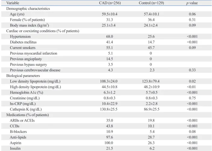
Table 1. Demographic and Clinical Variables of Control and CAD Patients
Variable CAD (n=256) Control (n=129) p value
Demographic characteristics
Age (yrs) 59.5±10.4 57.4±10.1 0.06
Female (% of patients) 31.3 36.4 0.31
Body mass index (kg/m2) 25.1±3.4 24.1±2.4 0.09
Cardiac or coexisting conditions (% of patients)
Hypertension 68.0 25.6 <0.001
Diabetes mellitus 41.4 14.7 <0.001
Current smokers 55.1 45.7 0.09
Previous myocardial infarction 5.1 0
Previous angioplasty 14.5 0
Previous bypass surgery 3.5 0
Previous cerebrovascular disease 4.3 2.3 0.33
Biological parameters
Low density lipoprotein (mg/dL) 108.3±24.0 123.8±79.4 0.02
High density lipoprotein (mg/dL) 44.5±10.8 48.2±10.9 <0.01
Hemoglobin A1c (%) 6.3±1.2 5.7±0.5 <0.001
Creatinine (mg/dL) 0.8±0.3 0.8±0.3 0.75
hs-CRP (mg/dL) 10.4±22.9 2.2±2.8 <0.001
Cathepsin K (ng/dL) 130.8±25.5 86.9±25.5 <0.001
Medications (% of patients)
ARBs or ACEIs 35.0 19.8 <0.001
CCBs 43.8 10.1 <0.001
B-blockers 10.9 5.4 0.08
Anti-lipids 97.6 28.7 <0.001
Aspirin 100.0 26.3 <0.001
Insulin 21.5 6.2 <0.001
hs-CRP, high sensitive C-reactive protein; ARBs, angiotensin II receptor blockers; ACEI, angiotensin converting enzyme inhibitor; CCBs, calcium channel blockers; CAD, coronary artery disease.
Values are expressed as mean±SD or number (%).
patients undergoing treatment with antilipid, antidiabetic, antihypertensive, or antiplatelet medications were higher than in the control subjects.
Table 2 shows the clinical characteristics of the UAP+
AMI (called hereafter the “acute coronary syndrome”) group and SAP group. There were also no significant differences in no significant differences in age, gender, or BMI (p>0.05 for
all comparisons). The patients with CAD had a significant- ly higher prevalence of diabetes and hypertension (p<0.01);
they were also more likely to have had cerebrovascular dis- ease or myocardial infarction or to have undergone a coro- nary bypass graft or angioplasty. The frequencies of CAD
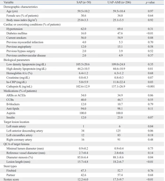
Table 2. Demographic and Clinical Variables of SAP and UAP-AMI
Variable SAP (n=50) UAP-AMI (n=206) p value
Demographic characteristics
Age (yrs) 59.5±10.2 59.5±10.4 0.97
Female sex (% of patients) 30.6 34.0 0.64
Body mass index (kg/m2) 25.0±3.3 25.1±3.5 0.92
Cardiac or coexisting conditions (% of patients)
Hypertension 62.0 69.4 0.31
Diabetes mellitus 16.0 47.6 <0.01
Current smokers 56.0 54.9 0.88
Previous myocardial infarction 4.0 5.3 0.70
Previous angioplasty 12.0 15.1 0.58
Previous bypass surgery 2.0 3.9 0.52
Previous cerebrovascular disease 2.0 4.9 0.37
Biological parameters
Low density lipoprotein (mg/dL) 105.5±20.6 109.0±24.8 0.35
High density lipoprotein (mg/dL) 44.2±10.5 44.6±10.9 0.81
Hemoglobin A1c (%) 6.4±1.2 6.3±1.2 0.68
Creatinine (mg/dL) 0.8±0.3 0.8±0.3 0.87
hs-CRP (mg/dL) 5.0±5.9 11.8±22.4 0.03
Cathepsin K (ng/mL) 102.6±12.9 137.1±26.9 <0.001
Medications (% of patients)
ARBs or ACEIs 34.0 34.9 0.86
CCBs 40.0 44.7 0.55
B-blockers 12.0 10.7 0.79
Anti-lipids 94.0 98.1 0.11
Aspirin 100.0 100.0
Insulin 12.0 23.8 0.07
Target lesion location
Left main artery 1 6 0.84
Left anterior descending artery 34 125 0.06
Left circumflex artery 12 83 0.18
Right coronary artery 16 91 0.48
QCA of target lesions
Minimal lumen diameter (mm) 0.9±0.2 0.9±0.4 0.75
Reference vessel diameter (mm) 2.7±0.4 2.8±0.6 0.14
Diameter stenosis (%) 85.8±6.4 88.1±8.6 0.04
Lesion length (mm) 15.7±4.8 18.2±6.7 <0.01
Stent types
Firebird 47.3 52.7 0.76
Partner 42.6 57.4 0.68
Syntax score 12.2±6.0 17.5±9.7 <0.01
QCA, quantitative coronary angiography; ARBs, angiotensin II receptor blockers; ACEI, angiotensin converting enzyme inhibitor; CCBs, calcium channel blockers; hs-CRP, high sensitive C-reactive protein; SAP, stable angina pectoris; UAP, unstable angina pectoris; AMI, acute myocardial infarction.
Values are expressed as mean±SD or number (%).
(r=0.32, p<0.001), whereas they were negatively correlated with the HDL levels (r=-0.29, p<0.001).
Independence of predictors of CAD
The results of the logistic regression analysis for CAD are shown in Table 3. In the single logistic regression analysis, diabetes mellitus, hypertension, cathepsin K, hs-CRP, LDL cholesterol, and HDL cholesterol were significantly associ- ated with CAD (Table 3). The multiple logistic regression analysis with age, BMI, diabetes mellitus, hypertension, ca- thepsin K, hs-CRP, LDL cholesterol, and HDL cholesterol revealed that the diabetes mellitus [odds ratio (OR), 7.69;
95% confidence interval (CI), 1.99 to 45.10; p<0.01], hy- pertension (OR, 9.88; 95% CI, 3.45 to 66.95; p<0.01), hs- CRP (OR, 4.23; 95% CI, 2.99 to 7.68; p<0.01), HDL (OR, 0.89; 95% CI, 0.81 to 0.96; p<0.05) and cathepsin K (OR, 1.76; 95% CI, 1.12 to 1.56; p<0.01) levels were significant- ly correlated with CAD (Table 3).
DISCUSSION
This study provides evidence that elevated levels of circulat- ing cathepsin K are independently associated with the preva- lence of CAD after adjusting for conventional CAD risk fac- tors including diabetes-related factors, blood pressure, dyslipidemia and BMI. Excess inflammation is strongly as- sociated with an increased risk of cardiovascular disease.26,27 Cathepsin K mRNA and proteins are highly expressed in hu- man and animal atherosclerotic plaques and injury-induced neointimal lesions,5,7,12 and the expression of the endoge- nous inhibitor of cysteine proteases, cystatin C, in the plas- age, gender, or BMI (p>0.05 for all comparisons). With the
exception of the prevalence of diabetes, there were no sig- nificant differences in clinical histories or medications be- tween the two groups (p>0.05 for all comparisons).
Atherosclerotic lesion location and characteristics As shown in Table 2, with the exception of the diameter stenosis, lesion length, and Syntax score, there were no sig- nificant differences in target lesion location or QCA results of the target lesions in the SAP and UAP+AMI groups (p>0.05 for all comparisons).
Circulating biomarkers
As shown in Table 1, compared to the control group, the pa- tients with CAD had significantly higher levels of serum ca- thepsin K (130.8±25.5 ng/mL vs. 86.9±25.5 ng/mL, p<
0.001). The levels of hs-CRP (10.4±22.9 mg/mL vs. 2.2±2.8 mg/mL, p<0.001) and HbA1c (6.3±1.2% vs. 5.7±0.5%, p<
0.001) were also significantly higher and the LDL cholester- ol (108.3±24.0 mg/mL vs. 123.8±79.4 mg/mL, p=0.02) and HDL cholesterol (44.5±10.8 mg/mL vs. 48.2±10.9 mg/mL, p<0.01) levels were significantly lower in the CAD group than in the control group, but there was no significant dif- ference in the creatinine levels. Moreover, patients with UAP+AMI had significantly higher hs-CRP (11.8±22.4 mg/mL vs. 5.0±5.9 mg/mL, p=0.03) and CatK (137.1±26.9 ng/mL vs. 102.6±12.9 ng/mL, p<0.001) levels than did the SAP patients. However, there were no significant differenc- es in the, LDL, HDL, HbA1c, or creatinine levels between the two CAD subgroups.
The linear regression analysis showed that the cathepsin K levels were positively correlated with the hs-CRP levels
Table 3. Independent Predictors of CAD According to Multivariable Logistic Regression Analysis
Single Multiple
Odds ratio
estimate 95% CI p value Odds ratio
estimate 95% CI p value
Age (yrs) 1.13 0.88--1.03 0.41 1.07 0.82--1.06 0.88
Gender 0.70 0.34--2.97 0.59
BMI (kg/m2) 0.69 0.54--0.83 <0.01 0.77 0.81--0.93 0.12
Diabetes mellitus (%) 6.08 2.32--69.16 0.02 7.69 1.99--45.10 <0.01
Hypertension (%) 8.96 1.78--54.15 <0.05 9.88 3.45--66.95 <0.01
LDL cholesterol (mg/dL) 0.67 0.68--1.11 0.78
HDL cholesterol (mg/dL) 0.92 0.79--0.92 <0.05 0.89 0.81--0.96 <0.05
hs-CRP 1.99 1.78--6.46 <0.05 4.23 2.99--7.68 <0.01
Cathepsin K 1.32 1.25--1.77 <0.001 1.76 1.12--1.56 <0.01
CI, confidence interval; CAD, coronary artery disease; BMI, body mass index; LDL, low-density lipoprotein; HDL, high-density lipoprotein; hs-CRP, high sen- sitive C-reactive protein.
Multiple regression model includes all variables at baseline with p<0.05 by univariable analysis.
the patients with UAP and AMI had higher levels of hs- CRP than did the SAP subjects. We detected higher cathep- sin K levels in patients with UAP and AMI compared to those with SAP. Moreover, our data revealed a significant positive correlation between cathepsin K and hs-CRP in all subjects. Another study highlighted the cathepsins-mediat- ed metabolism of the major components of the vascular ex- tracellular matrix, including the fibrous cap of atheroscle- rotic plaques.3
The activation of cathepsins S and K by monocyte/mac- rophages has been shown to promote plaque instability.18,39 Animal studies demonstrated that the genetic and pharma- cological inhibition of cathepsin K alleviates the extracellu- lar matrix metabolism of the atherosclerotic lesion and pre- vents plaque disruption.18,39,40 Thus, cathepsin K production by activated inflammatory cells and its release into the cir- culation appear to be strongly linked to the plaque instabili- ty and plaque rupture associated with local inflammatory processes within the vascular wall. On the other hand, the most extensively studied molecular candidates for rupture- producing proteases are the matrix metalloproteinases.41 It was shown that a plaque rupture being present in the culprit lesion was closely related to the high levels of metallopro- teinase-9 in patients with AMI and UAP.42 Therefore, an in- creased cathepsin K level together with the evaluation of the matrix metalloproteinase concentration may serve as a noninvasive method of documenting and monitoring coro- nary inflammatory atherosclerotic plaque vulnerability dur- ing acute coronary syndrome.
The involvement of cathepsin K in ApoB-100 proteolytic modification is likely to contribute to the extracellular LDL particle aggregation, lipid droplet formation, and LDL re- tention of arterial proteoglycans.43 Cathepsin K deficiency resulted in an increase in cholesterol ester storage in macro- phages of ApoE-/- bone marrow, which was stored in large lysosomal compartments.18 Our present data revealed a di- rect negative correlation between cathepsin K and HDL in all subjects. Cathepsin K has been shown to control choles- terol efflux by the degradation of preβ-HDL and apoA-1.44 Thus, cathepsin K-mediated cholesterol uptake and/or ef- flux could represent a common mechanism in the macro- phage-derived foam cell formation and plaque growth.
Study limitations
Here, we have to point out several our study limitations.
First, the sample size was too small to restrict the power for proving relationships and differences and to conduct the ma and the arterial tissues is decreased in atherosclerosis-
based arterial and vein disorders such as aneurysms and varicose veins.6 Thus, these observations indicate that ca- thepsin K could be a useful marker of CAD associated with inflammation.
Recent studies highlighted that cysteinyl cathepsin K is the most abundant and important protease synthesized by the cardiovascular cells and inflammatory cells, and that it is relevant to atherosclerosis-related cardiovascular disease and its implications.3,28 However, few studies have exam- ined peripheral blood cathepsin K levels in humans with or without CAD. Our present data show that the levels of se- rum cathepsin K were higher in the patients with CAD than in the control subjects. Our multivariable logistic regression analysis demonstrated that serum cathepsin K levels were independent predictors of CAD. Together with the finding that serum cathepsins S and L were increased in patients with coronary artery extasia or atherosclerotic stenosis,15,29,30 our results suggest that these cysteinyl cathepsins may partici- pate in coronary artery restenosis and aneurysm.
Elevated circulating cathepsin K CAD patients with se- vere stenosis is consistent with the notion that coronary ar- tery stenosis is a protease-mediated proteolysis involving such as cysteinyl cathepsins K, which is shear stress-sensi- tive and increased during atherosclerotic lesion and neointi- mal formation.31,32 Due to the characteristics of the coronary artery anatomy, i.e., plural branches and restriction, oscilla- tory blood flow during cardiac motion cycle, and collateral blood counter-flow during coronary vessel occlusion, it seems to be that cathepsin K might by secreted by the in- flammatory lesions of the coronary artery into the circula- tion. An increased expression of cathepsin K mRNA and/or protein in in vivo and in vitro cultured vascular endothelium and smooth muscles in response to inflammatory cytokines was reported,5,7,16 suggesting that these vascular cell types may also contribute to increased serum cathepsin K in pa- tients with CAD.
It is well established that atherosclerotic plaque instability and rupture induced by inflammation are the major mecha- nisms of acute coronary syndrome or an acute clinical event.33,34 Accumulating evidence indicates that elevated lev- els of CRP, an acute-phase protein widely used as a marker of inflammation, is predictive of the risk of first acute coro- nary syndrome and acute myocardial infarction.35,36 The se- verity of the superficial inflammation seen in atherosclerot- ic lesions has been implicated as a significant correlate of plaque instability and rupture.33,37,38 Here we observed that
opportunities. EMBO J 2001;20:4629-33.
2. Cheng XW, Shi GP, Kuzuya M, Sasaki T, Okumura K, Murohara T. Role for cysteine protease cathepsins in heart disease: focus on biology and mechanisms with clinical implication. Circulation 2012;125:1551-62.
3. Cheng XW, Huang Z, Kuzuya M, Okumura K, Murohara T. Cys- teine protease cathepsins in atherosclerosis-based vascular disease and its complications. Hypertension 2011;58:978-86.
4. Reiser J, Adair B, Reinheckel T. Specialized roles for cysteine ca- thepsins in health and disease. J Clin Invest 2010;120:3421-31.
5. Sukhova GK, Shi GP, Simon DI, Chapman HA, Libby P. Expres- sion of the elastolytic cathepsins S and K in human atheroma and regulation of their production in smooth muscle cells. J Clin Invest 1998;102:576-83.
6. Shi GP, Sukhova GK, Grubb A, Ducharme A, Rhode LH, Lee RT, et al. Cystatin C deficiency in human atherosclerosis and aortic aneurysms. J Clin Invest 1999;104:1191-7.
7. Cheng XW, Kuzuya M, Sasaki T, Arakawa K, Kanda S, Sumi D, et al. Increased expression of elastolytic cysteine proteases, ca- thepsins S and K, in the neointima of balloon-injured rat carotid arteries. Am J Pathol 2004;164:243-51.
8. Burns-Kurtis CL, Olzinski AR, Needle S, Fox JH, Capper EA, Kelly FM, et al. Cathepsin S expression is up-regulated following balloon angioplasty in the hypercholesterolemic rabbit. Cardio- vasc Res 2004;62:610-20.
9. Sun M, Chen M, Liu Y, Fukuoka M, Zhou K, Li G, et al. Cathep- sin-L contributes to cardiac repair and remodelling post-infarction.
Cardiovasc Res 2011;89:374-83.
10. Sun J, Sukhova GK, Zhang J, Chen H, Sjöberg S, Libby P, et al.
Cathepsin L activity is essential to elastase perfusion-induced ab- dominal aortic aneurysms in mice. Arterioscler Thromb Vasc Biol 2011;31:2500-8.
11. Sun J, Sukhova GK, Zhang J, Chen H, Sjöberg S, Libby P, et al.
Cathepsin K deficiency reduces elastase perfusion-induced ab- dominal aortic aneurysms in mice. Arterioscler Thromb Vasc Biol 2012;32:15-23.
12. Cheng XW, Murohara T, Kuzuya M, Izawa H, Sasaki T, Obata K, et al. Superoxide-dependent cathepsin activation is associated with hypertensive myocardial remodeling and represents a target for angiotensin II type 1 receptor blocker treatment. Am J Pathol 2008;173:358-69.
13. Li Z, Hou WS, Brömme D. Collagenolytic activity of cathepsin K is specifically modulated by cartilage-resident chondroitin sul- fates. Biochemistry 2000;39:529-36.
14. Novinec M, Grass RN, Stark WJ, Turk V, Baici A, Lenarcic B. In- teraction between human cathepsins K, L, and S and elastins:
mechanism of elastinolysis and inhibition by macromolecular in- hibitors. J Biol Chem 2007;282:7893-902.
15. Liu J, Sukhova GK, Yang JT, Sun J, Ma L, Ren A, et al. Cathepsin L expression and regulation in human abdominal aortic aneurysm, ath- erosclerosis, and vascular cells. Atherosclerosis 2006;184:302-11.
16. Cheng XW, Kuzuya M, Nakamura K, Di Q, Liu Z, Sasaki T, et al.
Localization of cysteine protease, cathepsin S, to the surface of vascular smooth muscle cells by association with integrin alphan- ubeta3. Am J Pathol 2006;168:685-94.
17. Lutgens SP, Cleutjens KB, Daemen MJ, Heeneman S. Cathepsin cysteine proteases in cardiovascular disease. FASEB J 2007;21:
3029-41.
18. Lutgens E, Lutgens SP, Faber BC, Heeneman S, Gijbels MM, de Winther MP, et al. Disruption of the cathepsin K gene reduces ath-
subgroup analysis of SAP and UAP+MI patients. Second, this study was not designed to determine the relationship of circulating cathepsin K to coronary plaque characteristics (including plaque volumes and fibrous volumes) by intravas- cular ultrasound. Third, it is well known that serum markers of cathepsin K and collagen turnover are not coronary-spe- cific. It is too difficult to separate cathepsin K and collagen markers from different arteries (carotid artery, peripheral ar- tery, or cerebral artery, etc.) and tissues (myocardium, bone, fat etc.). Additionally, it is unclear how their inclusion or ex- clusion would influence the present results. Fourth, our re- cent observations showed that long-term treatment with statins or angiotensin antagonist not only reduced plasma and tissues CatK levels but also prevented cardiovascular and re- nal injury in animal models.2,12,40 Moreover, the frequencies of patients with CAD under treatment with antihypertensive (angiotensin II receptor blockers or angiotensin converting enzyne inhibitors) and lipid-lowering drugs (statins) were 35.0% or 97.6%, respectively. It is well known that these drugs exert atherosclerotic regression effect in animal and humans.33,34 It might help to explain our unexpected observa- tion. Finally, it is necessary to investigate cardiovascular events as clinical outcomes in future follow-up studies.
A complete elucidation of the pathophysiology of cathep- sin K in human atherosclerosis requires further studies.
Therefore, the evaluation of circulating cathepsin K may pro- vide a noninvasive method of documenting and monitoring both the extent and mechanism of plaque rupture in CAD pa- tients and of evaluating pharmacologic measures designed to treat this disease. Further investigation and prospective clinical trials are needed to elucidate the exact role of ca- thepsin K-related proteolysis in CAD and to evaluate the importance and value of monitoring proteolytic activity and extracellular matrix turnover in clinical settings.
ACKNOWLEDGEMENTS
This work was supported in part by the Scientific Research Fund of the Chinese Ministry of Education (nos. 30960128, 82160068) and the Ministry of Education, Science and Technology of Korea (BioR&D program, 2010-0019913).
REFERENCES
1. Turk V, Turk B, Turk D. Lysosomal cysteine proteases: facts and
caps in pigs. Circulation 2011;123:621-30.
32. Platt MO, Ankeny RF, Shi GP, Weiss D, Vega JD, Taylor WR, et al. Expression of cathepsin K is regulated by shear stress in cul- tured endothelial cells and is increased in endothelium in human atherosclerosis. Am J Physiol Heart Circ Physiol 2007;292:
H1479-86.
33. Libby P. Mechanisms of acute coronary syndromes and their im- plications for therapy. N Engl J Med 2013;368:2004-13.
34. Nakamura K, Sasaki T, Cheng XW, Iguchi A, Sato K, Kuzuya M.
Statin prevents plaque disruption in apoE-knockout mouse model through pleiotropic effect on acute inflammation. Atherosclerosis 2009;206:355-61.
35. Hermus L, Lefrandt JD, Tio RA, Breek JC, Zeebregts CJ. Carotid plaque formation and serum biomarkers. Atherosclerosis 2010;
213:21-9.
36. Geluk CA, Post WJ, Hillege HL, Tio RA, Tijssen JG, van Dijk RB, et al. C-reactive protein and angiographic characteristics of stable and unstable coronary artery disease: data from the prospec- tive PREVEND cohort. Atherosclerosis 2008;196:372-82.
37. Liang J, Liu E, Yu Y, Kitajima S, Koike T, Jin Y, et al. Macro- phage metalloelastase accelerates the progression of atherosclero- sis in transgenic rabbits. Circulation 2006;113:1993-2001.
38. Libby P, Ridker PM, Maseri A. Inflammation and atherosclerosis.
Circulation 2002;105:1135-43.
39. Sukhova GK, Zhang Y, Pan JH, Wada Y, Yamamoto T, Naito M, et al. Deficiency of cathepsin S reduces atherosclerosis in LDL re- ceptor-deficient mice. J Clin Invest 2003;111:897-906.
40. Sasaki T, Kuzuya M, Nakamura K, Cheng XW, Hayashi T, Song H, et al. AT1 blockade attenuates atherosclerotic plaque destabili- zation accompanied by the suppression of cathepsin S activity in apoE-deficient mice. Atherosclerosis 2010;210:430-7.
41. Schwartz SM, Galis ZS, Rosenfeld ME, Falk E. Plaque rupture in humans and mice. Arterioscler Thromb Vasc Biol 2007;27:705-13.
42. Fukuda D, Shimada K, Tanaka A, Kusuyama T, Yamashita H, Ehara S, et al. Comparison of levels of serum matrix metallopro- teinase-9 in patients with acute myocardial infarction versus un- stable angina pectoris versus stable angina pectoris. Am J Cardiol 2006;97:175-80.
43. Oörni K, Sneck M, Brömme D, Pentikäinen MO, Lindstedt KA, Mäyränpää M, et al. Cysteine protease cathepsin F is expressed in human atherosclerotic lesions, is secreted by cultured macro- phages, and modifies low density lipoprotein particles in vitro. J Biol Chem 2004;279:34776-84.
44. Lindstedt L, Lee M, Oörni K, Brömme D, Kovanen PT. Cathep- sins F and S block HDL3-induced cholesterol efflux from macro- phage foam cells. Biochem Biophys Res Commun 2003;312:
1019-24.
erosclerosis progression and induces plaque fibrosis but accelerates macrophage foam cell formation. Circulation 2006;113:98-107.
19. Hua Y, Xu X, Shi GP, Chicco AJ, Ren J, Nair S. Cathepsin K knockout alleviates pressure overload-induced cardiac hypertro- phy. Hypertension 2013;61:1184-92.
20. Hua Y, Zhang Y, Dolence J, Shi GP, Ren J, Nair S. Cathepsin K knockout mitigates high-fat diet-induced cardiac hypertrophy and contractile dysfunction. Diabetes 2013;62:498-509.
21. Cheng XW, Obata K, Kuzuya M, Izawa H, Nakamura K, Asai E, et al. Elastolytic cathepsin induction/activation system exists in myocardium and is upregulated in hypertensive heart failure. Hy- pertension 2006;48:979-87.
22. Smith ER, Tomlinson LA, Ford ML, McMahon LP, Rajkumar C, Holt SG. Elastin degradation is associated with progressive aortic stiffening and all-cause mortality in predialysis chronic kidney disease. Hypertension 2012;59:973-8.
23. Liu J, Ma L, Yang J, Ren A, Sun Z, Yan G, et al. Increased serum cathepsin S in patients with atherosclerosis and diabetes. Athero- sclerosis 2006;186:411-9.
24. Thygesen K, Alpert JS, Jaffe AS, Simoons ML, Chaitman BR, White HD, et al. Third universal definition of myocardial infarc- tion. Eur Heart J 2012;33:2551-67.
25. Mintz GS, Nissen SE, Anderson WD, Bailey SR, Erbel R, Fitzger- ald PJ, et al. American College of Cardiology Clinical Expert Consensus Document on Standards for Acquisition, Measurement and Reporting of Intravascular Ultrasound Studies (IVUS). A re- port of the American College of Cardiology Task Force on Clini- cal Expert Consensus Documents. J Am Coll Cardiol 2001;37:
1478-92.
26. Libby P. Mechanisms of acute coronary syndromes. N Engl J Med 2013;369:883-4.
27. Libby P, Lichtman AH, Hansson GK. Immune effector mecha- nisms implicated in atherosclerosis: from mice to humans. Immu- nity 2013;38:1092-104.
28. Li X, Liu Z, Cheng Z, Cheng X. Cysteinyl cathepsins: multifunc- tional enzymes in cardiovascular disease. Chonnam Med J 2012;
48:77-85.
29. Zografos TA, Haliassos A, Korovesis S, Giazitzoglou E, Serelis J, Katritsis DG. Serum cathepsin levels in coronary artery ectasia.
Int J Cardiol 2010;145:606-7.
30. Liu Y, Li X, Peng D, Tan Z, Liu H, Qing Y, et al. Usefulness of se- rum cathepsin L as an independent biomarker in patients with cor- onary heart disease. Am J Cardiol 2009;103:476-81.
31. Chatzizisis YS, Baker AB, Sukhova GK, Koskinas KC, Papafaklis MI, Beigel R, et al. Augmented expression and activity of extra- cellular matrix-degrading enzymes in regions of low endothelial shear stress colocalize with coronary atheromata with thin fibrous