51
Print ISSN 1738-5520 / On-line ISSN 1738-5555 Copyright © 2011 The Korean Society of Cardiology IMAGES IN CARDIOVASCULAR MEDICINE
DOI 10.4070/kcj.2011.41.1.51
Open Access
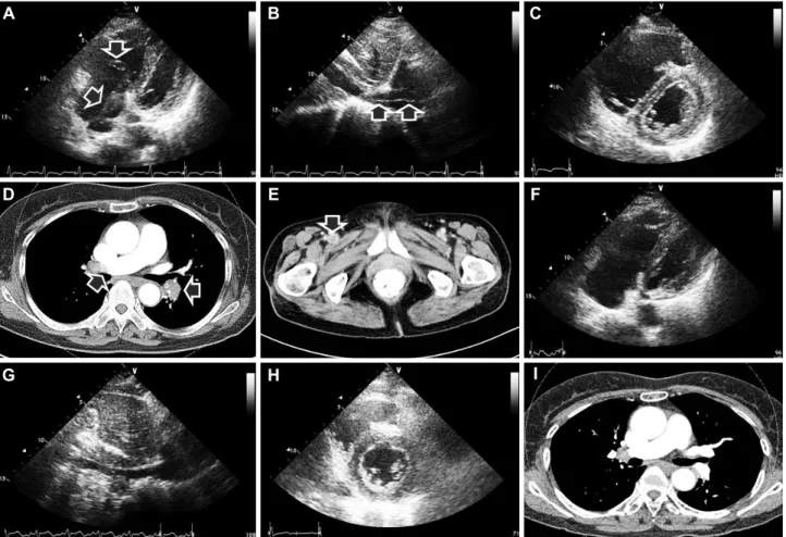
A Thief Caught in the Act
- Free Floating Venous Thrombus in the Right Heart Associated With Pulmonary Embolism - Seung-Pyo Lee, MD, Hyung-Kwan Kim, MD, Song-Yi Kim, MD, Il-Young Oh, MD,
Hyun-Jai Cho, MD, Yong-Jin Kim, MD and Dae-Won Sohn, MD
Department of Internal Medicine and Cardiovascular Center, Seoul National University College of Medicine, Seoul, Korea
Received: April 21, 2010 / Revision Received: May 10, 2010 / Accepted: May 31, 2010
Correspondence: Hyung-Kwan Kim, MD, Department of Internal Medicine and Cardiovascular Center, Seoul National University College of Medicine, 101 Daehak-ro, Jongno-gu, Seoul 110-744, Korea
Tel: 82-2-2072-0243, Fax: 82-2-2072-3757, E-mail: [email protected]
• The authors have no financial conflicts of interest.
cc