J O U R N A L O F
Veterinary Science
pISSN 1229-845X, eISSN 1976-555X J. Vet. Sci. (2012), 13(1), 99-101 http://dx.doi.org/10.4142/jvs.2012.13.1.99
Received: 1 Apr. 2011, Revised: 8 Jul. 2011, Accepted: 30 Jan. 2012
Short Communication
*Corresponding author: Tel: +91-9446283382; Fax : +91-4872370388; E-mail: [email protected]
ⓒ 2012 The Korean Society of Veterinary Science.
This is an Open Access article distributed under the terms of the Creative Commons Attribution Non-Commercial License (http://creativecommons.org/licenses/by-nc/3.0) which permits unrestricted non-commercial use, distribution, and reproduction in any medium, provided the original work is properly cited.
Evaluation and comparison of native and recombinant LipL21 protein-based ELISAs for diagnosis of bovine leptospirosis
Siju Joseph
1,*, Naicy Thomas
2, E. Thangapandian
1, Vijendra P Singh
1, Rishendra Verma
1, S. K. Srivastava
11
Indian Veterinary Research Institute, Bareilly 243122, U.P., India
2
Centre for Advanced Studies in Animal Genetics and Breeding, College of Veterinary and Animal Sciences, Kerala 680651, U.P., India
A 21-kDa leptospiral lipoprotein (LipL21) was evaluated for its diagnostic potential to detect bovine leptospirosis by ELISA. Both native LipL21 (nLipL21) and recombinant LipL21 (rLipL21) proteins were tested and compared regarding diagnostic efficiency, and no statistically significant difference was observed. The sensitivity of rLipL21 ELISA for 62 microscopic agglutination test (MAT) positive sera was 100% and the specificity with 378 MAT negative sera was 97.09%. Thus, rLipL21 protein-based ELISA could be used as an alternative to MAT for the diagnosis of bovine leptospirosis.
Keywords: ELISA, leptospirosis, microscopic agglutination test, nLipL21, rLipL21
Leptospirosis is an acute febrile zoonosis with a global distribution, widely recognized as an emergent or re- emergent disease [7]. The non-specific symptoms of the disease, fastidious nature of the organism, and complexities associated with the standard serological test-microscopic agglutination test (MAT) often make diagnosis difficult [9].
Recombinant protein-based ELISAs have been utilized as an alternative form of diagnostics, with high sensitivity and specificity [6]. Recombinant proteins that have been well characterized for their diagnostic potential include leptospiral lipoprotein (LipL) 32 [1], LipL41 etc [8].
Recently, a 21-kDa LipL21 was reported to be the second most abundant outer membrane protein in the Leptospira interrogans serovar Lai [2]. Though, this protein has been evaluated as a vaccine candidate [3]. Its diagnostic potential has not been explored. Previous analysis of the amino acid sequence of LipL21 protein in serovar Lai revealed a fatty acid incorporation site [3] which will be
absent in the recombinant LipL21. The present study evaluated and compared the potential of both native and recombinant LipL21 as antigens in ELISA for the serodiagnosis of leptospirosis in bovines to ascertain whether the presence of fatty acid influence the diagnostic potential of the protein significantly.
Native LipL21 (nLipL21) was isolated in pure form
following the protoco [10]. To achieve production of
recombinant LipL21 (rLipL21), the gene coding for the
21-kDa protein lipL21 was amplified using specific
primers, excluding the 54 bp long signal sequences from
the 5´ end of the lipL21 gene. The amplified product was
then cloned into pPRO.EX.HTc expression vector (Life
Technologies, USA) and transformed into competent
Escherichia coli M15 cells. The transformed cells were
plated on Luria- Bertani (LB) ampicillin (100 μg/mL) agar
plates (Sigma, USA) and confirmed by colony PCR. To
achieve expression of the cloned gene, the transformed
cells were subcultured onto LB broth and induced with 1 mM
concentration of isopropyl-beta-D-thiogalactopyranoside in
log growth phase and then further incubated for 37
oC for 7∼8
h. After the bacterial cells were pelleted by centrifugation
at 8,000 × g for 10 min, 7 mL of lysis buffer (100 mM
NaH
2PO
4, 10 mM Tris, 8 M urea, pH 8.0) was added to 0.5
g (wet wt.) of the bacterial pellet, which was kept at room
temperature for 1 h with intermittent vortexing. Cell debris
was removed by centrifugation at 8,000 × g for 15 min. The
supernatant was incubated with 700 μL of Ni-NTA resin
(Qiagen, USA) for 1 h in the presence of 20 mM imidazole
to induce binding of polyhistidine-tagged recombinant
protein to the affinity matrix. Then, the Ni-NTA resin cell
lysate mixture was packed into the column, and the
unbound proteins were allowed to pass through the column
and discarded. The column was washed thoroughly with
100 Siju Joseph et al.
Fig. 1. Western blot analysis. Lane 1-3: checking the identity and immunogenicity of rLipL21 using nLipL21 hyperimmune serum, Lane 1: induced Escherichia (E.) coli cell lysate, Lane 2:
non-induced E. coli cell lysate, Lane 3: purified rLipL21, Lanes 4-6: analysis of the immunoreactivity levels of rLipL21 and nLipL21 to MAT positive field cattle sera, Lane 4: purified rLipL21 protein, Lane 5: sarcosyl-soluble portion of OMP, Lane 6: sarcosyl-insoluble portion of OMP, Lanes 7-12: checking the cross-reactivity of rLipL21 hyperimmune sera to various bacteria, Lane 7: purified rLipL21, Lane 8: Brucella abortus culture lysate, Lane 9: Campylobacter jejuni culture lysate, Lane 10: Mycoplasma bovis culture lysate, Lane 11: Pasteurella multocida culture lysate, Lane 12: Salmonella typhimurium culture lysate.
Table 1. Comparison of rLipL21 ELISA with nLipL21 ELISA
Average optical density Positive samples
(n = 46)
Negative samples (n = 20) rLipL21 ELISA
nLipL21 ELISA
0.44 ± 0.03 0.463 ± 0.09
0.093 ± 0.009 0.102 ± 0.089
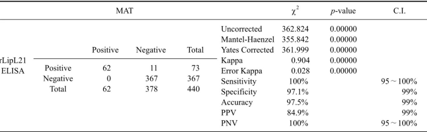
Table 2. Evaluation of rLipL21 ELISA for detection of anti-leptospiral antibodies in bovine sera as compared to MAT
MAT χ
2p-value C.I.
rLipL21 ELISA
Positive Negative Total
Uncorrected 362.824 0.00000 Mantel-Haenzel 355.842 0.00000 Yates Corrected 361.999 0.00000
Kappa 0.904 0.00000
Error Kappa 0.028 0.00000
Sensitivity 100% 95∼100%
Specificity 97.1% 99%
Accuracy 97.5% 99%
PPV 84.9% 99%
PNV 100% 95∼100%
Positive Negative Total
62 0 62
11 367 378
73 367 440 15 mL of wash buffer (100 mM NaH
2PO
4, 10 mM Tris, 8 M urea, pH 6.3), and the fusion protein was eluted with 3 mL of elution buffer (100 mM NaH
2PO
4, 10 mM Tris, 8 M urea, pH 4.5). The eluted rLipL21 protein was then dialyzed against phosphate- buffered saline (pH 7.4) overnight at 4
oC, after which the concentration was estimated to be 3 mg/mL.
The rLipL21 was analyzed by Western blotting using the hyperimmune serum raised against nLipL21 following standard protocol [12]. A strong reaction was observed (Fig.
1, lane 1∼3), proving the identity and immunogenicity of rLipL21. The immunoreactivity levels of rLipL21 and nLipL21 to the sera of bovines affected with leptospirosis were also tested (Fig. 1, lane 4∼6), and reactivity was observed. Further, the specificity of rLipL21 was tested by further Western blot analysis employing different bacterial organisms viz., Brucella abortus, Campylobacter jejuni,
Mycoplasma bovis, Pasteurella multocida, and Salmonella typhimurium, using the hyperimmune sera raised against rLipL21, and no cross-reactivity was observed (Fig. 1, lane 7∼12).
A total of 440 sera samples collected from cattle suspected to have leptospirosis from different states of India were screened by employing MAT according to standard procedures [5]. The sera samples were collected from unvaccinated cattle between 2∼4 weeks after showing symptoms of blood in milk as well as reproductive problems such as repeat breading, high number of services per conception, early embryonic loss, and irregular estrus cycles. Nine different leptospiral serovars viz., Leptospira (L.) interrogans serovars Canicola, Hardjo, Pomona, Pyrogenes, and Icterohaemorrhagiae along with L.
borgpetersenii serovars Tarassovi, Javanica, Sejroe, and Ballum, were used to screen the sera samples using MAT.
Sixty-two samples (14.1%) with a titer of 1:100 or above were designated as positive for the presence of leptospiral antibodies [13]. In addition to the 440 sera samples tested, negative control sera (n = 45) obtained from healthy calves (n = 20) and cattle with diseases other than leptospirosis (n
= 25), which were found to be sero-negative for leptospiral antibodies using MAT at a 1:50 dilution, were used in this study for the standardization of ELISA.
ELISAs were standardized for both rLipL21 and nLipL21
according to standard procedures [4], and the optimum
LipL21 protein-based ELISA for diagnosis of bovine leptospirosis 101