365
서 론
담관 폐쇄 시 일반적으로 관찰되는 담관의 확장은 현재 까지는 담관 내의 압력 증가 때문으로만 알려져 있다. 담관 압력의 증가가 어떠한 기전으로 담관의 평활근을 이완시키 는지는 알려져 있지 않으며, 연구 또한 전무한 상태이다. 그 리고 모든 폐쇄성 황달 환자에서 담관의 확장이 일어나는 것도 아니다. 이러한 임상적인 양상은 단순히 담관 압력의 증가가 담관의 확장을 유발한다고 설명하기는 어렵다.
최근 흰쥐 담관 결찰에 의한 간 섬유화 실험 모델에서 담 관을 결찰 하기 전 중농도의 에탄올을 담관 내에 주입하여 담관 상피 세포를 파괴하면 담관의 확장이 억제되고 단순 담관 결찰보다 더 효과적인 간 섬유화 모델이 될 수 있다는 것이 보고되었다.(1) 본 연구에서도 반복 실험을 통해, 담관 의 상피세포가 담관 확장에 중요한 역할을 함을 확인하였 다(Fig. 1, 2). 이러한 관찰은 담관 결찰 후 담관의 확장은 담관 내의 증가된 압력뿐 아니라 다른 요인에 의해 담관 상피세포에서 어떤 물질이 분비되어 담관의 평활근을 이완 시켜 담관의 확장이 일어나는 것을 시사한다고 하겠다.
1990년 초부터 평활근 이완 물질로서 연구되어온 일산화 질소(이하 NO)는 체내 거의 모든 부위에 존재하며, 대표적 인 평활근 이완 물질이다.(2,3) NO는 nitric oxide synthase(이 하 NOS)에 의해 arginine을 citrulline으로 전환시키면서 만들 어진다.(4) NOS는 3개의 isoenzyme이 알려져 있는데, 혈관 the dilated bile duct epithelium and the iNOS antagonist partially inhibited bile duct dilatation. iNOS induction in the epithelium is partly responsible for the dilatation of the bile duct after duct ligation. (J Korean Surg Soc 2003;64:
365-371)
Key Words: Bile duct ligation, Bile duct epithelial cell, iNOS 중심 단어: 담관 결찰, 담관 상피 세포, iNOS ꠏꠏꠏꠏꠏꠏꠏꠏꠏꠏꠏꠏꠏꠏꠏꠏꠏꠏꠏꠏꠏꠏꠏꠏꠏꠏꠏꠏꠏꠏꠏꠏꠏꠏꠏꠏꠏꠏꠏꠏꠏꠏꠏꠏꠏꠏꠏꠏꠏꠏ Departments of Surgery and 1Pathology, College of Medi- cine, Chungbuk National University, Cheongju, Korea
Expression of Inducible Nitric Oxide Synthase (iNOS) in the Epithelial Cells of the Distended Bile Duct by the Bile Ductal Ligation
Sang-Su Park, M.D., Guang-Bi Jin, M.D., Jin-Woo Park, M.D., Lee-Chan Jang, M.D., Ro-Hyen Sung, M.D.1 and Jae-Woon Choi, M.D.
Purpose: Recent studies in an obstructive jaundice rat model showed that the bile duct epithelium is also very important in the bile duct dilatation besides the increased luminal pressure. This study evaluated the role of iNOS in the bile duct epithelium in a rat obstructive jaundice model.
Methods: Bile duct ligations were performed in male Sprague-Dawley rats. The bile ducts were harvested on seven consecutive days. Immunohistochemical staining in the bile duct was performed using anti-iNOS polyclonal antibodies. Aminoguanidine (an iNOS antagonist) was injected intraperitoneally after bile duct ligation (0, 100, and 200 mg/kg/day, n=6 in each group). One week after surgery, the diameter of bile duct was measured and bile was collected for NO analysis by 280NOA (Silvers).
Results: The iNOS expression level was increased in the dilated ductal epithelium after the bile duct ligation but not in the normal epithelium. Aminoguanidine decreased the mean diameter of the bile duct after the bile duct ligation:
11±2.3 mm in the duct ligation only group; 7.5±0.75 mm in the 100 mg/kg/day aminoguanidine; 6±0.82 mm in the 200 mg/kg/day of aminoguanidine group (mean±SE, P<
0.05). The NO concentration in the bile was decreased by aminoguanidine: 16±4.2 mM in the sham operation group;
40±4.5 mM in duct ligation only group; 34±6.4 mM in the 100 mg/kg/day of aminoguanidine group; 11±1.2 mM in the 200 mg/kg/day of aminoguanidine group (mean±SE).
Conclusion: Bile duct ligation induced iNOS expression in
담도 결찰로 인한 담도 확장 시 담도 상피 세포에서의 Nitric Oxide Synthase 발현
충북대학교 의과대학 외과학교실, 1병리학교실
박상수․김광필․박진우․장이찬․성노현1․최재운
책임저자:최재운, 충북 청주시 흥덕구 개신동 62번지 ꂕ 361-711, 충북대학교병원 외과학교실 Tel: 043-269-6358, Fax: 043-266-6037 E-mail: [email protected]
접수일:2003년 2월 17일, 게재승인일:2003년 4월 2일
ꠏꠏꠏꠏꠏꠏꠏꠏꠏꠏꠏꠏꠏꠏꠏꠏꠏꠏꠏꠏꠏꠏꠏꠏꠏꠏꠏꠏꠏꠏꠏꠏꠏꠏꠏꠏꠏꠏꠏꠏꠏꠏꠏꠏꠏꠏꠏꠏꠏꠏꠏꠏꠏꠏꠏꠏꠏꠏꠏꠏꠏꠏꠏꠏꠏꠏꠏꠏꠏꠏꠏꠏꠏꠏꠏꠏꠏꠏꠏꠏꠏꠏꠏꠏꠏꠏꠏꠏꠏꠏꠏꠏꠏꠏꠏꠏꠏꠏꠏꠏꠏꠏꠏꠏꠏꠏꠏꠏꠏꠏꠏꠏꠏꠏꠏ
의 확장과 관련되어서 혈관 상피 세포에 존재하는 eNOS가 처음 발견되었으며, 신경전달물질로서 NO를 생성하는 bNOS, interferon-γ, TNF-α, endotoxin 등에 의해 발현되는 iNOS가 있다.(5,6) iNOS는 macrophage,(7) 섬유 세포,(8) 상 피 세포,(9) chondrocyte,(10) Kuppfer cell,(11) 혈관 평활 근,(12) 등에서 발현되며, NOS isoenzyme 중에서 NO를 가장 많이 생성하며, 그 역할도 면역 기능에서부터 평활근 이완 에까지 다양하다.(2,3)
소화기관에서 NO의 역할은 매우 다양하게 보고되는데, 소화기관의 평활근 운동,(13) 위 점막 혈류 증진,(14) 위 점막 보호,(15) 위장 근육 이완의 최종 매개체,(16) 담낭 운동(17) 등 모든 소화기관에 관련이 있어 보인다. NO 분비의 결핍이나 과잉 분비는 여러 가지 질환에서 동반되어 관찰된다.
신생아에서 흔히 볼 수 있는 질환으로서 선천성 유문 협 착증(hypertrophic pyloric stenosis)은 유문부에 정상으로 존 재하는 NOS가 존재하지 않아 유문부의 이완이 일어나지 않아서 협착이 오는 것으로 설명되며,(18) 식도 무이완증 (esophageal achalasia) 역시 식도하부에 NOS의 결핍으로 이 완이 일어나지 않아 식도 폐쇄가 나타난다.(19) 또한 Hirschsprung씨 질환에서도 신경절이 없는 대장에 NOS가 결핍된 것이 발견되었다.(20) 그 외에도 전체 위장관의 운 동에 NO가 관련이 있는 것으로 보고되고 있으며, 췌장 염,(21) 염증성 장염(inflammatory bowel disease) 등에서는 NOS가 중요한 역할을 한다.(22)
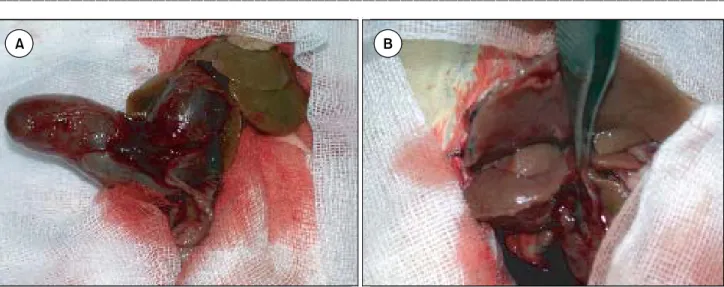
iNOS는 정상적으로는 발현되지 않으나 거의 모든 세포 에서 발현될 수 있는 효소로서 많은 양의 NO를 생산하는 Fig. 1. Operative findings of the bile duct one week after bile duct ligation. Marked dilatation of the bile duct (8∼15 mm) was observed in the rat without ethanol infusion into the bile duct before bile duct ligation (A). No dilatation of the bile duct was observed in the rat with 100 ul of 60% ethanol infusion into the bile duct before bile duct ligation (B).
A B
A
A BB
Fig. 2. Microscopic findings of the extrahepatic bile duct one week after bile duct ligation. Flattened and destructed bile duct epithelium were observed in the bile duct with 100 ul of 60% ethanol infusion into the bile duct before bile duct ligationb (A). Well preserved tall columnar epithelium was observed in the dilated bile duct with the simple ligation of the bile duct (B).
A B
A
A BB
것이 특징적이다. iNOS의 발현으로 발생하는 과다한 평활 근 이완과 관련된 임상적 질환으로서는 패혈성 쇼크에서 혈압의 저하를 들 수 있다.(23,24)또한 간경변 환자에서 볼 수 있는 비정상적인 혈역학의 증가 및 식도 정맥의 확장에 도 iNOS의 발현이 중요한 역할을 한다.(25,26) 이러한 소견 은 비정상적인 장기의 확장 혹은 평활근 이완에는 NO가 관 련 될 수 있음을 보여주며, iNOS의 발현이 이러한 것을 설 명할 수 있는 가능성이 있다고 하겠다.
본 연구에서는 흰쥐 담관 결찰 후 발생하는 담관의 확장 과 NO의 관계를 규명하기 위해 고안되었다. 연구의 가설은 담관 결찰에 의해 상피세포에서 iNOS가 발현되고 담관의 평활근을 이완하는 물질인 NO가 분비되며, 이를 통해 담관 확장이 진행될 것이라는 것이다. 이것을 검증하기 위해 확 장된 담관에서 iNOS의 발현을 관찰하고, iNOS 길항제의 투 여가 담관 확장을 억제하는지를 밝히는 데 있다.
방 법
1) 실험 시약 및 기자제
iNOS antibody (Transduction lab.), aminoguanidine, sodium nitrite (Sigma) Xylane, Ketamine, 280NOA (Silvers)
2) 실험동물 사육 및 수술 방법
7 주령의 수컷 Sprague-Dawley 흰쥐를 사용하였다. 실험 동물은 설치류 사료를 사용하여 키웠으며, 사육실 온도는 20∼25oC로 유지하고 사료 및 물의 공급은 자유롭게 섭취하도 록 하였다. 실험 당시 실험동물의 체중은 200 gm 전후였다.
실험동물의 마취는 xylane과 ketamine을 사용하였으며, 담관 결찰 수술은 정중 절개를 가한 후 담관이 십이지장에 접하는 부위에서 5 mm 상방에서 4-0 흑색 실크를 이용하여 이중 결찰하였다. 실험동물의 사육 및 수술에 관한 모든 사 항은 충북대학교 병원 임상연구소 실험 동물 윤리위원회의 규정을 준수하였다.
3) 면역형광 검사
절제한 담관 조직을 급냉동한 뒤 전기 동결 박절기 (crystat)로 10 mm 두께로 박절하여 알부민이 부착된 슬라이 드에 부착시킨 후 아세톤 용액에서 20분간 고정한다. Phos- phate buffered saline (PBS)으로 세척하고, 형광 염료와 poly- clonal iNOS 항체를 1:50으로 희석하여 암실에서 실온 상 태로 1시간 반응시킨 후 PBS로 3회 세척하고 crystal mount 로 봉입한다. 반응 여부는 형광 현미경(Olympus BH2- RFCA)으로 관찰하고 사진을 찍어 보관하였다.
4) Aminoguanidine에 의한 iNOS 억제
iNOS 길항제인 aminoguanidine이 담관 결찰 후 발생하는 담관 확장을 억제할 수 있는지를 확인하기 위해 단순 결찰
군(n=6), 담관 결찰 후 aminoguanidine 100 mg/kg을 매일 복 강내에 주입한 군(n=6), 200 mg/kg를 주입한 군(n=6)으로 나 누어 1주일 후 담관의 직경을 측정하여 담관 확장 정도를 비교하였다. 담관의 직경을 측정한 후 담즙 내의 NO양을 측정하기 위해 담즙을 채취하여 -70oC에 보관하였다.
5) 담즙내 NO의 측정
담관 결찰하지 않은 정상 쥐의 담관을 천자하여 수집한 정상 담즙, 단순 결찰 후 1주일 후 채취한 담즙, aminogua- nidine을 투여한 군의 담즙을 대상으로 하였다. 채취한 담즙 0.5 ml를 취하여 0oC 99% ethanol을 넣은 후 잘 혼합한 후 14,000 rpm으로 원심 분리하여 담즙 내 단백질을 제거한 뒤, 시료를 280NOA (Silvers)를 이용하여 담즙 내의 NO 대사물 질인 nitrite/nitrate를 NO로 환원하여 측정하였다.
6) 통계 처리 방법
모든 통계 수치는 표준±표준오차로 표시하였다. 본 연 구에서 분석한 담관의 직경과 담즙 내의 NO 대사물의 분석 은 Wilkoxon Rank Sum으로 처리하였으며 P-value가 0.05 이 하일 때 유의성이 있는 것으로 판정하였다.
결 과
1) 면역형광 검사
iNOS에 대한 항체를 이용한 면역 형광 검사에서 정상 담 관 상피 세포는 형광 발현이 거의 없고, 담관 결찰 후의 늘어 난 담관 상피 세포에서는 증가된 형광이 관찰되었으며, 이러 한 증가된 면역 형광은 결찰 후 1주일까지 지속되었다(Fig. 3).
2) iNOS 길항제인 aminoguanidine의 효과
단순 담도 결찰 군, 담도 결찰 후 aminoguanidine 100 mg/kg 복강 내 투여 군, 200 mg/kg 복강 내 투여 군으로 나 누었으며 복강 내 주입하는 용매의 양은 같은 양으로 하였 다. 단순 담도 결찰 군은 생리 식염수 동일 양을 매일 복강 내에 주사하였다.
1주일 후 담관의 최대 직경을 측정하였는데, 단순 담관 결찰 군은 11±2.3 mm (mean±SE), 100 mg/kg 군은 7.5±
0.75 mm, 200 mg/kg 군은 6±0.82 mm로, 통계적으로 유의 하게 직경이 감소되었으며, aminoguanidine의 양의 증가에 따라 직경의 감소도 증가하였다(Fig. 4).
3) 담즙 내의 NO 양의 변화
정상 담즙, 결찰 후 1주일째의 담즙, 결찰 후 aminog- uanidine을 100 mg/kg, 200 mg/kg를 복강내에 투여한 군의 담즙을 각각 1주일째 채취하여, 담즙 내에 존재하는 NO의 대사물인 nitrite 및 nitrate를 측정하였다. 280NOA (Silvers) 를 이용하여 분석하였는데, sodium nitrate를 대조군으로 사
ꠏꠏꠏꠏꠏꠏꠏꠏꠏꠏꠏꠏꠏꠏꠏꠏꠏꠏꠏꠏꠏꠏꠏꠏꠏꠏꠏꠏꠏꠏꠏꠏꠏꠏꠏꠏꠏꠏꠏꠏꠏꠏꠏꠏꠏꠏꠏꠏꠏꠏꠏꠏꠏꠏꠏꠏꠏꠏꠏꠏꠏꠏꠏꠏꠏꠏꠏꠏꠏꠏꠏꠏꠏꠏꠏꠏꠏꠏꠏꠏꠏꠏꠏꠏꠏꠏꠏꠏꠏꠏꠏꠏꠏꠏꠏꠏꠏꠏꠏꠏꠏꠏꠏꠏꠏꠏꠏꠏꠏꠏꠏꠏꠏꠏꠏ
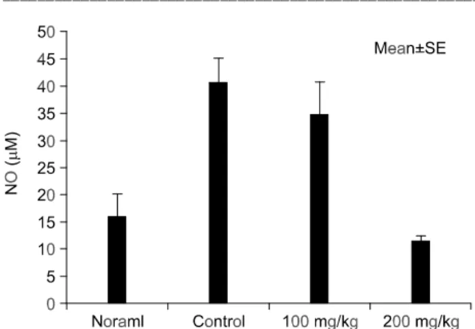
용하고, peak의 면적을 계산하여 NO의 양으로 표시하였다 (Fig. 5). 각 군의 NO 측정량은 정상 담즙의 경우 16±9.3 mM (mean±SE), 단순 담도 결찰군은 40±10 mM, aminog- uanidine 100mg/kg 투여군은 32±12 mM, 200mg/kg 투여군 은 11±2.6 mM이었다(Fig. 6). NO의 양은 단순 결찰군에서 가장 높았으며, aminoguanidine 투여군은 용량에 반비례하 게 NO의 양이 감소하였다. 이러한 소견은 담관 확장 정도 와 비례하였다.
고 찰
폐쇄성 황달 시 발생하는 담관의 확장은 단순히 담관 내 의 압력 증가에 의한 수동적인 확장으로 생각되어왔으며 특별한 연구의 대상이 되지 않았다. 그러나 수술 시 담관
내부에 높은 압력을 주더라도 급격한 담관의 확장을 관찰 하기 어렵다. 따라서 담관 폐쇄 후 발견하는 담관의 확장은 담도 내부의 압력의 증가에 따른 담관 조직에서 이차적인 반응, 예를 들면 담관 근육을 이완시키는 물질이 분비될 수 있다는 것을 가정하게 한다. 담관 압력 증가에 일차적으로 노출되는 세포는 담관의 상피 세포이다. 따라서 담관의 상 피세포에서 담관 평활근을 이완시키는 물질의 분비를 가정 할 수 있다. 실제로 Fig. 1, 2에서 보듯이, 담관 상피세포가 파괴된 담관은 결찰하더라도 담관의 확장이 일어나지 않는 다. 이러한 소견은 담관 확장에 있어서 담관 상피 세포가 중요한 역할을 하고 있다는 것을 시사한다.
소화기 평활근 이완에 가장 중요한 매개체로 알려진 물 질은 NO이며, NO의 결핍 및 증가는 선천성 유문 협착 증,(18) 식도 무이완증,(19) 간 경화시 발생하는 혈류 역학 Fig. 3. Immunofluorescence image of iNOS expression in the bile duct epithelium in control and bile duct ligation. No immunofluorescence activity was observed in normal bile duct (A). Increased immunofluorescence activity was observed in the bile duct epithelium one week after bile duct ligation (B).
Fig. 4. Diameter of bile duct treated with aminoguanidine after ligation. Intraperitoneal administration of aminoguanidine inhibited dilatation of bile duct after the bile duct ligation.
Inhibition of bile duct dilatation by aminouanidine was dose dependent (n=6 in each group).
Fig. 5. Diagram of NO analysis by 280NOA (Silvers). Prior to analysis, NO metabolites such as nitrite and nitrate, which were dissolved in the bile sample, were reduced to NO.
변화(25,26) 등 소화기 질환과도 깊은 관련이 있다.
본 연구에서 면역 형광 검사를 통하여 확장된 담관 상피 세포에서 iNOS 항체에 의한 면역 형광의 증가를 관찰하였 지만, 무엇이 iNOS의 발현을 유발하는지는 밝히지 못하였 다. 일반적으로 iNOS를 발현시키는 물질로서 알려진 것은 endotoxin, interferon-γ, TNF-α(5,6) 등 염증성 물질이며, 밝 혀지지 않은 부분도 있으나, 담즙과 관련된 것은 문헌에서 찾아보기 어려웠다. 담관 결찰이 물리적으로는 담관 내부 의 압력을 증가시키는 것은 뚜렷하지만, 증가된 압력이 상 피세포를 자극하여 iNOS의 발현을 유발하는지, 아니면 담 관 결찰 후 발생하는 담관의 확장과 담즙의 정체로 담관 내의 담즙에 세균이 자라고, 상피세포에서 cytokine 등이 분 비되면서 iNOS가 발현되는지는 밝혀져야 할 문제이다. 본 연구에서도 이것을 규명하기 위해 담관 상피 세포를 배양 하여 in vitro 실험을 시도하려 하였으나, 흰쥐의 담관 구조 가 주위에 있는 췌관과 연결되어 있어 순수한 담관 상피 세포를 분리 배양하기 힘들어 실패하였다. 그러나 본 실험 에서 담즙의 육안적 관찰에서는 담즙이 화농되었거나, 담 즙의 색깔이 변화한 것은 관찰하지 못하였다. 또 다른 가능 성으로는 간에서 생긴 iNOS 발현 유도 물질이 담즙 속에 함유되어 담관의 상피 세포에서 iNOS를 유도할 수도 있다 고 생각한다. 간 손상 시 iNOS의 발현은 일반적으로 잘 알 려진 사실이다.(11,27) 담관 결찰 후 발생하는 간 손상 시에 도 간에서 iNOS가 발현된다.(11) 본 연구에서 관찰된 담관 상피 세포에서의 iNOS 발현도 간손상에 의한 이차적인 발 현의 가능성이 없는지 향후 검증되어야 하겠다.
담도 결찰에 의한 담관 상피 세포의 iNOS 발현이 담관
확장의 원인이라면, iNOS 길항제의 투여가 담관 결찰 후 발생하는 담관 확장을 억제할 수 있을 것이다. 본 연구 결과 길항제의 양에 비례하여 담관의 확장이 억제되는 것이 관 찰되었으나, 완전한 억제는 이루어지지 않았다. 이것은 투 여한 iNOS의 양이 불충분하거나, iNOS 역할 외에 담관의 압력증가 자체가 중요한 역할을 함을 의미한다고 하겠다.
그러므로 iNOS 길항제인 aminoguanidine이 담즙의 분비 및 간에 미치는 영향을 완전히 배제하여야 하겠다. 본 연구에 서는 aminoguanidine이 담관 내 압력에 미치는 영향을 관찰 하지는 못하였지만, 문헌 보고에 의하면, 분리된 간 세포에 서는 NO의 이차 매개체인 cGMP가 담즙의 분비를 증가시 키나, 분리한 담관의 단위에서는 영향을 미치지 못한다.(28) 또한 endotoxin으로 간을 처리하여 iNOS의 발현을 증가시 키면, 간내 담즙의 정체가 생기며 NO 양과 세포 내 cGMP의 양이 증가하며, 이때 iNOS 길항제인 aminoguanidine을 투여 하면 담즙의 정체가 감소하고 담즙 분비가 증가하는 것으 로 보고된다.(29) 따라서 본 연구에서 투여한 iNOS 길항제 가 담관의 압력을 감소시키기보다는 오히려 증가시켰을 가 능성이 높으므로, aminoguanidine의 투여에 의한 결찰 담관 확장의 억제는 담즙 분비 감소에 의한 것이라기 보다는 담 관 상피 세포에서 발생하는 NO를 억제한 효과로 추정할 수 있겠다.
마지막으로, 담관 결찰 후 담관 상피에서 iNOS가 발현하 여 NO의 분비량이 증가한다면, NO의 대사물인 nitrite/
nitrate가 담즙내에 증가되어 있을 것이며, iNOS 길항제인 aminoguanidine을 투여하면, nitrite/nitrate의 양이 감소될 것 이다. 세포에서 분비된 NO의 반감기는 수초에서 30 초까지 로 알려져 있으며, 분비량도 매우 적어 생체 내에서 NO를 직접 측정하기는 매우 어렵다. 대부분의 실험에서는 NO의 대사물인 nitrite, nitrate를 측정함으로써 NO의 양을 평가한 다.(30) 본 실험에서도 담즙 내의 nitrite/nitrate를 측정하여 NO 량을 간접적으로 평가하였다. 실험 결과 단순 담관 결 찰군에서 NO의 양이 가장 높았으며, aminoguanidine을 투여 한 군에서는 aminoguanidine의 양에 비례하여 NO의 양이 감 소하였고, 담관 확장도 비례하여 억제되었다. 이러한 소견 은 담관 결찰 후 발생되는 담관 확장에 NO가 부분적으로 관련이 있음을 보여주는 직접적인 증거라고 하겠다. 담관 결찰 후 관찰되는 담관 상피세포에서의 iNOS 면역 형광 증 가와 함께 생각해볼 때, 담관 결찰 후 발생하는 담관의 확장 은 NO가 중요한 매개체로 작용하며, NO는 상피세포에서 발현하는 iNOS에서 유래하는 것으로 생각된다.
본 연구에서 저자들은 흰쥐 담관 결찰 후 발생하는 확장 된 담관의 상피 세포에서 iNOS의 발현을 면역 형광 방법으 로 확인하였으며, iNOS 길항제의 투여가 담관의 확장을 억 제하는 것을 관찰하였다. 이러한 발견은 담관의 확장과 관 련된 여러 질환의 병인 규명과 치료에 도움을 줄 수 있다고 생각한다.
Fig. 6. The amount of NO metabolite in the bile. One week after Sham operation or dile duct ligation, NO concentration of bile in the group of Sham operation, duct ligation only, duct ligation with 100 mg/kg/day of aminoguanidine, and duct ligation with 200 mg/kg/day of aminoguanidine were measured. NO concentration was the highest in the duct ligation only group and it was decreased by amino- guanidine administration.
ꠏꠏꠏꠏꠏꠏꠏꠏꠏꠏꠏꠏꠏꠏꠏꠏꠏꠏꠏꠏꠏꠏꠏꠏꠏꠏꠏꠏꠏꠏꠏꠏꠏꠏꠏꠏꠏꠏꠏꠏꠏꠏꠏꠏꠏꠏꠏꠏꠏꠏꠏꠏꠏꠏꠏꠏꠏꠏꠏꠏꠏꠏꠏꠏꠏꠏꠏꠏꠏꠏꠏꠏꠏꠏꠏꠏꠏꠏꠏꠏꠏꠏꠏꠏꠏꠏꠏꠏꠏꠏꠏꠏꠏꠏꠏꠏꠏꠏꠏꠏꠏꠏꠏꠏꠏꠏꠏꠏꠏꠏꠏꠏꠏꠏꠏ
결 론
흰쥐 담관 결찰 후 확장된 담관 상피세포에서 면역 형광 검사를 통하여 담관 상피세포에서 iNOS가 발현됨을 확인 하였고, iNOS 길항제인 aminoguanidine의 투여로 담관 확장 이 부분적으로 억제되는 것을 관찰하였으며, 담즙내의 NO 양의 변화도 담관의 확장과 연관이 있음을 확인하였다. 이 상의 결과는 흰쥐 담관 결찰 후 발생하는 담관의 확장은 단순한 담관 내압의 증가로만 이루어지는 것이 아니라, 담 관 상피 세포에서 발현하는 iNOS가 부분적인 역할을 하고 있음을 보여준다고 하겠다.
본 연구의 결과는 임상에서 발견되는 담관 확장의 기전을 밝히는데 기여할 것으로 생각되며, 또한 담관 확장과 연관 된 질환의 규명에도 도움이 될 것으로 기대된다.
REFERENCES
1) Song JH, Sung RH, Jang LC, Park SM, Chae HB, Choi JW.
Liver fibrosis by bile duct sclerosis with ethanol: A new experimental model for liver fibrosis. Korean Journal of HBP Surgery 2002;6:42-7.
2) Bredt DS, Snyder SH. Nitric oxide a novel neuronal messen- ger. Neuron 1992;8:3-11.
3) Lowenstein CJ, Dinerman JL, Snyder SH. Nitric oxide: A physiologic messenger. Ann Intern Med 1994;120:227-37.
4) Bredt DS, Snyder SH. Isolation of nitric oxide synthase, a calmodulin-requiring enzyme. Proc Natl Axad Sci USA 1990;
87:682-5.
5) Marletta MA. Nitric oxide synthase structure and mechanism.
J Biol Chem 1993;268:12231-4.
6) Chartrain NA, Geller DA, Koty PP, Sitrin NF, Nussler AK, Hoffman EP, et al. Molecular cloning, structure, and chro- mosomal localization of the human inducible nitirc oxide synthase gene. J Biol Chem 1994;269:6765-72.
7) Xie QW, Cho HJ, Calaycay J, Mumford RA, Swiderek KM, Lee TD, et al. Cloning and characterization of inducible nitric oxide synthase from mous macrophages. Science 1992;256:
225-8.
8) Willis RA, Nussler AK, Fries KM, Geller DA, Phipps RP.
Induction of nitric oxide synthase in subsets of murine pulmonary fibroblasts: Effects on fibroblast interleukin-6 production. Clinical Immunology and Immunopathology 1994;71:231-9.
9) Robbins RA, Springall DR, Warren JB, Kwon OJ, Buttery LDK, Wilson AJ, et al. Inducible nitric oxide synthase is in- creased in murine lung epithelial cells by cytokine stimulation.
Biochemical and Biophysical Research Communications 1994;
198:835-43.
10) Mair R, Bilbe G, Rediske J, Lotz M. Inducible nitric oxide synthase from human articular chondrocytes: cDNA cloning
and analysis of mRNA expression. Biochimica et Biophysica Acta 1994;1208:145-50.
11) Rockey DC, Chung JJ. Regulation of inducible nitric oxide synthase and nitric oxide during hepatic injury and fibro- genesis. Am J Physiol 1997;273:G124-30.
12) Schini VB, Busse R, Vanhoutte PM. Inducible nitric oxide synthase in vasculat smooth muscle. Arzneim-Forsch/Drug Res 1994;44:432-5.
13) Starl ME, Szurzewski JH. Role of nitric oxide in gastrointes- tinal and hepatic function and disease. Gastroenterology 1992;
103:1928-49.
14) Tanaka T, Guth P, Tache Y. Role of nitric oxide in gastric hyperemia induced by central vagal stimulation. Am J Physiol 1993;264:G280-4.
15) MacNaughton WK, Cirino G, Walace JL. Endothelium-derived relaxing factor (nitric oxide) has protective actions in the stomach. Life Sci 1989;45:1869-76.
16) Grider JR, Murthy KS, Jin JG, Makhlouf GM. Stimulation of nitric oxide from muscle cells by VIP: Prejunctional enhan- cement of VIP release. Am J Physiol 1992;262:G774-8.
17) Mourelle M, Guarner F, Molero X, Moncada S, Malagelada JR. Regulation of gallbladder motility by the arginine-nitric oxide pathway in guinea pigs. Gut 1993;34:911-5.
18) Vanderwinden JM, Mailleux P, Schiffmann SS. Nitric oxide synthase activity in infantile hypertrophic pyloric stenosis. N Engl J Med 1992;327:511-5.
19) Mearin F. Absence of nitric oxide synthase in the gastroe- sophageal junction of patients with achalasia. Gastroenterology 1993;104:A550.
20) Vanderwiden GM, De laet MH, Schiffmann SM, Mailleux P, Lowenstein CJ, Snyder S, et al. Nitric oxide distribution in the enteric nervous system of Hirschsprung's disease. Gastroe- nterology 1993;105:969-73.
21) Al-Mufti RA, Williamson RC, Mathie RT. Increased nitric oxide activity in a rat model of acute pancreatitis. Gut 1998;
43:564-70.
22) Rachmilewitz D, Stamler JS, Bachwich D, Karmeli F, Acker- man Z, Loscalzo J, et al. Enhanced colonic NO generation and stimulated NO synthase activity in experimental colitis and in active inflammatory bowel disease. Gastroenterology 1993;
104:A766.
23) Sako K, Miyakawa K, Takeya M, Hattori R, Yui Y, Sunamoto M, et al. Immunohistochemical expression of inducible nitric oxide synthase (iNOS) in reversible endotoxic shock studied by a novel monoclonal antibody against rat iNOS. Journal of Leukocyte Biology 1955;57:36-44.
24) Buttery LD, Evans TJ, Springall DR, Carpenter A, Cohen J, Polk JM. Immunochemical localization of inducible nitric oxide synthase in endotoxin-treated rats. Laboratory Inves- tigation 1994;71:755-64.
25) Morales-Ruiz M, Jimenez W, Perez-Sala D, Ros J, Leivas A, Lamas S, et al. Incresed nitric oxide synthase expression in
arterial vessels of cirrhotic rats with ascites. Hepatology 1996;24:1481-7.
26) Whittle BJR, Moncada S. Nitric oxide: The elusive mediator of the hyperdynamic circulation of cirrhosis? Hepatology 1992;16:1089-92.
27) Taylor BS, Alarcon LH, Billiar TR. Inducible nitric oxide synthase in the liver:regulation and function. Biochemistry 1998;63:766-81.
28) Trauner M, Mennone A, Gigliozzi A, Fraioli F, Boyer JL.
Nitric oxide and guanosine 3',5'-cyclic monophosphate stim- ulate bile secretion in isolated rat hepatocyte couplets, but not in isolated bile duct units. Hepatology 1998;28:1621-8.
29) Shiomi M, Wakabayashi Y, Sano T, Shinoda Y, Nimura Y, Ishimura Y, et al. Nitric oxide suppression reversibly attenu- ates mitochondrial dysfunction and cholestasis in endotoxemic rat liver. Hepatology 1998;27:108-15.
30) Archer S. Measurement of nitric oxide in biological models.
The FASEB Journal 1993;7:349-60.