Radical Scavenging Activities of Phellinus pini
Byung Hyouk Nam, Wol Soon Jo1, Yong Cui, Yoo Jin Choi1, Jae Dong Lee* and Min Ho Jeong1* Department of microbiology, College of Natural Sciences, Pusan National University, Busan 609-735, Korea
1Department of parasitology, College of Medicine, Dong-A University, Busan 602-714, Korea Received December 23, 2009 /Accepted December 30, 2009
The concentration of phenolics in Phellinus pini (CY001) extracts, expressed as mg of GAEs per g of P. pini fractions, and the EtOAc fraction (436.5 mg GAEs/g) of P. pini had a higher phenolic content than other fractions. Several biochemical assays were used to screen antioxidant properties such as reducing power, 2, 2-diphenyl-1-picrylhydrazyl (DPPH) radical scavenging capacity, NBT/XO super- oxide system and inhibition of DCF/AAPH peroxyl radicals. Among the six mushroom extracts, the EtOAc fraction from P. pini (CY001) showed the most potent DPPH radical, superoxide radical, and peroxyl radical scavenging activities, with IC50 values of 11.49 μg/ml, 8.32 μg/ml, and 1.91 μg/ml, respectively. The EtOAc fraction ofP. pini (CY001) significantly inhibited enzymatic lipid peroxidation and effectively attenuated LPS-induced NO production of RAW 264.7 cells without cytotoxicity. We also found that the EtOAc fraction had a significant hepato-protectant effect on tacrine-induced cyto- toxicity in HepG2 cells. These findings suggest thatP. pini (CY001) may have potential as a natural antioxidant, which contains compound(s) with radical scavenging activity.
Key words : Phellinus pini, free radicals, phenol content, nitric oxide, hepatoprotection
*Corresponding author
*Tel:+82-51-240-2865, Fax:+82-51-242-7265
*E-mail : [email protected]
Introduction
The genus Phellinus is taxonomically classified into Aphyllophorales in Hymenochaetceae of Basidiomycota.
These fungi are widely distributed in subtropical and trop- ical regions of Asia including China, Japan, southeastern USA, Maxico, and India and include both annual and per- ennial forms [26]. The mushroom of the Phellinus family when are also parasitic on the mulberry,P. linteus, P. ignirius, P. baumi, P. gilvus, P. nigricans etc. are belonging to a similar family. Among them, P. pini is a well-known fungus of the genus Phellinus in the family of Hymenochaetaceae [30].
Phellinus species are known to cause white pocket rot and sever plant diseases such as root rot, canker, or heart rot in living trees, as well as destroying and other woody resi- dues [22]. Also P. pini is a white-rot fungus that fructifies over the stem of Pinus pinaster. They damage lignin from wood, thus inducing its decay and giving a red color over the attacked surface. This process drastically reduces the me- chanical properties of wood and consequently its economic value and especially, the most economically important coni- fer decay fungus in western [30]. The genusPhellinus gen-
erally has been used as a traditional herb medicine for years in oriental countries. In folk medicine, it has been known to possess a curing effect against stomach aches, in- flammation, tumors and so on. It is also used to improve overall health and prevent various diseases, such as gastro- enteric disorders, lymphatic diseases, and cancers [9].
AmongPhellinus, a few pharmacological actions of P. linteus very well known to be elucidated in the last decade.
Especially, the active polysaccharide, isolated from mycelial culture ofP. linteus, stimulates humoral and cell-mediated immunity, and exhibits a wider range of immunostimulation and anti-tumor activity than other polysaccharides isolated from Basidiomycetes [21]. Their polysaccharide showed the hypoglycemic effect, which was investigated in streptozoto- cin-induced diabetic rats, and also decreased total cholester- ol, triacylglycerol and aspartate aminotransferase activity [23]. Most of all mushrooms inclusive of the genus Phellius had abundant biological active compounds and the phenolic compound was representative. Mushrooms accumulate a va- riety of secondary metabolites, including phenolic com- pounds, polyketides, terpenes, and steroids. A mushroom phenolic compound has been found to be an excellent anti- oxidant and synergist which is not mutagenic. Phenolics are compounds possessing one or more aromatic rings with one or more hydroxyl groups and are generally categorized as phenolic acids, flavonoids, stilbenes, coumarins, and tannins
[20]. The bioactivity of phenolics may be related to their abil- ity to chelate metals, inhibit lipoxygenase (LOX), and scav- enge free radicals [11]. LOX are sensitive to antioxidants, and the most common way of their action may consist in inhibition of lipid hydroperoxide formation due to scaveng- ing of lipidoxy or lipid peroxy-radicals formed in course of enzymic peroxidation. This LOXs comprise a family of non-heme iron-containing dioxygenases, representing the key enzymes in the biosynthesis of leukotrienes that have been postulated to play an important role in the pathophysi- ology of several inflammatory and allergic diseases [3].
Inflammation is a complex pathophysiological process medi- ated by a variety of signaling molecules produced by leuko- cytes, macrophages, mast cells, platelets, etc [17]. In various inflammatory conditions, LOX and secretory phospholipase A2 (sPLA2), as well as iNOS, are induced in vascular and/or macrophages [36]. LOX enzymes have been shown to be ex- pressed in macrophage-rich areas of atherosclerotic lesion [35]. In particular, nitric oxide (NO) is constitutively gen- erated in normal liver, and levels increase markedly in re- sponse to liver injury from diverse insults [28], including hepatotoxins, endotoxemia, and ischemia-reperfusion.
Increased NO production results from enhanced expression of inducible nitric oxide synthase (iNOS) in hepatocytes and Kupffer cells [42]. Prolonged inflammation contributes to the pathogenesis of many inflammatory diseases, including hep- atitis [28], bronchitis [47], gastritis [41], inflammatory bowel disease (IBD) [13], multiple sclerosis (MS) [25], and rheuma- toid arthritis (RA) [48]. P. pini is not well known to anti- oxidant and immunostimulating activity so far and improve overall health and prevent various diseases so far. In the present study, we elucidated the possible contribution of the radical scavenging effect to the LOX inhibitory mechanism of P. pini (CY001) fractions, which containing phenolic compound. AlsoP. pini (CY001) fractions were investigated relation between inhibition of lipoxgenase and the regu- latory mechanisms of stimulus-induced NO production. In the search for more effective antioxidant agents against oxi- dative stress-induced cell damage, the use ofP. pini (CY001) fractions to antagonize oxidative cell injury was explored in HepG2 cell subjected to tacrine-induced oxidative stress.
Materials and Methods
Materials and reagentsRPMI Medium 1640, DMEM, FBS (fetal bovine serum), pen- icillin and streptomycin were purchased from Gibco Ltd. (NY,
USA). DCF (2,7-dichlorofluorescindiacetate) was purchased from Calbiochem (CA, USA). DPPH (diphenylpicrylhydrazyl), NBT (ntiroblue tetrazolium), HPX (hypoxanthine), XOD (xanthine oxidase), AAPH (2,2’-azobis (2-amidinopropane) dihydro chloride), L-ascorbic acid, LPS (lipopolysaccharide, Escherichia coli 0111: B4), MTT (3-(4,5-dimethylthiazol-2-yl)- 2,5-diphenyl tetrazolium bromide) and Tacrine (1,2,3,4-tetra- hydroacridin-9-amine) were purchased from Sigma Chemicals Co. (St. Louis, MO, USA). All other solvents/chemicals used were reagent or analytical grade.
Identification of P. pini (CY001)
P. pini (CY001) was obtained from China (Yanbian Shenglin Fungus industry. Co., Ltd, Xingan Village, Yanji, Jilin, Yong Cui) and then later recognized by DNA sequence identification in the department of Microbiology, College of Natural Sciences, Pusan National University. Total DNA was extracted from fruit body of P. pini (CY001) with an extraction kit (QIAGEN, CA, USA). Oligonucleotide sense and anti-sense universal primers based on Internal Transcribed Spacers (ITS) were used for PCR amplification (ITS 5F;
5-GGAAGTAAAAGTCGTAACAAGG-3, ITS 4R; 5-TCCTCC GCTTATTGATATGC-3). The primers that ITS 5F and ITS 4R detectedPhellinus species were amplified conserved re- gions in ITS1, 5.8S and ITS2 ribosomal DNA gene. They were performed by using the Bigdye Terminator Cycle DNA se- quencing v2.0 Kit (PE Applied Biosystems, NJ, USA). For identification ofP. pini (CY001), we analysed the determined sequencing data betweenP. pini (CY001) and Phellinus spe- cies in a genebank.
Preparation of P. pini (CY001) fractions
PowderedP. pini (CY001) (120 g) was extracted overnight with 70% ethanol at room temperature, and then fractio- nated different solvents (PE, chloroform, EtOAc, n-BuOH and water) were used to fractionate the soluble compounds in ascending polarity. The 70% ethanolic fraction was evapo- rated and reduced to half its volume and then added equal volume of D.W. and the precipitated fraction was then freeze-dried (Lipophilic fraction, 0.88%). The aqueous phase was fractionated with PE, chloroform, EtOAc andn-BuOH to give a PE fraction (yield: 0.11%), chloroform fraction (yield: 0.54%), EtOAc fraction (yield: 0.78%),n-BuOH frac- tion (yield: 0.64%) and a residue H2O fraction (yield: 0.44%).
Each fraction was then pre-solubilized in dimethyl sulph- oxide (Merck, USA) for carrying out the in vitro radical scav-
enging activities. The final concentration of dimethyl sulph- oxide was maintained at a level of 0.01~0.1% (v/v).
Folin-Ciocalteu assay for total phenolics
Total phenolic constituents of the aforementioned frac- tions were determined by using literature methods involving the Folin-Ciocalteu reagent and gallic acid as the standard [4]. Briefly, 100 μl of sample solution (final Conc. 25~50 μg /ml) was mixed with 100 μl of Folin-Ciocalteu reagent. After 3 min of incubation at room temperature, 100 μl of saturated Na2CO3(35% aqueous solution) was then added to the mix- ture, followed by 700 μl addition of distilled water. The mix- ture was kept in the dark for 90 min and its absorbance rate was then measured at 725 nm. The amount of total phe- nolics was expressed as gallic acid equivalents (GAEs, mg gallic acid/g sample) through the calibration curve of gallic acid. The calibration curve range was 2~20 μg/ml (R2= 0.9986).
Radicals scavenging activity assay
The free radical scavenging capacity of P. pini (CY001) fractions was determined by using DPPH, superoxide radi- cals, and peroxyl radicals [24,33,46]. The reactivity ofP. pini (CY001) fractions with DPPH was estimated according to fol- low [33]. Various concentrations of the stock solutions (diluted to final concentrations of 50, 25, 5 and 1 μg/ml) were mixed with 0.25 mM DPPH in ethanol, to produce a final DPPH concentration of 0.1 mM. The mixture was then vigorously shaken and left to stand for 10 min in the dark, and its absorbance was measured at 517 nm. L-ascorbic acid was used as the control. As previously described the scav- enging potential of the mushroom extract for superoxide radicals was analyzed using a HPX-XOD generating system coupled with NBT reduction [24], with a slight modification involving the use of 96-well plates. The reaction mixture con- tained 136 μl buffer (50 mM KH2PO4/KOH, pH 7.4), 10 μl of 20 mM Na2EDTA, 10 μl of 6 mM HPX, 2 μl of 10 mM NBT, and 10 μl of extract. The microplates were read 2.5 min after adding 32 μl of xanthine oxidase (1 unit per 10 ml buffer) at 550 nm using an ELISA reader (TECAN, Salzburg, Austria). The superoxide scavenging activity was expressed as the percentage inhibition compared to the blank (buffer instead of fraction). L-ascorbic acid was used as a positive control. An azo initiator, AAPH, was used to produce peroxyl radicals, and the scavenging activity of each fraction was monitored via the spectrophotometric analysis
of DCF [6]. The activation of DCF was achieved by mixing DCF (3.41 μl of 50 μg/ml solution) and NaOH (1.75 ml of 0.01 N solution) and allowing the mixture to stand for 20 min before adding 18.25 ml of sodium phosphate buffer (25 mM, pH 7.2). The reaction mixture contained 10 μl of extract (diluted to final concentrations of 50, 25, 5, and 1 μg/ml), 170 μl activated DCF solution and 20 μl of 600 mM AAPH (adjusted to a final concentration of 60 mM). This reaction was ance was read at 490 nm using an ELISA reader (TECAN, Salzburg, Ainitiated by adding the previously de- scribed AAPH solution. After 10 min, the absorbustria). The inhibition rate was determined by comparing it to L-ascorbic acid.
Lipoxygenase (LOX) assay
As previously described the LOX activity was measured in borate buffer solutions (0.2 M, pH 9.00) as previously de- scribed [32] the increase in absorbance at 234 nm was re- corded at 30s intervals for 5 min at 25oC after the addition of the enzyme, using linoleic acid (134 μM) as a substrate.
The final enzyme concentration was 167 U/ml. Each frac- tions (25~50 μg/ml) ofP. pini (CY001) was added as DMSO solutions (final DMSO conc. 0.05%) and the DMSO alone was added in uninhibited control experiments.
Inhibition of NO production in LPS-stimulated RAW264.7 cells
RAW264.7 cell was obtained from the KCLB (Korean Cell Line Bank) and cultured in plastic dishes containing a RPMI 1640 medium supplemented with 10% fetal bovine serum (FBS) in a CO2 incubator (5% CO2in air) at 37oC. After 20 passages, each cell was no longer used for the assay. Cell viability (cytotoxicity) was evaluated by the MTT assay as previously described [38]. RAW 264.7 cells were pretreated with vehicle, isolated lipophilic fraction, PE, choloroform, EtOAc, n-BuOH and water fractions from P. pini (CY001) at concentrations ranging from 0.001-0.1 mg/ml for 4 hr, and cells incubated for 24 hr in both the absence or presence of 1 μg/ml LPS. Nitrite accumulation in medium was an indicator of NO production, this was measured using the Griess reagent [18].
The hepatoprotective activity on tacrine-induced cytotoxicity in HepG2 cells
A HepG2 human hepatoma cell line was obtained from the ATCC (American Type Culture Collection) and was cul-
tured in complete DMEM (containing 10% FBS, 100 units/ml of penicillin, 100 mg/ml of streptomycin, pH 7.4) at 37oC in 5% CO2. Briefly, HepG2 cells were maintained at 3ⅹ104 cells /well in a complete medium consisting of DMEM sup- plemented with 10% FBS and then incubated at 37oC in a humidified atmosphere containing 5% CO2 for 12 hr.
Cytotoxicity was then assessed by using MTT assay [38], in- cubating cells for 2 hr in the corresponding medium in the presence of 1 mM tacrine and isolated the EtOAc fraction from P. pini (CY001) was also tested at different concen- trations from IC50values in triplicate at the same time. EC50
values for hepatoprotective effects (defined as percentage vi- abilityversus the respective control) were calculated by line- ar regression using mean values, and are expressed as means S.D. of three separate independent experiments. Silymarin (100 μg/ml) was used as the positive control.
Statistical analysis
The significance of the differences between the results was assessed using the Student’st-test, and this significance was accepted for p-values <0.05.
Results
Alignment sequence of P. pini (CY001)For the identification ofP. pini (CY001), we searched alig- ment between determined ITS region sequence data of P.
pini (CY001) and nucleotide sequence data of Phellinus spe- cies in a genebank. This result showed thatP. pini (CY001) was the same nucleotide sequences of ITS region as AF250930 (Accession No)P. pini and had 99% of similarity in nucleotide sequences of ITS region of AY089743 (Accession No)P. pini (Table 1). Therefore, P. pini (CY001) was equal to P. pini strain of genebank.
Total phenolic content ofP. pini (CY001) fractions
It had been reported that the radicals scavenging activites Table 1. Alignment sequence of ITS regionP. pini(CY001) and
phellinus species
Accession Strain Total
score Maximum
identification (%) AF250930
AY089743 AY089744 AM269798 AY558636
Phellinus pini Phellinus pini Phellinus pini Porodaedalea pini Porodaedalea pini
1284 12491243 12321232
100 9998 9898
The alignment was performed using CLUSTALW.
of mushroom extracts are correlated with the content of their phenolic compounds [4]. So, it is important to consider the effect of the total phenolic content on the antioxidant activity of mushroom extracts. The concentration of phenolics in these fractions, expressed as mg of GAEs per g of P. pini (CY001) fractions, was dependent on the solvent used in the fractionation, as shown in Table 2. The EtOAc fraction (436.5 mg GAEs/g) ofP. pini (CY001) had a higher phenolic con- tent than the other fraction, followed by the chloroform frac- tion (204.9 mg GAEs/g). Both the lipophilic fraction (203.8 mg GAEs/g) and the n-BuOH fraction (107 mg GAEs/g) had similar with total phenolic contents. But PE fraction (8.3 mg GAEs/g) and H2O fraction (28.4 mg GAEs/g) had a com- paratively small of total phenolic contents. Therefore, the EtOAc fraction ofP. pini (CY001) contained the highest phe- nolic content.
Radicals scavenging activites of P. pini (CY001) fractions
In order to assess the radical scavenging potential of P.
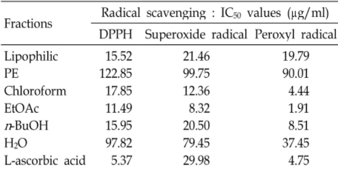
pini (CY001) fractions, we performed DPPH, superoxide rad- icals and peroxyl radicals assay, and these results were shown in Table 3. These different fractions showed variable DPPH radical-scavenging activities (Table 3). The EtOAc, chloroform, n-BuOH, and lipophilic fraction exerted free radical scavenging effects in a dose dependent manner (data not shown). In particular, the EtOAc, chloroform,n-BuOH, and lipophilic fraction showed strong antioxidant activity (IC50: 11.49; 17.85; 15.95; 15.52), but its effect was less than that of L-ascorbic acid (IC50: 5.37). The PE and water fraction had comparatively small of antioxidant effect (IC50: 122.85;
97.82). These antioxidant activities had positive correlations with their total phenolic content in the different fractions ofP. pini (CY001). With respect to superoxide radicals scav- enging, the results indicated that different fractions showed Table 2. Total phenolic contents of P. pini (CY001) extracts
Fractions Gallic acid equivalents (mg/g) Lipophilic
PE Chloroform EtOAc n-BuOH H2O
203.8±0.65 8.3±0.23 204.9±1.97 436.5±1.53 107±0.78 28.4±0.60
The total phenolic contents were expressed as mg of GAEs per g of extracts (means±S.D. of three independent measurements done in triplicate). Each extract was prepared fromP. pinias described in the Materials and Methods.
Table 3. Radical scavenging activity and lopoxygenase in- hibition of P. pini (CY001) extracts
Fractions Radical scavenging : IC50 values (μg/ml) DPPH Superoxide radical Peroxyl radical Lipophilic
PE Chloroform EtOAc n-BuOH H2O
L-ascorbic acid 15.52 122.85 17.85 11.49 15.95 97.82 5.37
21.46 99.75 12.36 20.508.32 79.45 29.98
19.79 90.01 4.44 1.918.51 37.45 4.75 Data shown were the mean IC50 values (μg/ml) as 50% in- hibition concentration compared to the blank (buffer instead of fractions) in activity of three independent assays done in triplicate. L-ascorbic acid was used as a positive control.
similar tendency to inhibit this free radical. In particular, the ability to scavenge superoxide radicals was higher for EtOAc, chloroform, n-BuOH, and lipophilic fraction (IC50: 0.32; 1.23; 5.5; 11.46) than for L-ascorbic acid (IC50: 29.98).
Similar responses were observed for peroxyl radicals. These results had also indicated that both the EtOAc and chloro- form fraction (IC50: 1.91; 4.44) was similar than L-ascorbic acid (IC50: 4.75). Therefore, the EtOAc fraction of P. pini (CY001) showed the most potent the capacity to scavenge free radicals and these antiradical reactivity may be attrib- uted to the total phenolic contents constituting EtOAc fraction.
LOX inhibitory activity ofP. pini (CY001) fractions
In this study, we elucidated the possible contribution to the LOX inhibitory activity ofP. pini (CY001) fractions. The LOX activity was monitored as an increase in the absorbance at 234 nm, which reflects the formation of hydro- peroxylinoleic acid. When tested for inhibition of the en- zyme LOX in vitro, which peroxidizes polyunsaturated fatty acids, such as linoleic acid to their respective hydroperoxy derivatives, P. pini (CY001) fractions showed inhibitory activity. The results were shown in Table 4. Clearly, lip- ophilic fraction (22.17%), chloroform (33.80%), and EtOAc fraction (70.97%) significantly inhibited the LOX-catalyzed oxidation of linoleic acid at 50 μg/ml dose in vitro, and the highest inhibitory effect was obtained for EtOAc fraction.
In conclusion, these data were presented that the EtOAc frac- tion contained the highest total phenolic content and showed potent radicals scavenging activities and LOX inhibitory ac- tivity amongP. pini (CY001) fractions. Therefore, we used
Table 4. Effects ofP. pini(CY001) extracts of LOX activity and LPS-induced NO production of RAW 264.7 cells Fractions aInhibition LOX
activity (%)
.bInhibition of NO production (%) Lipophilic 25 μg/ml
50 μg/ml 10.33±1.61
22.17±2.56 31.55±1.74 41.57±1.23
PE 25 μg/ml
50 μg/ml 1.23±0.75
3.13±0.91 8.05±0.57 8.50±1.18 Chloroform 25 μg/ml
50 μg/ml 8.20±1.08
33.80±3.52 15.57±1.08 35.67±3.55
EtOAc 25 μg/ml
50 μg/ml 15.13±1.10
70.97±3.62 34.13±2.80 55.51±2.73 n-BuOH 25 μg/ml
50 μg/ml 5.70±1.14
10.27±1.46 10.15±1.21 12.23±0.91
H2O 25 μg/ml
50 μg/ml 4.83±1.53
6.07±0.24 2.71±1.21 5.88±1.20 L-ascorbic acid 25 μg/ml
50 μg/ml 6.80±1.40
10.80±2.71 -
-
abExpressed as percent inhibition compared to control (vehicle).
bRAW264.7 cells were incubated with each extract for 4 hr and followed by LPS stimulation (1 μg/ml) for 24 hr.
the EtOAc fraction in RAW 264.7 cell and HepG2 cell assay related to inflammation.
Inhibition of P. pini (CY001) fractions on LPS- induced NO production
To determine anti-inflammatory roles of P. pini (CY001) fractions between LOX activity and NO production in the LOX pathway, we were treated with different fractions of P. pini (CY001) in LPS-induced NO production in RAW264.7 cells. After a 24 hr incubation, each fraction was produced a dose-dependent decrease in nitrite levels (Table 4). In this result, treatment with water fractions ofP. pini (CY001) did not show any inhibition in the preliminary test and among P. pini (CY001) fractions, the EtOAc fraction was found to be most effective in the inhibition of NO production in RAW 264.7 cell. When the EtOAc fraction was also treated at different concentrations, it provided anti-inflammatory activity on LPS-induced NO production in dose-dependent manner and the LPS-untreated cells had no effect on NO release. L-MMNA, well known as a NOS inhibitor, was used as a positive control, and showed inhibition of 35%
in NO production at 100 μM (Fig. 1). In addition to, we have confirmed cell viability of RAW 264.7 cells in a dose-de- pendent manner. The results of the cell viability assay ap- peared in Fig. 1. The data showed no cytotoxicity with all
Fig. 1. Inhibitory effect ofP. pini(CY001) ethyl acetate fraction (EtOAc) on LPS-induced nitric oxide secretion of macro- phages. RAW 264.7 cells were treated with LPS (1 μg /ml) in the presence or absence of ethyl acetate fraction for 24 hr. L-NMMA was used as a positive control. The viability of cells was determined by MTT assay and ni- trite level of culture supernatant was determined by us- ing Griess reagents. Three independent assays were per- formed in triplicate and the data shown are the mean±
S.D. of the percent inhibition of nitrite and cell viability compared with the control. (*p<0.05, vs. control).
concentrations up to 100 μg/ml of the EtOAc fraction ofP.
pini (CY001). In the Table 4, the EtOAc fraction (50 μg/ml) of P. pini (CY001) reduced by 70.97% in LOX activity and by 55.51% in NO production. Taken together, the EtOAc fraction of P. pini (CY001) possessed strong anti-in- flammatory activity.
Hepatoprotection of P. pini (CY001) fractions on tacrine-induced cytotoxicity in HepG2 cells
In the present study, we investigated hepatoprotective ac- tivity of the EtOAc fraction of P. pini (CY001) in the liver damage related to inflammation. The EtOAc fraction was treated on tacrine-induced cytotoxicity in HepG2 cells and we identified the cytotoxicity in a dose-dependent manner by us- ing the MTT assay method. The cytotoxicity of the EtOAc fraction didn’t detect concentrations of 1-500 μg/ml (data not shown). Also the value of the 50% effective concentration (EC50) of the EtOAc fraction was 500 μg/ml and showed hep- atoprotection of 5~50% at each concentration (1~500 μg/ml).
Silymarin, well known for its hepatoprotective efficiency, was used as a positive substance, and showed a protective effect of 46% at 100 μg/ml (Fig. 2).
Discussion
Many natural antioxidants have already been isolated
Fig. 2. Hepatoprotective effect ofP. piniethyl acetate (EtOAc) fraction on tacrine-induced cytotoxicity in HepG2 cells.
HepG2 cells were treated with tacrine (1 mM) in the presence or absence of ethyl acetate fraction for 2 hr.
Silymarin was used as a positive control. Cytotoxicity was measured by MTT assay. Three independent as- says were performed in triplicate and data shown are the mean±S.D. of the percent inhibition of nitrite and cell viability compared with the control. (*p<0.05, vs.
control).
from different kinds of plant materials, such as oilseeds, ce- real crops, vegetables, leaves, roots, spices, and herbs.
Scavenging effects of mushrooms may serve as a significant indicator of its potential antioxidant activity as a radical scavenger and phenols are also important mushrooms con- stituents because of their radical scavenging ability [34]. A correlation between the concentration of their phenolics and the total antioxidant capacity has been some reported [31].
P. linteus is a well-known fungus of the genus Phellinus and extracts of them could be attributed, at least partially, to its ability of inducing antioxidant enzyme activities and increas- ing the glutathione (GSH) level. Some components of the PL preparation showed protection against reactive oxygen species (ROS) [15,39,43,44]. ButP. pini is little known many research report as yet in pharmacological action. Indeed, for the measurement of the antioxidant activity,P. pini (CY001) were fractionated by different organic solvent and we have investigated the total phenolic contents in the presence of these fractions. Among them, the highest concentration (436.5 mg/g) of the total phenolics was present in the EtOAc fraction ofP. pini (CY001), whereas the lowest concentration (8.3 mg/g) of phenolics was recorded in the PE fraction (Table 2). The antioxidant properties were evaluated though DPPH (2, 2-diphenyl-1-picrylhydrazyl) radical scavenging activity, superoxide radicals scavenging activity, oxidative inhibition of 2, 20-azobis (2-amidinopropane) dihydro chlor- ide (AAPH) and inhibition of lipid peroxidation by LOX
acitity. In the table 3 and 4, the EtOAc fraction amongP.
pini (CY001) fractions showed the most potent the capacity to scavenge free radicals. These results were presented that the EtOAc fraction amongP. pini (CY001) was contained the most phenolic contents and all these antioxidant activity pa- rameters were also correlated to the phenolic content.
Antioxidant compounds reduced the action of reactive oxygen species (ROS) in tissue damage. Once formed, ROS depletes antioxidant defenses, rendering the affected cells and tissues more susceptible to oxidative damage [49]. The oxidative damage caused by free radicals may be related to aging and diseases, such as atherosclerosis, diabetes, cancer and cirrhosis [14]. Especially, oxidative damage is a major pathophysiological event in a broad range of inflammatory states, including the early stages of atherosclerosis [7,14,37].
Tissue damage and adverse effects due to excess in- flammation may therefore be reduced by the use of suitable anti-oxidants which prevent the formation of oxygen free radicals, or scavenge them once they are formed and before they react with sites such as unsaturated lipids in the cell membranes [38-40]. The processes associated with the in- flammatory response are complex but important aspects which have been exploited for screening for anti-in- flammatory compounds are the various functions of macro- phage, the metabolic products of arachidonic acid and the role played by reactive oxygen species (ROS) [27]. Arachidonic acid is released from cell membranes by phospholipase A2 (PLA2) under the stimulus of several factors associated with inflammation. The products of metabolism of arachidonic acid are collectively known as eicosanoids and the two most important groups are the prostaglandins and leukotrienes, formed by the actions of cyclo-oxygenases (COX) and LOX, respectively [50]. In particular, LOXs comprise a family of non-heme iron-containing dioxygenases, representing the key enzymes in the biosynthesis of leukotrienes that have been postulated to play an important role in the pathophysi- ology of several inflammatory and allergic diseases [51]. In this study, as a preceding step to identify active principle(s) of P. pini (CY001), this was successively fractionated with different organic solvents.P. pini (CY001) fractions appeared to significantly inhibit enzymatic lipid peroxidation medi- ated by the LOX activity at 50 μg/ml and in particular, lip- ophilic fraction (22.15%), chloroform fraction (33.31%), and EtOAc fraction (70.95%) seems to be the more active fraction (Table 4). Among the fractions, the EtOAc fraction appeared to be most effective inhibition in lipid peroxidation, imply-
ing that P. pini (CY001) would contain active anti-in- flammatory component(s) related to LOX pathway.
The enzymes PLA2, LOX and COX are involved in IL-1 β-induced NO production and iNOS protein expression [16].
Nitric oxide (NO) synthesis by inducible nitric oxide syn- thase (iNOS) is increased in inflammatory diseases and leads to cellular injury [1,17]. Macrophages play a crucial role in the generation of pro-inflammatory molecules like nitric ox- ide (NO). After stimulation with bacterial lipopolysaccharide (LPS), many cells including macrophages express the iNOS which is responsible for the production of large amount of NO [40]. Also the aberrant release of NO can lead to amplifi- cation of inflammation, as well as tissue injury such as hep- atotoxicity [2,12,51,52]. For example, Kupffer cells in liver produce large amounts of the free radical nitric oxide (NO·) from the inducible form of nitric oxide synthase (iNOS) when stimulated by cytokines or lipopolysaccharide [4,19,28, 42,45]. Indeed, inhibition of NO production by antioxidant is a very important therapeutic target in the development of anti-inflammatory agents. These antioxidative properties have also been examined in foods or plants, however, it has yet to be examined whether theP. pini have such activities.
In the present study, six fractions of P. pini (CY001) were checked for their inhibitory effect on nitric oxide production from macrophages (RAW 264.7 cells) induced by LPS. The EtOAc fraction among these fractions showed strong in- hibitory activity on nitric oxide production in induced cells (Table 3). Also this fraction produced a dose-dependent de- crease in nitrite and the results were supported by the cell viability experiment (cell viability >90%) (Fig. 1). In addi- tion, the EtOAc fraction of P. pini (CY001) showed hep- atoprotection on tarcine-inudced cytotoxicty in HepG2 cell (Fig. 2). Therefore, intake of oxygen radical scavengers (antioxidants and phytochemicals) fromP. pini (CY001) may be a good defense mechanism for hepatoprotection. Many related studies have shown that sesquiterpenes, coumarins, diarylheptanoids and phenolic compounds exhibit hep- atoprotective activities on the tacrine-induced cytotoxicity of HepG2 cells [29]. Despite the important number of past in- vestigations, the relationships between antioxidative effect and related to inflammation disease remained obscure.
Consequently, many researchers have attempted to find the mechanisms that govern these radical scavenging activities and the correlations between them.
In conclusion, we suggested that the crude fractions iso- lated fromP. pini (CY001), which contained abundant phe-
nolic compound as potent radical scavenging activities showed hepatoprotection by anti-inflammatory activity.
Further study will be required to investigate the detailed mechanisms of the relationship between the antioxidative and hepatoprotective acitivities of liver damage by in- flammation from the EtOAc fraction of P. pini (CY001) in vitro and in vivo model.
Acknowledgement
This study was supported by research funds from Dong-A University.
References
1. Acquisto, F., M. J. May, and S. Ghosh. 2002. Inhibition of nuclear factor kappa B (NF-κ B): an emerging theme in an- ti-inflammatory therapies. Mol. Interv. 2, 22-35.
2. Adachi, Y., B. U. Bradford, W. Gao, H. K. Bojes, and R.
G. Thurman. 1997. Inactivation of Kupffer cells prevents early alcohol-induced liver injury. Hepatology20, 453-460.
3. Bezakova, L., P. Mueaji, E. Eisenreichova, M. Haladova, I.
Paulikova, and M. Obložinsky. 2004. Effect of different com- pound from Lilium candidumL. on lipoxygenase activity.
Acta. Facult Pharm Univ. Comenianae 51, 45-50.
4. Bonino, F. and M. R. Brunetto. 2003. Chronic hepatitis B e antigen (HBeAg) negative, anti-HBe positive hepatitis B:
an overview. J. Hepatol. 39, 160-163.
5. Boyer, C. S., G. L. Bannenberg, E. P. Neve, A. Ryrfeldt, and P. Moldeus. 1995. Evidence for the activation of the sig- nal-responsive phospholipase A2 by exogenous hydrogen peroxide. Biochem. Pharmacol. 50, 753-761.
6. Cathcart, R., E. Schwiers, and B. N. Ames. 1983. Detection of picomole levels of hydroperoxides using a fluorescent dichlorofluorescein assay. Anal. Biochemistry134, 111-116.
7. Chakraborti, S. and J. R. Michael. 1993. Role of protein kin- ase C in oxidant-mediated activation of phospholipaseA2 in rabbit pulmonary arterial smooth muscle cells. Mol. Cell.
Biochem. 122, 9-15.
8. Chen, X., A. Gresham, A. Morrison, and A. Pentland. 1996.
Oxidative stress mediated synthesis of cytosolic phospholi- pase A2 after UVB injury. Biochim. Biophys. Acta1299, 23-33.
9. Cho, J. H., S. D. Cho, H. Hu, S. H. Kim, S. K. Lee, Y. S.
Lee, and K. S. Kang. 2002. The roles of ERK1/2 and p38 MAP kinases in the preventive mechanisms of mushroom P. linteus against the inhibition of gap junctional inter- cellular communication by hydrogen peroxide.Carcinogenesis 23, 1163-1169.
10. Cuzzocrea, S., D. P. Riley, A. P. Caputi, and D. Salvemini.
2001. Antioxidant therapy: a new pharmacological approach in shock, inflammation and ischemia/reperfusion injury.
Pharmacol. Revi. 53, 135-159.
11. Decker, E. A. 1997. Phenolics: prooxidants or antioxidants.
Nutri. Revi. 55, 396-398.
12. Evans, C. H. 1995. Nitric oxide: what role does it play in inflammation and tissue destruction.Agents Actions. Suppl.
47, 107-116.
13. Fichtner-Feigl, S., I. J. Fuss, J. C. Preiss, W. Strober, and A.
Kitani. 2005. Treatment of murine Th1- and Th2-mediated inflammatory bowel disease with NF-kappa B decoy oligonucleotides. J. Clin. Invest15, 3057-3071.
14. Halliwell, B. and J. M. C. Gutteridge. 1998. Free Radicals in Biology and Medicine.Endocrinology 16, 241-248.
15. Han, S. B., C. W. Lee, Y. J. Jeon, N. D. Hong, I. D. Yoo, K. H. Yang, and H. M. Kim. 1999. The inhibitory effect of polysaccharides from P. linteus on tumor growth and metastasis. Immunopharmacology41, 157-164.
16. Hashimoto, T., M. Kihara, K. Yokoyama, T. Fujita, and S.
I. Kobayashi. 2003. Lipoxygenase Products regulate nitric oxide and inducible nitric oxide synthase production in in- terleukin-1β stimulated vascular smooth muscle cells.
Hypertens. Res. 26, 177-184.
17. Heras, B. D. L., M. J. Abad, A. M. Silvan, R. Pascual, P.
Bermejo, B. Rodriguez, and A. M. Villar. Effects of six di- terpenes on macrophage eicosanoid biosynthesis.Life Sci.
70, 269-278.
18. Hibbs, J. B., R. R. Taintor, Z. Vavrin, and E. M. Rachlin.
1988. Nitric oxide: a cytotoxic activated macrophage effector molecule. Bioch. Biophy. Res. Comm. 157, 87-94.
19. Iida, S., H. Ohshima, S. Oguchi, T. Hata, H. Suzuki, H.
Kawasaki, and H. Esumi. 1992. Identification of inducible calmodulin-dependent nitric oxide synthase in the liver of rats.J. Biol. Chem. 267, 25385-25388.
20. Ishikawa, Y., K. Morimoto, and T. Hamasaki. 1984.
Flavoglaucin, a metabolite of Eurotium chevalieri, its anti- oxidation and synergism with tocopherol.J. Am. Oil. Chem.
Soc. 61, 1864-1868.
21. Kim, H. M., S. B. Han, G. T. Oh, Y. H. Kim, D. Hong, N.
D. Hong, and I. D. Yoo. 1996. Stimulation of humoral and cell mediated immunity by polysaccharide from mushroom P. linteus. Int. J. Immunopharmacol. 18, 295-303.
22. Kim, M. O., G. Y. Kim, B. H. Nam, C. Y. Jin, K. W. Lee, J. M. Park, S. J. Lee, and J. D. Lee. 2005. Development of species-specific primers for rapid detectionP.linteusandP.
baumii. Mycobiology 33, 104-108.
23. Kim, Y. K., R. G. Kim, S. J. Park, J. H. Ha, J. W. Choi, H.
J. Park, and K. T. Lee. 2002. In vitro anti-inflammatory activ- ity of kalopanaxsaponin A isolated fromKalopanax pictus in murine macrophage RAW 264.7 cells.Biol. Pharmaceut.
Bull.25, 472-476.
24. Kirby, A. J. and R. J. Schmidt. 1997. The antioxidant activity of Chinese herbs for eczema and of placebo herbs-I. J.
Ethnopharmacol.56, 103-108.
25. Klotz, L., M. Schmidt, T. Giese, M. Sastre, P. Knolle, T.
Klockgether, and M. T. Heneka. 2005. Proinflammatory stimulation and pioglitazone treatment regulate peroxisome proliferator-activated receptor gamma levels in peripheral blood mononuclear cells from healthy controls and multiple sclerosis patients. J. Immunol. 175, 4948-4955.
26. Larsen, M. J. and L. A. Cobb-Poulle. 1990. Phellinus (Hymenochaetaceae): A survey of the world taxa. Synop.
Fung. 3, 1-206.
27. Laupattarakasem, P., P. J. Houghton, J. R. Hoult, and A.
Itharat. 2003. An evaluation of the activity related to in- flammation of four plants used in Thailand to treat arthritis.
J. Ethnopharmacol. 85, 207-215.
28. Li, J. and T. R. Billiar. 1999. Nitric oxide: IV. Determinants of nitric oxide protection and toxicity in liver.Am. J. Physiol.
276, 1069-1073.
29. Lin, J. M., C. C. Lin, M. F. Chen, and T. A. Ujiie. 1995.
Takata. Radical scavenging and antihepatotoxic activity of Ganoderma formosanum, G.lucidum, G. lucidum and G.
neo-japonicum. J. Ethnopharmacol. 47, 33-41.
30. Lourenço, A., A. M. Lobo, B. Rodríguez, and M. L. Jimeno.
1996. Ceramides from the fungusP. pini.Phytochemistry43, 617-620.
31. Madsen, H. L., B. R. Nielsen, G. Bertelsen, and L. H.
Skibsted. 1996. Screening of antioxidative activity of spices.
Food Chemistry 57, 331-337.
32. Maiga, A., K. E. Malterud, D. Diallo, and B. S. Paulsen. 2006.
Antioxidant and 15-lipoxygenase inhibitory activities of the Malian medicinal plants Diospyros abyssinica (Hiern) F.
White (Ebenaceae), Lannea velutina A. Rich (Anacardiaceae) and Crossopteryx febrifuga (Afzel) Benth. (Rubiaceae). J.
Ethnopharmacol. 104, 132-137.
33. McCune, L. M. and T. Johns. 2002. Antioxidant activity in medicinal plants associated with the symptoms of diabetes mellitus used by the indigenous peoples of the North American boreal forest. J. Ethnopharmacol. 82, 197-205.
34. Meir, S., J. Kanner, B. Akiri, and S. P. Hadas. 1995.
Determination and Involvement of Aqueous Reducing Compounds in Oxidative Defense Systems of Various Senescing Leaves. J. Agric. Food. Chem. 43, 1813-1819.
35. Natarajan, R., R. G. Gerrity, J. L. Gu, L. Lanting, L. Thomas, and J. L. Nadler. 2002. Role of 12-lipoxygenase and oxidant stress in hyperglycaemia-induced acceleration of athero- sclerosis in a diabetic pig model. Diabetologia45, 125-133.
36. Natarajan, R., W. Bai, V. Rangarajan, N. Gonzales, J. L. Gu, L. Lanting, and J. L. Nadler. 1996. Platelet-derived growth factor BB mediated regulation of 12-lipoxygenase in porcine aortic smooth muscle cells. J. Cell. Physiol. 169, 391-400.
37. Ross, R. 1993. The pathogenesis of atherosclerosis.Nature 362, 801-809.
38. Page, M., N. Bejaoui, B. Cinq-Mars, and P. Lemieux. 1988.
Optimization of the tetrazolium-based colorimetric assay for the measurement of cell number and cytotoxicity.Int.
J. Immunopharmacology 10, 785-793.
39. Park, J., B. R. Lee, L. H. Jin, C. K. Kim, and K. S. Choi.
2001. The stimulatory effects of Garnoderma lucidum and P.linteuson the antioxidant enzyme catalase.J. Biochem. Mol.
Biol. 34, 144-149.
40. Saha, K., N. H. Lajis, D. A. Israf, A. S. Hamzah, S. Khozirah, S. Khamis, and A. Syahida. 2004. Evaluation of antioxidant and nitric oxide inhibitory activities of selected Malaysian medicinal plants. J. Ethnopharmacol. 92, 263-267.
41. Sakagami, T., J. Vella, M. F. Dixon, J. Rourke, F. Radcli, P. Sutton, T. Shimoyama, K. Beagley, and A. Lee. 1997. The endotoxin ofHelicobacter pyloriis a modulator of host-de- pendent gastritis.Infect. Immun. 65, 3310-3316.
42. Sass, G., K. Koerber, R. Bang, H. Guehring, and G. Tiegs.
2001. Inducible nitric oxide synthase is critical for immune- mediated liver injury in mice.J. Clin. Invest.107, 439-447.
43. Shon, Y. H. and K. S. Nam. 2001. Antimutagenicity and in- duction of anticarcinogenic phase II enzymes by basidiomycetes. J. Ethnopharmacol. 77, 103-109.
44. Song, Y. S., S. H. Kim, J. H. Sa, C. Jin, C. J. Lim, and E.
H. Park. 2003. Antiangiogenic, antioxidant and xanthine ox- idase inhibition activities of the mushroom P. linteus. J.
Ethnopharmacol.88, 113-116.
45. Thurman, R. G. 1998. Mechanisms of hepatic toxicity II.
Alcoholic liver injury involves activation of Kupffer cells by endotoxin. Am. J. Physiol. 275, 605-611.
46. Valkonen, M. and T. Kuusi. 1997. Spectrophotometric assay for total peroxyl radical-trapping antioxidant potential in human serum.J. Lipid Res. 38, 823-833.
47. Vernooy, J. H., M. A. Dentener, R. J. van Suylen, W. A.
Buurman, and E. F. Wouters. 2002. Long-term intratracheal lipopolysaccharide exposure in mice results in chronic lung inflammation and persistent pathology.Am. J. Respir. Cell.
Mol. Biol.26, 152-159.
48. Walsh, N. C., T. N. Crotti, S. R. Goldring, and E. M.
Gravallese. 2005. Rheumatic diseases, the effects of in- flammation on bone.Immunol. Rev. 208, 228-251.
49. Wettasinghe, M. and F. Shahidi. 2000. Scavenging of re- active-oxygen species and DPPH free radicals by extracts of borage and evening primrose meals.Food Chemistry70, 17-26.
50. Williams, C. S., M. Mannand, and R. N. DuBois. 1999. The role of cyclooxygenases in inflammation, cancer, and development.Oncogene18, 7908-7916.
51. Xie, B. J. and H. Shi. 1994. Antioxidant properties of frac- tions andpolyphenol constituents from green, oolong and black teas. Nat. Prod. Res.6, 19-25.
52. Yu, B. P. 1994. Cellular defenses against damage from re- active oxygen species. Physiol. Rev. 74, 139-162.
초록:상황버섯(Phellinus pini)의 라디칼 소거작용 남병혁․조월순1․최 영․최유진1․이재동*․정민호1*
(부산대학교 자연과학대학 미생물학과, 1동아대학교 의과대학 기생충학교실)
Phellinus pini (CY001) 추출물에서의 페놀류성분의 농도는 P. pini (CY001)분획 g 당 GAEs mg으로 나타내었고, P. pini 의 EtOAc 분획(436.5 mg GAEs/g)이 다른 분획에 비해 가장 높은 함량을 나타내었다. 환원력과 같은 항산 화 활성을 screening 하기 위해 몇 가지 생화학적 시험이 수행되었고, 그것은 2, 2-diphenyl-1-picrylhydrazyl (DPPH) 라디칼 소거작용, NBT/XO superoxide system, DCF/AAPH peroxyl radicals 저해능 등이다. 여섯개의 버섯추출물 분획 중에서 EtOAc 분획이 DPPH, superoxide 라디칼, peroxyl 라디칼 소거작용이 뛰어났고, IC50
values는 각각 11.49 μg/ml, 8.32 μg/ml, and 1.91 μg/ml이었다.P. pini (CY001)의 EtOAc 분획은 중요한 효소적 지질과산화 저해와 cytotoxicity 없이 RAW 264.7 macrophages의 LPS 유도 NO 생성을 효과적으로 감소시켰다.
또한 EtOAc 분획이 HepG2 cell의 타크린 유도 cytotoxicity에서 간 보호 작용을 나타내었다. 이러한 결과를 바탕
으로P. pini (CY001)가 라디칼 소거작용을 하는 성분을 함유한 천연 항산화제로써의 잠재력을 갖고 있는 것으로
사료된다.