ans의 부착에 미치는 영향
2
단국대학교 치과대학 치과보철학교실 박수정, 송영균*
의치상레진의 3D 프린팅 출력 각도가
Candida albicans의 부착에 미치는 영향
The build angle of 3D printing denture base resin on candida albicans adhesion.
Department of prosthodontics, College of Dentistry, Dankook University, Cheonan, Republic of korea Su-Jung Park, Young-Gyun Song*
Purpose: The purpose of this study is to compare the adhesion of Candida albicans according to build angle in 3D printing denture base resin.
Methods: The 3D printing was performed by setting the build angle of the disk type specimen designed by CAD program at 0 degree, 30 degrees, 60 degrees, and 90 degrees. Surface roughness was measured using a non-contact 3D microsurface profiler. The specimens were incubated in Candida albicans suspension for 24 hours. The attached Candida albicans were detached by cell scraper. The suspension of detached C. albicans was serially diluted and plated on Trypticase soy broth.
After 48 hours of incubation, total colony forming unit was counted.
Results: There was no significant difference in surface roughness(Sa) between the test groups, but the interlayer boundary was observed. There was no statistically significant difference in total colony forming units of Candida albicans between the test groups.
Conclusion: There was no difference in the average surface roughness and adhesion of Candida albicans between the specimens. It is considered that the setting of the build angle should be set considering the accuracy or strength rather than the roughness of the surface.
Keywords: Build angle, 3D printing, Candida albicans
Corresponding Author Young-Gyun, Song
Department of prosthodontics, College of Dentistry, Dankook University,
119, Dandae-ro, Dongnam-gu, Cheonan-si, Chungnam, 31116, Republic of Korea.
Tel: +82-41-550-1932, E-mail: [email protected]
ABSTRACT
ORIGINAL ARTICLE
Ⅰ. 서론
보철분야에서 폴리머는 1930년대부터 의치를 제작하 기 위한 재료로 널리 사용되어 왔으며, polymethylme- thacrylate(PMMA)를 비롯한 여러 재료들이 현재 사용 되어 지고 있다1). 의치상 재료로서의 폴리머는 강도, 색 조 안정성, 열적 성질 등 여러 조건을 만족해야 한다. 임 상적인 측면에서 보았을 때 위와 같은 성질을 모두 충 족 시키는 것 이외에도 세균 부착에 대한 고려도 필요하 며, 특히, 가철성 보철물을 장착한 환자에서 많이 발생하 는 의치성 구내염의 주된 원인균인 Candida albicans 에 대한 고려가 필요하다2, 3). 의치의 경우, 구강 내로 노 출된 조직면은 연마를 통해 거칠기를 조절하여 세균의 부착율을 낮추어 줄 수 있으나, 구강조직과 닿는 조직면 의 경우, 의치의 적합성을 이유로, 연마가 불가능하기 때 문에, 재료나 제작방식에 따른 표면의 특성 매우 중요 한 부분이다.
3D 프린팅 기술을 이용한 의치상 제조방법은 기존의 제작방식에 비해, 기공 과정이 간단하며, 동일한 보철물 을 재제작할 수 있다는 장점 때문에, 최근 많이 연구되는 방법이다4). 3D 프린팅의 대표적인 방식에는 FDM(Fused deposition modeling), SLA(Stereolithographic ap- paratus), DLP(Digital light processing)등이 있으며, 치과영역에서는 속도와 정밀성등으로 고려하여, SLA 방 식과 DLP방식이 주로 사용되고 있다5). 이 중에, SLA 방 식은 정밀도는 높으나 적층시간이 상대적으로 많이 소 요되기 때문에, 의치상을 만드는 방법에는 DLP 방식이 많이 선호되고 있다. DLP 방식은 광중합레진을 광원을 digital micromirror을 이용하여 원하는 위치에 비추어 적층하는 방식으로 출력을 위해서는 출력 각도를 임의 대로 설정할 수 있다6, 7). 출력 각도는 3D 프린팅시에 술 자의 임의대로 설정할 수는 있지만, 출력각도에 따라 정 확도와 강도에 유의한 차이가 있어, 출력각도를 설정할
때에는 이러한 요소를 고려해야 한다는 보고가 있다8, 9). 이번 연구에서는 3D 프린팅으로 제작된 의치상레진 에서 출력각도에 따른 Candida albicans의 부착 정도 를 비교분석하고자하며, 귀무가설은 ‘3D 프린팅으로 제 작된 의치상레진에서 출력각도에 따른 C. albicans의 부착정도는 차이가 없다’이다.
Ⅱ. 재료 및 방법
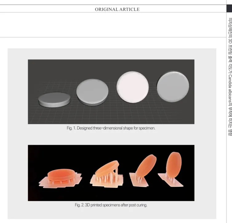
시편의 제작을 위해 직경 20mm, 두께 3mm의 디스 크를 CAD프로그램(meshmixer, Autodesk, San Ra- fael, CA, USA)을 이용하여, 디자인하였다(Fig. 1). 디자 인한 디스크를 3D 프린팅하기 위해 출력각도를 0도, 30 도, 60도, 90도로 설정하여 서포트를 설계하였고, 3D 프 린팅 의치상레진(Nextdent base, Nextdent BV, Soes- terberg, Netherlands)을 이용하여, DLP 기반의 3D 프 린터(Phrozen Shuffle, phrozen, Hsincu city, taiwon) 로 출력하여 isoprophylalcohol을 이용하여 비경화 레 진을 세척하였으며, UV 후경화를 통해 잔류모노머를 최 소화시켜 각 각도당 16개의 시편을 제작하였다(Fig. 2).
또한 실험 전에 36도 증류수에 24시간 보관하였다.
출력된 시편의 서포트를 제거하고, 표면 분석을 위해, 비접촉 3차원 미세 형상 측정기(NV-2000, Nano Sys- tem Co., Daejeon, Korea)를 이용하여 표면을 분석하 였다. 표면분석은 서포트가 위치하지 않은 면을 기준으 로 측정하였다.
본 실험에서 의치구내염 관련 세균으로 Candida al- bicans ATCC 10231을 사용하였으며, 단순 진균 증식 을 위해서 Trypticase soy broth (TSB; BD bioscience, San Jose, CA, USA)를 이용하여 24시간 동안 37℃ 호 기상태에서 180 rpm 설정으로 교반기(Mini shaker, JEIO TECH, Daejeon, Korea)를 이용하여 배양하였
ans의 부착에 미치는 영향
다. C. albicans의 균사막 (biofilm)을 시편표면에 형성 시키기 위해서 시편을 ethylene oxide 가스멸균기(HS- 3241EO, Hanshin medical, Incheon, Korea)를 이용 하여 멸균시켰다. 구강내 진균 형태(hyphae)의 균사막 형성을 위해 시편을 12-well cell culture plate(Nunc™
Cell-Culture Treated Multidish, Thermo Fisher Sci- entific inc., Waltham, MA, USA)위치 시킨 후, Ham’s F-12 nutrient 배지 (Hyclone, Rockford, IL, USA)에 침적시키고 C. albicans를 접종한 후, 37℃ 호기상태에 서 24시간 배양하였다(Fig. 3). 배양 후 부착되지 않은 C.
albicans를 제거하기 위해 phosphate buffered saline 으로 시편을 3회 세척하였다. 디스크의 측면을 멸균된 핀셋을 이용하여 고정한 뒤 Cell scraper(Nunc™ Cell Scraper, Thermo Fisher Scientific inc., Waltham, MA, USA)를 이용하여, 프린팅시 서포트가 위치하지 않 은 면에 부착된 C. albicans를 분리한 뒤, Trypticase soy broth (TSB; BD bioscience, San Jose, CA, USA) 배지에 침적시킨 후 37℃ 호기성 배양기(WIG01105, DAIHAN SCIENTIFIC CO, Seoul, Korea)에서 12시간 동안 배양하였다. 이후 배양액을 10배, 100배, 1000배 Fig. 1. Designed three-dimensional shape for specimen.
Fig. 2. 3D printed specimens after post curing.
ORIGINAL ARTICLE
희석하여, Tryptic soy agar (TSA; BD Difco, Detroit, MI, USA) 배지에 접종하여, 확인하였다. 1000배 희석한 배양액은 Streptomycin (20 ug/ml)이 된 Sabouraud's dextrose agar 표면에 접종 후 spreader(SPL Life Sci- ences Co., Ltd, Pocheon, Korea)를 이용하여 넓게 펴 준 후, 37℃ 호기상태에서 24시간 배양한 뒤, 집락수 (CHU; Colony forming unit)를 측정하였다.
배양액의 총 집락수 분석은 IBM SPSS V25 (IBM corp., Amonk, NY, USA)를 이용하여, one-way anal- ysis of variance(ANOVA)로 분석하였다. 유의 수준은 95%로 정하였고, Tukey test를 이용하여 사후 검정을 시행하였다.
Ⅲ. 결과
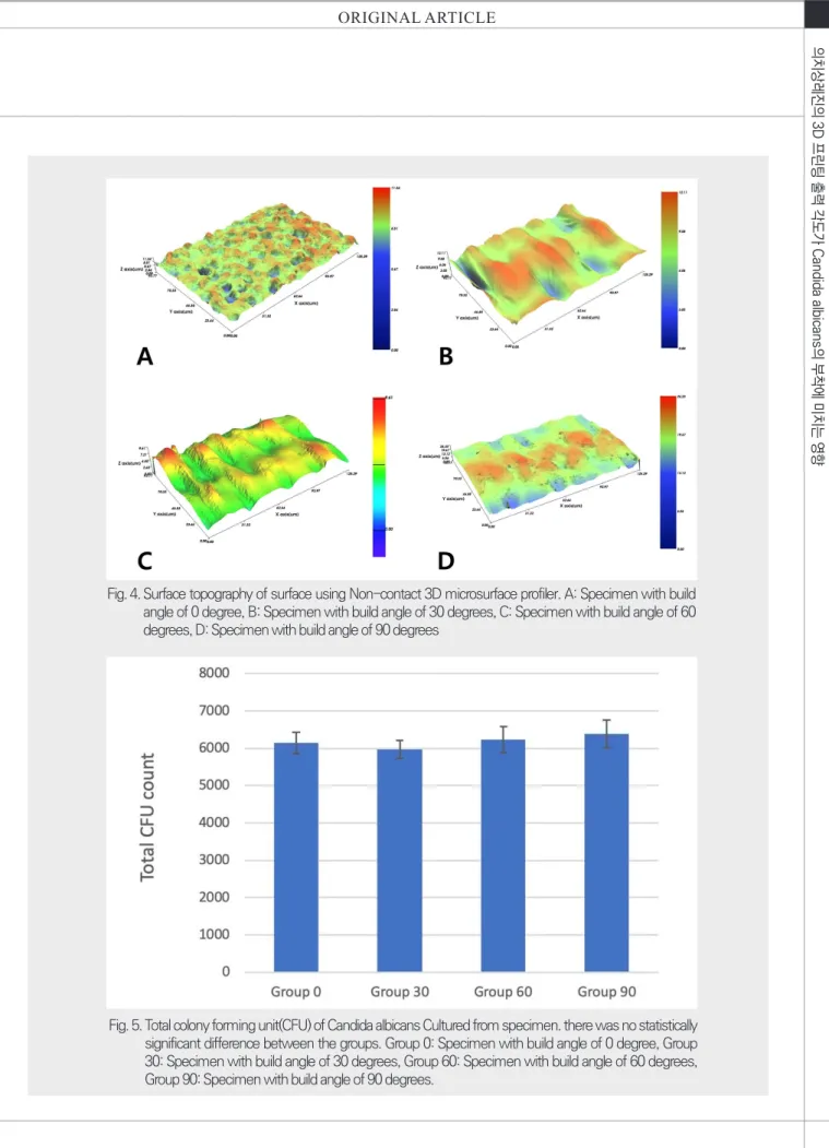
측정된 표면 거칠기(Sa)의 평균값은 Table I. 과 같이 나타났다. 표면 거칠기는 그룹간 유의한 차이는 없었으
나, Surface topography에서 관찰시 적층에 의한 층간 경계의 형상이 서로 다르게 관찰되었다(Fig. 4).
각 시험군의 colony forming unit(CFU)는 Table II.와 같이 나타났다. C. albicans의 부착에 있어서 시 험군간의 통계적인 유의차는 나타나지 않았다(Fig. 5).
Ⅳ. 고찰
디지털을 이용한 치과 보철물 제작이 최근에 널리 이 용되면서, 관련된 재료들에 대한 연구들이 많이 이루 어지고 있다. 특히 초창기에 주를 이루었던 절삭방식 (subtractive manufacturing)에 비해, 효율성과 경제성 면에서 더 우수하다고 평가를 받는 적층방식(additive manufacturing)이 최근에는 더 관심의 대상이 되고 있 다10). 적층방식의 3D 프린팅으로 제작한 폴리머 재료 에 대한 연구는 현재까지 주로 정확성이나, 물성에 대한 연구가 대부분이었으며, 구강내에서의 세균과의 관계를 Fig. 3. Specimens in 12-well cell culture plate for candida albicans adhesion
ans의 부착에 미치는 영향
Fig. 4. Surface topography of surface using Non-contact 3D microsurface profiler. A: Specimen with build angle of 0 degree, B: Specimen with build angle of 30 degrees, C: Specimen with build angle of 60 degrees, D: Specimen with build angle of 90 degrees
Fig. 5. Total colony forming unit(CFU) of Candida albicans Cultured from specimen. there was no statistically significant difference between the groups. Group 0: Specimen with build angle of 0 degree, Group 30: Specimen with build angle of 30 degrees, Group 60: Specimen with build angle of 60 degrees, Group 90: Specimen with build angle of 90 degrees.
ORIGINAL ARTICLE
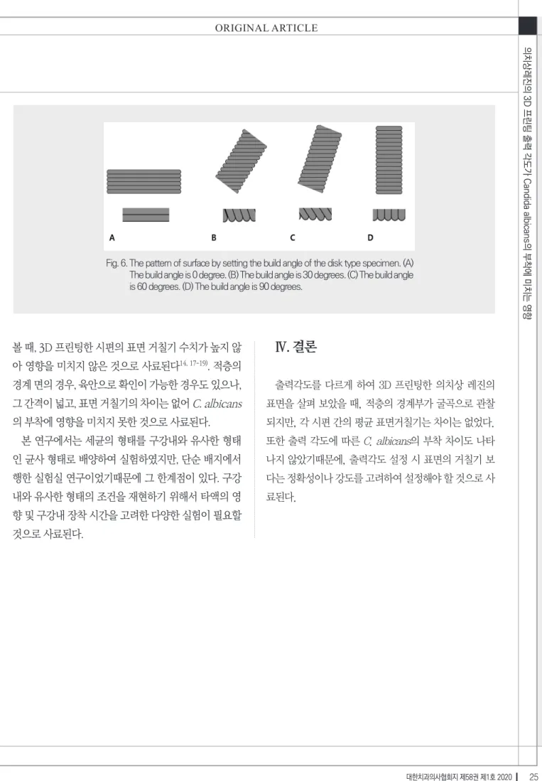
연구한 것은 아직 불충분한 실정이다6, 11-13). 이번 연구에 서는 적층재료에 대한 특성을 살펴보고자 하였으며, 특 히, 출력 각도와의 관계를 살펴보고자 하였다. 출력 각도 와 평균 표면 거칠기 간의 관계는 통계학적으로 유의한 차이는 보이지 않았다. 다만, 표면의 형상은 출력각도에 따라 상이하게 나타났다(Fig. 6). 특히, 출력 각도를 0도 로 설정한 시편은 표면이 micromirror의 크기에 따라 특정 모양의 패턴을 보이는 것이 특징적이었다. 출력각 도를 90도로 설정한 표면에서는 다른 출력각도와는 다 르게 적층의 경계가 세밀하게 나타나지 않고, 부드럽게 나타났다. 따라서, 의치상 레진의 3D 프린팅 시에 출력 각도 설정은 강도나 정확성을 더 중요한 요소로 보고 설 정해야 할 것으로 사료된다.
C. albicans의 부착은 의치성 구내염 발생에서 초기 에 중요한 역할을 한다. 의치상 레진의 표면 거칠기는 세균의 부산물 등에 의한 비가역적인 부착이 일어나기 이전에 매우 중요한 요소로 작용하며, 이는 표면적의 증
가로 인한 효과로 보고되고 있다14). 출력각도에 따른 C.
albicans의 부착은 각 실험군간의 유의한 차이가 없었 다. 선행된 연구에서 C. albicans는 표면의 거칠기 뿐만 아니라, 표면의 에너지, 재료의 소수성여부등과 같은 다 양한 요인의 영향을 받지만, 표면에너지와 소수성의 영 향은 재료의 표면이 타액 또는 배지의 피막이 게재되면 서 감소되기 때문에 표면의 거칠기가 중요한 요소라고 보고하였다2, 15). 최근 한 연구에서는 3D 프린팅용 레진 에 TiO2를 첨가하여, 의치상 자체에 향균성을 높이는 연구가 진행되었다16). 하지만, 이 연구는 실험적으로 의 치상면을 연마한 뒤에 시행한 연구였기 때문에 표면거 칠기와 연관된 이번 연구와는 연구의 방향이 다르다고 할 수 있다. 이번 연구에서 C. albicans의 부착이 실험군 별간의 차이를 보이지 않았다. 이는 평균 표면 거칠기가 0.2 ㎛ 이하의 경우에서는 미생물의 부착이 비교적 억제 되며, 이 이상의 표면거칠기에서는 표면 거칠기와 세균 의 부착 정도가 상관관계를 보인다는 연구를 바탕으로 Table I. Mean surface roughness(Sa) of each tested group (unit: ㎛)
Group 0 Group 30 Group 60 Group 90
Mean±SD 0.19±0.02 0.23±0.09 0.24±0.04 0.3±0.10
SD : Standard deviation, Group 0: Specimen with build angle of 0 degree, Group 30: Specimen with build angle of 30 degrees, Group 60 : Specimen with build angle of 60 degrees, Group 90: Specimen with build angle of 90 degrees. there was no statistically significant difference between the groups.
Table II. Mean colony forming unit (CFU) of each tested group (unit: CFU)
Group 0 Group 30 Group 60 Group 90
Mean±SD 6145±293.5 5970±243.4 6230±353.6 6387±374.8
SD: standard deviation, Group 0: Specimen with build angle of 0 degree, Group 30: Specimen with build angle of 30 degrees, Group 60: Specimen with build angle of 60 degrees, Group 90: Specimen with build angle of 90 degrees. there was no statistically significant difference between the groups
ans의 부착에 미치는 영향
볼 때, 3D 프린팅한 시편의 표면 거칠기 수치가 높지 않 아 영향을 미치지 않은 것으로 사료된다14, 17-19). 적층의 경계 면의 경우, 육안으로 확인이 가능한 경우도 있으나, 그 간격이 넓고, 표면 거칠기의 차이는 없어 C. albicans 의 부착에 영향을 미치지 못한 것으로 사료된다.
본 연구에서는 세균의 형태를 구강내와 유사한 형태 인 균사 형태로 배양하여 실험하였지만, 단순 배지에서 행한 실험실 연구이었기때문에 그 한계점이 있다. 구강 내와 유사한 형태의 조건을 재현하기 위해서 타액의 영 향 및 구강내 장착 시간을 고려한 다양한 실험이 필요할 것으로 사료된다.
Ⅳ. 결론
출력각도를 다르게 하여 3D 프린팅한 의치상 레진의 표면을 살펴 보았을 때, 적층의 경계부가 굴곡으로 관찰 되지만, 각 시편 간의 평균 표면거칠기는 차이는 없었다.
또한 출력 각도에 따른 C. albicans의 부착 차이도 나타 나지 않았기때문에, 출력각도 설정 시 표면의 거칠기 보 다는 정확성이나 강도를 고려하여 설정해야 할 것으로 사 료된다.
Fig. 6. The pattern of surface by setting the build angle of the disk type specimen. (A) The build angle is 0 degree. (B) The build angle is 30 degrees. (C) The build angle is 60 degrees. (D) The build angle is 90 degrees.
ORIGINAL ARTICLE
1. Murray MD, Darvell BW. The evolution of the complete denture base. Theories of complete denture retention--a review. Part 3. Aust Dent J. 1993;38(5):389-93.
2. Klotz SA, Drutz DJ, Zajic JE. Factors governing adherence of Candida species to plastic surfaces. Infect Immun. 1985;50(1):97-101.
3. Webb BC, Thomas CJ, Willcox MD, Harty DW, Knox KW. Candida- associated denture stomatitis. Aetiology and management: a review.
Part 3. Treatment of oral candidosis. Aust Dent J. 1998;43(4):244-9.
4. Lima JM, Anami LC, Araujo RM, Pavanelli CA. Removable partial dentures: use of rapid prototyping. J Prosthodont. 2014;23(7):588- 5. Laverty DP, Thomas MBM, Clark P, Addy LD. The Use of 3D Metal 91.
Printing (Direct Metal Laser Sintering) in Removable Prosthodontics.
Dent Update. 2016;43(9):826-8, 31-2, 34-5.
6. Stansbury JW, Idacavage MJ. 3D printing with polymers: Chal- lenges among expanding options and opportunities. Dent Mater.
2016;32(1):54-64.
7. Congalton RG. A review of assessing the accuracy of classifica- tions of remotely sensed data. Remote Sensing of Environment.
1991;37(1):35-46.
8. Osman RB, Alharbi N, Wismeijer D. Build Angle: Does It Influence the Accuracy of 3D-Printed Dental Restorations Using Digital Light- Processing Technology? Int J Prosthodont. 2017;30(2):182-8.
9. Chockalingam K, Jawahar N, Chandrasekar U, Ramanathan KN. Es- tablishment of process model for part strength in stereolithography.
Journal of Materials Processing Technology. 2008;208(1):348-65.
10. Ishida Y, Miyasaka T. Dimensional accuracy of dental casting pat- terns created by 3D printers. Dent Mater J. 2016;35(2):250-6.
11. Saleh WK, Ariffin E, Sherriff M, Bister D. Accuracy and reproduc- ibility of linear measurements of resin, plaster, digital and printed
study-models. J Orthod. 2015;42(4):301-6.
12. Alharbi N, Osman R, Wismeijer D. Effects of build direction on the mechanical properties of 3D-printed complete coverage interim dental restorations. J Prosthet Dent. 2016;115(6):760-7.
13. Alharbi N, Osman RB, Wismeijer D. Factors Influencing the Di- mensional Accuracy of 3D-Printed Full-Coverage Dental Res- torations Using Stereolithography Technology. Int J Prosthodont.
2016;29(5):503-10.
14. Teughels W, Van Assche N, Sliepen I, Quirynen M. Effect of mate- rial characteristics and/or surface topography on biofilm develop- ment. Clin Oral Implants Res. 2006;17 Suppl 2:68-81.
15. Kuhar M, Funduk N. Effects of polishing techniques on the sur- face roughness of acrylic denture base resins. J Prosthet Dent.
2005;93(1):76-85.
16. Totu EE, Nechifor AC, Nechifor G, Aboul-Enein HY, Cristache CM.
Poly(methyl methacrylate) with TiO2 nanoparticles inclusion for stereolitographic complete denture manufacturing - the fututre in dental care for elderly edentulous patients? J Dent. 2017;59:68-77.
17. Bollen CM, Papaioanno W, Van Eldere J, Schepers E, Quirynen M, van Steenberghe D. The influence of abutment surface roughness on plaque accumulation and peri-implant mucositis. Clin Oral Im- plants Res. 1996;7(3):201-11.
18. Nevzatoglu EU, Ozcan M, Kulak-Ozkan Y, Kadir T. Adherence of Candida albicans to denture base acrylics and silicone-based resil- ient liner materials with different surface finishes. Clin Oral Investig.
2007;11(3):231-6.
19. Verran J, Maryan CJ. Retention of Candida albicans on acrylic resin and silicone of different surface topography. J Prosthet Dent.
1997;77(5):535-9.
참 고 문 헌