933
"J. Korean Soc. Radiol., Vol. 13, No. 7, December 2019"
Measurement of Radiation Dose of HR CT and Low Dose CT by using Anthropomorphic Chest Phantom and Glass Dosimetry
Dae Cheol Kweon*
Department of Radiological Science, College of Bioecological Health, Shinhan University
Received: October 22, 2019. Revised: December 27, 2019. Accepted: December 31, 2019
ABSTRACT
The purpose of this study is to provide basic clinical data by evaluating images, measuring absorbed dose and effective dose by using high resolution CT and low dose CT by using anthropomorphic chest phantom and glass dosimeter. Tissue dose was measured by inserting a glass dosimeter into the anthropomorphic chest phantom. A 64-slice CT system (SOMATOM Sensation 64, Siemens AG, Forchheim, Germany) and CARE Dose 4D were used, and the parameters of the high resolution CT were 120 kVp, Eff. Scan parameters of mAs 104, scan time 7.93 s, slice 1.0 mm (Acq. 64 × 0.6 mm), convolution kernel (B60f sharp) were used, and low dose CT was 120 kVp, Eff. mAs 15, scan time 7.41 s, slice 3.0 mm (Acq. 64 × 0.6 mm), scan of convolution kernel B50f medium sharp. CTDIvol was measured at 8.01 mGy for high resolution CT and 1.18 mGy for low dose CT. Low dose CT scans showed 85.49% less absorbed dose than high resolution CT scans.
Keywords: Absorbed dose, Anthropomorphic phantom, Computed tomography, Effective dose, Glass dosimetry
Ⅰ. INTRODUCTION
CT (computed tomography) 검사로 의료영상을 획 득하기 위해 방사선 노출을 수반하고 있지만, 단순 엑스선 검사에 비해 공간 해상력 (spatial resolution) 및 움직이는 장기를 스캔하는데 유리하게 사용되 는 시간 해상력 (temporal resolution)이 우수하고 입 체적인 영상처리가 가능하여 흉부질환을 진단하는 데 우수한 검사이다.[1,2] 그러나 이러한 CT검사 스 캔에 필연적으로 환자에게 선량이 노출되어 피검 사자가 피폭을 받게 되므로 가능한 방사선량을 합 리적으로 획득 가능한 범위 내에서 가능한 피폭 방 사선 선량을 낮게 유지하는 ALARA (as low as reasonably possible) 원칙에 의거하여 피폭을 줄이는 노력이 절대적으로 필요하다.[3,4]
의료 영상을 획득하기 위한 CT검사에서는 방사 선 노출이 문제가 되기 때문에 가능한 선량을 감소
하는 스캔 프로토콜로 대체하여 스캔하는 방법을 추천한다. 그러나 이러한 저선량 (low dose)의 스캔 은 간단한 작업이 아니며 스캔에 따라 필수적으로 영상의 질적 저하를 수반하여 진단하는데 영향을 줄 수 있다.
저선량 흉부 CT는 공기와 폐결절 사이의 고대조 도로 인해 영상의 질을 유지하면서 방사선 선량을 여러 파라메터인 관전압, 관전류를 이용하여 선량 을 낮출 수 있다. 이러한 검사로 선량은 2 mSv의 평균유효선량에서 스캔할 수 있다.[5]
고해상력 (high resolution) CT는 비침습적 검사로 흉부의 폐기종, 섬유화 및 기관지확장 질환 등에서 유용하게 임상에서 적용되고 있다.
흉부CT 검사에서 저선량 CT와 함께 고해상력 CT의 선량의 비교는 주로 임상환자를 대상으로 하 여 비교하여 연구한 보고가 있었고, 또한 관전류자 동조절기법 (automatic exposure control)과 관전류고
* Corresponding Author: Dae Cheol Kweon E-mail: [email protected] Tel: +82-31-870-3411 https://doi.org/10.7742/jksr.2019.13.7.933
정기법 (fixed tube current)을 이용하여 비교한 보고 가 있으나 인체등가형 흉부팬텀 안에 유리선량계 를 삽입하여 측정한 연구의 보고는 전무하였다.[6]
이번 연구에서는 인체등가형 흉부팬텀을 이용하여 선량과 함께 영상을 비교하고자 하였다.
본 연구는 MDCT에서 인체등가형 흉부팬텀과 유 리선량계를 이용하여 고해상력 및 저선량 CT를 스 캔하여 영상의 평가 및 흡수선량 (absorbed dose) 및 유효선량 (effective dose)을 측정하여 기초자료 및 데이터를 제공하여 임상에서 적용하는데 목표를 두고자한다.
Ⅱ. MATERIAL AND METHODS
1. 재료 및 기기
흉부 CT 스캔을 위해 인체등가형 흉부팬텀 Alderson Rando Phantom (Alderson Research Laboratories, Inc., Long Island City, NY, USA)을 이용하여 고해상력 및 저선량 CT를 스캔하여 선량을 평가하였다. 유리 선량계를 이용하여 선량을 평가하기 위해 인체등 가형 흉부팬텀 내부에 유리선량계를 삽입하여 조 직선량을 측정하였고, Fig. 1은 유리선량계가 위치 한 흉부인체등가팬텀의 level 16의 영상이다.
Fig. 1. Acquisition using by anthropomorphic chest phantom CT.
유리선량계는 137Cs의 표준선원을 이용하여 6 mGy의 선량으로 조사된 유리소자를 이용하여 calibration을 하여 CT스캔의 선량을 측정하였다. 유 리선량계는 유리소자 (GD-353M, Chiyoda Technol, Tokyo, Japan)를 사용하였고, 선량 판독은 판독기
사용하여 흡수선량을 측정하였다.
방사선 선량 측정을 위해 유리선량계의 소자 (element)는 고해상력 CT 및 low dose CT 스캔 전에 annealing을 하였다. 이러한 annealing 과정은 400 ℃ 에서 1시간의 가열 후에 냉각을 하고 자연방사선량 을 측정하고 CT 스캔의 선량에서 감산하여 선량을 산출하였다.
고해상력 CT 및 저선량 CT 스캔 후에 방사선 선량 의 판독은 CT 스캔을 하고 예열 (pre- heating)을 70
℃에서 1시간을 가열하고 냉각한 소자를 판독기를 이용하였고, CT 스캔한 유리소자에 판독기의 레이 저로 10회 조사하여 흡수선량의 평균값과 표준편 차를 산출하였다.[7]
2. 방법
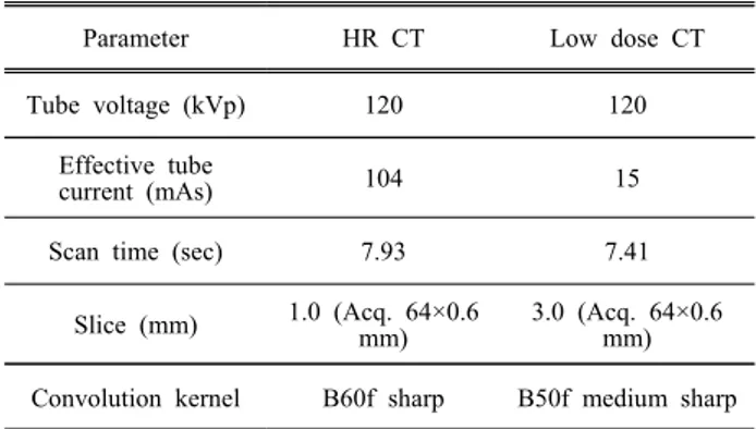
환자를 64-slice CT system (SOMATOM Sensation 64, Siemens AG, Forchheim, Germany)과 CARE Dose 4D와 파라메터를 Table 1과 같이 이용하였다.
Table 1. Parameter of HR and low dose CT
Parameter HR CT Low dose CT
Tube voltage (kVp) 120 120
Effective tube
current (mAs) 104 15
Scan time (sec) 7.93 7.41
Slice (mm) 1.0 (Acq. 64×0.6 mm)
3.0 (Acq. 64×0.6 mm)
Convolution kernel B60f sharp B50f medium sharp
고해상력 CT에서의 파라메터는 관전압 120 kVp, Eff. mAs 104, scan time 7.93 sec, slice 1.0 mm (Acq.
64×0.6 mm), convolution kernel B60f sharp의 스캔 파라메터가 사용되었고, 저선량 CT는 120 kVp, Eff.
mAs 15, scan time 7.41 sec, slice 3.0 mm (Acq.
64×0.6 mm), convolution kernel B50f medium sharp 의 스캔 파라메터를 이용하였다.
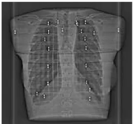
Fig. 2은 CT 스캔을 위한 topogram으로 유리선량 계를 인체등가형 흉부팬텀에 삽입하고 고해상력 CT 및 저선량 CT 스캔하여 영상을 획득하였다.
"J. Korean Soc. Radiol., Vol. 13, No. 7, December 2019"
Fig. 2. Topogram of HR CT and low dose CT image using anthropomorphic chest phantom.
3. 선량 및 영상평가
인체등가 흉부팬텀을 이용하여 고해상력 CT 및 저선량 CT로 스캔하여 흡수선량을 측정하고 유효 선량을 아래의 식으로 산출하였다.
× (1)
유효선량 (E)은 ICRP 103을 적용하여 폐의 조직 가중계수 (WT)인 0.12와 흡수선량 (DT,R)를 곱하여 산출하였다.
인체등가 흉부팬텀으로 스캔하여 획득한 데이터 의 영상을 ImageJ (NIH, Bethesda, MD, USA) 프로 그램에서 좌우 폐의 상부, 중부, 하부에 ROI (region of interest) 방법을 이용하여 평균과 노이즈 (표준편 차)를 3회 측정하여 비교 분석하였다.
4. 통계분석
스캔한 영상을 이용하여 고해상력 CT 및 저선량 CT스캔에서 방사선 선량을 비교하기 위해 범용적 인 통계 프로그램 IBM SPSS (version 22, IBM Corp., Armonk, NY, USA)를 이용하였고, 대응표본 t-검증 (paired t-test)으로 통계처리 하였고, 통계적 유의수준은 p < .05로 설정하여 통계적 유의성을 검정하였다.
Ⅲ. RESULTS
인체등가형 흉부 팬텀을 이용하여 고해상력 CT 및 저선량 CT 프로토콜로 스캔한 영상은 Fig. 3과 같다.
(a)
(b)
Fig. 3. (a) HR CT image using anthropomorphic phantom. (b) Low dose CT image using anthropomorphic chest phantom.
인체등가형 흉부팬텀을 이용하여 스캔에 따른 CTDIvol은 고해상력 CT에서 8.01 mGy, 저선량 CT 는 1.18 mGy로 Table 2와 같이 흡수선량이 측정되 었다.
Table 2. CTDIvol of HR and low dose CT HR CT (mGy) Low dose CT (mGy)
8.01 1.18
유리선량계를 이용한 고해상력 CT 및 저선량 CT 스캔에서 선량을 측정하였고, 고해상력 CT에서 흡 수선량 11,569 uGy, 유효선량은 1.38 mSv, 저선량 CT에서 흡수선량 1,678 uGy, 유효선량 0.2 mSv로 Table 3과 같이 측정되었다.
Table 3. Radiation dose of HR and low dose CT HR CT Low dose CT Absorbed dose (uGy) 11,569.14±1,404.77 1,678±151.13
Effective dose (mSv) 1.38 0.2
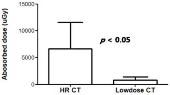
CT 스캔에 따른 저선량 CT 스캔의 선량은 고해 상력 CT 스캔에 비해 85.49%의 감소하였고, 선량 에 대한 비교는 Fig. 4와 같다. 흉부인체등가팬텀을 이용하여 고해상력 CT 및 저선량 CT스캔에서의 선 량은 고해상력 CT가 저선량 CT에 비해 유의하게 높은 값을 나타냈다 (p < .05).
Fig. 4. Absorbed dose measured of HR CT and low dose CT using anthropomorphic chest phantom.
흉부인체등가팬텀을 이용하여 고해상력 CT에서 는 평균으로 나타내는 CT감약계수 (HU)는 – 606.6.4 HU, 노이즈는 179.04 HU이었고, 저선량 CT 스캔에서의 CT감약계수는 –417.61 HU, 노이즈는 137.06 HU으로 Table 4와 같이 산출되었다.
Table 4. CT coefficient and noise calculated of HR and low dose CT (unit: HU)
HR CT Low dose CT
Average -606.34 -417.61
SD 179.04 137.06
Ⅳ. DISCUSSION
흉부 CT검사에서 임상에서 적용하여 검사하고 있는 저선량 CT 및 고해상력 CT는 폐의 해부학적 구조 및 정밀한 폐의 질환을 진단하는데 적용되어 스캔하고 있다. 이러한 흉부 CT 검사에서 영상의 질을 결정하는 요소는 CT감약계수, 노이즈, 공간분
한다.
고해상력 CT와 저선량 CT의 검사에서 영상의 노 이즈는 관전류, 슬라이스 두께 등에 영향을 받으며 픽셀에서 CT의 HU가 동일하지 않고 분산된 형태 를 나타내는 표준편차로 표시한다. 인체등가형 흉 부팬텀을 이용한 폐의 CT감약계수와 노이즈 측정 에서 고해상력 CT는 179.04 HU, 저선량 CT의 노이 즈는 137.06 HU로 측정되어 고해상력 CT가 저선량 CT에 비해 높게 측정되었다.
흉부팬텀을 이용하여 자동관전류기법 (CARE Dose 4D)으로 스캔한 노이즈 측정에서 저선량 CT 가 고해상력 CT에 비해 노이즈가 낮게 측정되어 본 연구와 일치한 결과를 나타내었다.[6,9]
유리선량계를 인체등가형 흉부팬텀에 삽입하여 측정한 고해상력 CT에서 흡수선량 11,569 uGy, 유 효선량은 1.38 mSv, 저선량 CT에서는 흡수선량 1,678 uGy, 유효선량 0.2 mSv로 측정되어 기존에 보고한 저선량 CT의 유효선량이 0.7 ± 0.2 mSv로 본 연구와 선량측정 값은 근사한 차이를 보였
고,[10,11] 이러한 선량의 차이는 환자를 대상으로
DLP (dose length product)를 이용하여 계산한 선량 으로 측정한 선량으로 생각된다.[11]
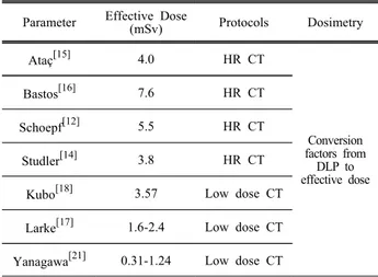
기존에 보고된 고해상력 CT의 성인의 유효선량 3.8-7.6 mSv에 비해 선량이 적게 측정되었다.[12-16] 저 선량 CT에서도 기존 보고는 1.6-2.4 mSv,[17] 3.57 mSv[18]로 보고하여 본 연구에 비해 높게 조사되었다.
이러한 선량의 차이는 기존에 유효선량에 대한 측정 은 CT 스캔에 따른 DLP 값에 변환계수를 곱하여 계 산한 선량에 따른 차이로 생각된다.[12-19]
AAPM 팬텀을 이용한 국내 연구의 보고에서도 고해상력 CT는 1.95 mGy, 저선량 CT는 0.35 mGy로 본 연구에 비해 낮은 선량을 보고하였고, 노이즈에 대한 측정에서도 고해상력 CT는 40.1 HU, 저선량 CT는 99.6 HU로 차이가 있었다.[19] 이러한 노이즈 의 차이의 배경은 환자를 대상으로 측정한 연구이 고, 본 연구는 팬텀을 측정한 연구로 차이가 존재 한 것으로 생각된다.
본 연구에서는 고해상력 CT 스캔에 비해 저선량 CT 스캔의 선량이 85.49% 감소하였고 통계적으로
"J. Korean Soc. Radiol., Vol. 13, No. 7, December 2019"
고해상력 CT가 저선량 CT에 유의한 차이가 있다 (p < .05). 이러한 연구는 CT검사에서 방사선 선량 에 대한 진단참고준위 (DRL, diagnostic reference levels)에 적용하여 활용할 수 있다. 또한 기존의 스 캔에 비해 선량이 적은 ultra 고해상력 및 다양한 재구성 방법을 이용한 고해상력 CT와 저선량 CT를 임상에서 응용하여 적용하도록 한다.[20,21]
Table 5. Summary of effective dose of HR and low dose CT
Parameter Effective Dose
(mSv) Protocols Dosimetry
Ataç[15] 4.0 HR CT
Conversion factors from
DLP to effective dose
Bastos[16] 7.6 HR CT
Schoepf[12] 5.5 HR CT
Studler[14] 3.8 HR CT
Kubo[18] 3.57 Low dose CT
Larke[17] 1.6-2.4 Low dose CT
Yanagawa[21] 0.31-1.24 Low dose CT
본 연구는 인제등가형 흉부 팬텀을 이용하여 고 해상력 및 저선량 CT검사에서 유리선량계를 이용 한 선량 및 영상을 확인하였으나 실제 적용하기 위 해서는 임상적인 방법의 추가적인 연구가 필요하 고 다양한 연구의 제한점을 가지고 있다. 첫째는, 임상 환자를 대상으로 하지 않고 인체등가팬텀을 적용한 한계를 가지고 있다. 두 번째는 추후에는 다양한 CT검사 프로토콜 및 bolus tracking technique를 포함하여 CT 스캔하여 영상평가 및 유 효선량을 측정하여 연구할 필요가 있다.[22,23] 세 번 째는, 다양한 선량계측기기를 이용하여 영상 및 선 량을 측정하여 정성적 및 물리적인 정량적 평가에 대한 추가적인 연구가 필요하다. 네 번째는 임상에 적용할 수 있는 프로토콜을 위해 피검사자의 정성 적이고 정량적인 후향적 연구가 추가적인 연구가 필요하다.
Ⅴ. CONCLUSION
해상력 CT 및 저선량 CT의 스캔에서 인체등가형 흉부 팬텀과 유리선량계를 이용하여 흡수선량을
측정하고 영상을 평가하였다. 저선량 CT 검사는 고 해상력 CT 검사에 비해 흡수선량이 85.49%가 감소 하였고 영상의 차이는 없어 임상에서 유용하게 적 용할 수 있다.
Reference
[1] M. K. Kalra, M. M. Maher, S. Rizzo, D. Kanarek, J.
A. Shepard, "Radiation exposure from chest CT:
issues and strategies," Journal of Korean Medical Science, Vol. 19, No. 2, pp. 159-166, 2004.
[2] M. M. Maher, M. K. Kalra, T. L. Toth, C. Wittram, S. Saini, J. Shepard, "Application of rational practice and technical advances for optimizing radiation dose for chest CT," Journal of Thoracic Imaging, Vol. 19, No. 1, pp. 16-23, 2004.
[3] F. A. Jr. Mettler, M. Bhargavan M, K. Faulkner, et al., "Radiologic and nuclear medicine studies in the United States and worldwide: frequency, radiation dose, and comparison with other radiation
sources-1950–2007," Radiology, Vol. 253, No. 2, pp.
520-531. 2009.
[4] D. A. Schauer, O. W. Linton, "National Council on Radiation Protection and Measurements report shows substantial medical exposure increase," Radiology, Vol. 253, No. 2, pp. 293-296, 2009.
[5] R. Cristiano, O. Daniela, B. Massimo, "Low-dose CT: technique, reading methods and image
interpretation Cancer Imaging," Cancer Imaging, Vol.
12, No. 3, pp. 548-556, 2012.
[6] T. S. Park, J. H. Han, S. Y. Jo, E. L. Lee, K. W.
Jo, D. C. Kweon, "Comparison of noise and doses of low dose and high resolution chest CT for automatic tube current modulation and fixed tube current technique using glass dosimetry," Journal Radiation Industry, Vol. 11, No. 3, pp. 131-137, 2017.
[7] M. Y. Jung, D. C. Kweon, S. I. Kwon,
"Effectiveness of bismuth shield to reduce eye lens radiation dose using the photoluminescence dosimetry in computed tomography," Journal of Radiological Science and Technology, Vol. 32, No. 3, pp.
307-312, 2009.
[8] B. G. Yoo, D. C. Kweon, J. S. Lee, K. J. Jang, S.
H. Yang, Y. S. Kim, "Comparison radiation dose os
z-axis automatic tube current modulation technique with fixed tube current multi-detector row CT scanning of lower extremity venography," Journal of Radiation Protection and Research, Vol. 32, No. 3, pp. 123-133, 2007.
[9] J. S. Lee, D. C. Kweon, B. G. You, "Radiation dose reducing effect during the AEC system in the chest and abdomen of the MDCT scanning," The Journal of the Korea Contents Association, Vol. 9, No. 3, pp. 225-231, 2009.
[10] H. J. Lim, M. J. Chung, K. S. Shin, H. S. Hwang, K. S. Lee, "The impact of iterative reconstruction in low-dose computed tomography on the evaluation of diffuse interstitial lung disease," Korean Journal of Radiology, Vol. 17, No. 6, pp. 950-960, 2016.
[11] AAPM, The measurement, reporting, and management of radiation dose in CT. Alexandria, VA: AAPM; 2008.
[12] U. J. Schoepf, R. D. Bruening, C. Hong, R. Eibel, S. Aydemir, A. Crispin, C. Becker, M. F. Reiser,
"Multislice helical CT of focal and diffuse lung disease: comprehensive diagnosis with reconstruction of contiguous and high-resolution CT sections from a single thin-collimation scan," American Journal of Roentgenology, Vol. 177, No. 1, pp. 179-184, 2001.
[13] D. M. Kelly, I. Hasegawa, R Borders, H. Hatabu, P.
M. Boiselle, "High-resolution CT using MDCT:
comparison of degree of motion artifact between volumetric and axial methods," American Journal of Roentgenology, Vol. 182, No. 3, pp. 757-759, 2004.
[14] U. Studler, T. Gluecker, G. Bongartz, J. Roth, W.
Steinbrich, "Image quality from high-resolution CT of the lung: comparison of axial scans and of sections reconstructed from volumetric data acquired using MDCT," American Journal of Roentgenology, Vol. 185, No. 3, pp. 602-607, 2005.
[15] G. K. Ataç A. Parmaksız, T. İnal, E. Bulur, F.
Bulgurlu, T. Öncü, S. Gündoğdu, "Patient doses from CT examination in Turkey," Diagnostic and Interventional Radiology, Vol. 21, No. 5, pp.
428-434, 2015.
[16] Md. Bastos, E. Y. Lee, K. J. Strauss, D.
Zurakowski, D. A. Tracy, P. M. Boiselle, "Motion artifact on high-resolution CT images of pediatric
methods," American Journal of Roentgenology, Vol.
193, No. 5, pp. 1414-1418, 2009.
[17] F. J. Larke, R. L. Kruger, C. H. Cagnon, M. J.
Flynn, M. M. McNitt-Gray, X. Wu, P. F. Judy, D.
D. Cody, "Estimated radiation dose associated with low-dose chest CT of average-size participants in the National Lung Screening Trial," American Journal of Roentgenology, Vol. 197, No. 5, pp.
1165-1169, 2011.
[18] T. Kubo, Y. Ohno, M. Nishino, Pei-Jan Lin, S.
Gautam, Hans-Ulrich K, H. Hatabu, iLEAD study group, "Low dose chest CT protocol (50 mAs) as a routine protocol for comprehensive assessment of intrathoracic abnormality," European Journal o Radiology Open. Vol. 3, pp. 86-94, 2016.
[19] W. J. Lee, "Usefulness evaluation of low-dose CT for emphysema: compared with high-resolution CT,"
Journal of Radiological Science and Technology, Vol. 39, No. 3, pp. 329-336, 2016.
[20] A. Hata, M. Yanagawa, O. Honda, N. Kikuchi, T.
Miyata, S. Tsukagoshi, A. Uranishi, N. Tomiyama,
"Effect of Matrix Size on the Image Quality of Ultra-high-resolution CT of the Lung: Comparison of 512 × 512, 1024 × 1024, and 2048 × 2048," Academic Radiology, Vol. 25, No. 7, pp. 869-876, 2018.
[21] M. Yanagawa, T. Gyobu, A. N. Leung, M. Kawai, Y. Kawata, H. Sumikawa, O. Honda, N. Tomiyama,
"Ultra-low-dose CT of the lung: effect of iterative reconstruction techniques on image quality,"
Academic Radiology, Vol. 21, No. 6, pp. 695-703, 2014.
[22] D. C. Kweon, J. W. Choi, "Analysis of radiation dose for the bolus tracking technique using the CTDI phantom of CT study," Journal of Radiation Industry, Vol. 13, No. 3, pp. 229-234, 2019.
[23] D. C. Kweon, J. E. Jung, Y. H. Ryu, W. K.
Chung, "Comparison radiation dose with upper and lower limbs of automated injection of contrast media with a bolus tracking system in pulmonary
embolism MDCT," Journal of Radiation Industry, Vol. 3, No. 4, pp. 323-328, 2009.
"J. Korean Soc. Radiol., Vol. 13, No. 7, December 2019"
인체등가형 흉부팬텀과 유리선량계를 이용한 고해상력 및 저선량 CT의 선량측정
권대철*
신한대학교 바이오생태보건대학 방사선학과
MDCT에서 인체등가형 흉부팬텀과 유리선량계를 이용하여 고해상력 및 저선량 CT로 검사하여 영상의 평가 및 흡수선량 및 유효선량을 측정하여 임상 기초자료를 제공하는데 목표를 두고자한다. 인체등가형 흉 부팬텀내부에 유리선량계를 삽입하여 조직선량을 측정하였다. 64-slice CT system (SOMATOM Sensation 64, Siemens AG, Forchheim, Germany)과 CARE Dose 4D를 이용하였고, 고해상력 CT에서의 파라메터는 관전압 120 kVp, Eff. mAs 104, scan time 7.93 sec, slice 1.0 mm (Acq. 64×0.6 mm), convolution kernel (B60f sharp)의 스캔 파라메터가 사용되었고, 저선량 CT는 120 kVp, Eff. mAs 15, scan time 7.41 sec, slice 3.0 mm (Acq. 64×
0.6 mm), convolution kernel B50f medium sharp의 스캔하였다. 인체등가형 흉부팬텀을 이용하여 스캔에 따른 CTDIvol은 고해상력 CT에서 8.01 mGy, 저선량 CT는 1.18 mGy로 측정되었다. 저선량 CT 검사는 고해상력 CT 검사에 비해 흡수선량이 85.49%가 감소하였고 영상의 차이는 없어 임상에서 유용하게 적용할 수 있다.
중심단어: 컴퓨터단층영상, 유리선량계, 인체등가형 흉부팬텀, 흡수선량, 유효선량
성명 소속 직위
(단독저자) 권대철 신한대학교 바이오생태보건대학 방사선학과 교수
연구자 정보 이력