Corresponding author: Sung Hyuk Lim
Department of Neurology, Samsung Medical Center, 81 Ilwon-ro, Gangnam-gu, Seoul 06351, Korea
E-mail: [email protected]
ORCID: https://orcid.org/0000-0002-5344-6837
CASE REPORT
An Effective Transcranial Electric Motor-Evoked Potentials Method in Spinal Dural Arteriovenous Fistula Ligation Surgery
Min Hwan Jang, In Seok Lee, Sung Hyuk Lim
Department of Neurology, Institute of Neuroscience Center, Samsung Medical Center, Seoul, Korea
척수경막동정맥루 결찰술에서의 효과적인 경두개운동유발전위 검사방법
장민환, 이인석, 임성혁
삼성서울병원 뇌신경센터 신경과
ARTICLE INFO ABSTRACT
Received April 29, 2021 Revised May 26, 2021 Accepted May 26, 2021
The purpose of spinal dural arteriovenous fistula (SDAVF) ligation is to prevent neurological injury and the poor blood supply through ligation of arteriovenous fistula. Therefore, intraoperative neurophysiological monitoring (INM) is required via multimodal neurological examination for minimizing the side effects after surgery based on the patient’s symptoms. Transcranial electric motor-evoked potentials (TceMEP) help to check the condition of the corticospinal tract. Whenever ligation is performed, TceMEP should be performed every minute to check for abnormalities.
However, an examiner’s lack of knowledge about the operation procedure and examination and also poor communication between the examiner and surgeon can cause incorrect timing of the stimulation of TceMEP that interferes with the procedure and causes side effects such as paralysis and motor weakness. As a result of this SDAVF ligation survey, it is believed that for proper INM, case reports will be needed along with further research and the examiner will also have to work closely with the surgeon to minimize neurological damage to patients.
Copyright Ⓒ 2021 The Korean Society for Clinical Laboratory Science. All rights reserved.
Key words INM Neurosurgery SDAVF TceMEP
서 론
척수경막동정맥루(spinal dural ateriovenous fistula, SDAVF)는 척수에서 발생하는 가장 흔한 혈관기형으로 경막내 에서 동맥과 정맥간의 비정상적인 연결로 인해 울혈성 척수병증 을 유발하는 질환이다[1]. 척수경막동정맥루는 동정맥 단락의 경막, 척수와의 위치관계, 공급혈관의 혈류 특성에 따라 척수경
막 동정맥루, 척수주변 동정맥루(perimedullary arteriovenous fistula), 그리고 척수내 동정맥기형(intramedullary arterio- venous malformation)으로 구분할 수 있다. 척수 동정맥기형 은 증상을 일으키면 진행되는 신경학적 결손을 유발하고 수명의 단축을 초래하는 것으로 알려져 비가역적 척수손상이 오기 전에 적절한 치료가 요구된다[2].
척수경막동정맥루의 치료는 동정맥루를 차단하는 혈관색전 술과 클립을 이용한 외과적 결찰술이 있는데 혈관색전술로 치료 하기 어려운 경우 외과적 결찰을 진행한다[3]. 척수경막동정맥 루 결찰술은 동정맥루를 결찰하여 혈액공급을 원활하게 하는 수 술법이다. 하지만 동정맥루의 분지가 신경혈관으로 가게 되면
Korean Society for Clinical Laboratory Science
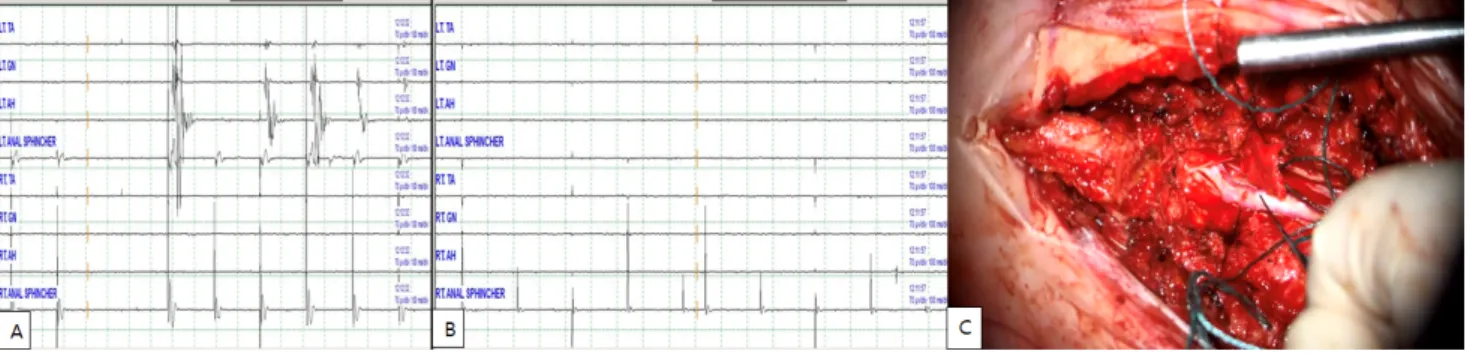
Figure 1. Waves of direct nerve electrical stimulation.
결찰 후 다양한 신경학적 손상이 발생할 수 있기 때문에 검사자 는 복합(multimodal)적인 수술중신경계감시(intraoperative neurophysiological monitoring, INM)를 효율적으로 진행 해야 한다. 국내에서는 수술 중의 경두개운동유발전위(Tran- scranial electric motor-evoked potential, TceMEP)는 근 수축으로 수술공간의 움직임을 유발하기 때문에 연속적으로 시 행하기 힘들다는 제한점이 보고되었다[4].
본 증례는 결찰로 인한 TceMEP의 감소 및 소실을 예방하고 자 수술 전 집도의와 소통해 결찰 후 수술을 잠시 중단한 후 TceMEP의 이상 여부를 확인하기로 하였다. 결찰 후 수술을 잠 시 중단한 상태에서 최대 5분 동안 검사를 진행하여 TceMEP의 감소 및 소실을 확인해 외과적 결찰술에서 하지마비와 위약감과 같은 부작용의 발생을 없애 본 증례를 보고하는 바이다.
증 례
1. 방법
INM 검사기기는 Xltek Protektor IOM system (Natus Medical Incorporated, Plesanton, CA, USA)을 사용해 TceMEP, 체성감각유발전위(somatosensory evoked poten- tials, SSEPs), 직접신경검사법(direct nerve stimulation, DNS), 구해면체 반사(bulbocavernous reflex, BCR), 자발근 전도(free running electromyography)를 시행하였다. TceMEP 는 자극을 위해 국제 10∼20 뇌파체계(10∼20 system)에 따라 C3와 C4에 각각 활성전극과 기준전극을 삽입하고 양극자극 (anodal stimulation)법을 통해 자극을 주어 각 말초근육에서 복합근육활동전위(compound muscle action potential, CMAP)를 기록하였다. TceMEP를 기록하는 근육은 상지 근육 에서는 짧은엄지벌림근(abductor pollicis brevis, APB)과 새 끼벌림근(abductor digiti quinti, ADQ)에서 기록하고, 하지 근육에서는 앞정강근(tibialis anterior, TA)과 엄지벌림근
(abductor hallucis, AH)에서 기록을 하였고, 수술하는 척추 레벨에 따라 추가적으로 근육을 선택하여 기록한다.
본 검사에서는 가쪽넓은근(vastus lateralis, VL)과 비복근 (gastrocnemius, GN) 그리고 외항문괄약근(external anal sphincter)을 추가적으로 기록하였다. 본 검사에서 TceMEP의 설정값은 range ±2.5 mV, reject threshold ±1.5 mV, low-frequency filter 10 Hz, high-frequency filter 1 kHz, notch filter off로 설정하였다. 자극은 450 V, 5 pulses로 설 정하였다. TceMEP의 경고 기준은 기준파형(baseline)에서 50% 이상의 진폭감소를 경고 기준으로 삼았다. SSEPs는 상지 는 짧은 엄지벌림근에서 손목으로 5 cm 정도 거리에서 긴손바 닥근(palmaris longus)과 노쪽손목굽힘근(flexor carpi radialis) 사이 정중신경(median nerve)을 자극부위로 두고 하지는 안쪽복사뼈(medial malleolus)와 아킬레스힘줄(Achilles tendon) 사이 후경골신경(posterior tibial nerve)을 자극부 위로 두고 각각 활성전극이 근위부, 기준전극은 원위부로 향하 도록 삽입하여 시행한다.
기록전극으로는 국제 10∼20 뇌파체계(10∼20 system)에 따라 C3’, Cz’, C4’와 C5s에 활성전극, Fpz에 기록전극을 삽입 하여 검사를 진행하였다. SSEPs의 기본 설정값은 range ±250 μV, reject threshold ±80 μV, low-frequency filter 30 Hz, high-frequency filter 200 Hz, notch filter off로 설정하였다.
자극은 정중신경 SSEPs는 15 mA, 0.2 duration, 후경골신 경 SSEPs는 30 mA, 0.2 duration으로 설정하였다. SSEPs의 경고 기준은 기준파형 대비 50%의 진폭감소와 10%의 잠복기 증가를 경고 기준으로 삼았다. BCR은 음경의 근위부와 원위부 에 평판전극을 이용해 음부신경(pudendal nerve)을 자극하였 고 기록전극은 양측 외항문괄약근에서 파형을 기록하였다.
EMG는 가쪽넓은근, 외항문괄약근, 앞정강근, 엄지벌림근에 서 neurotonic dischage의 관찰되는 경우 경고기준으로 삼았 다. DNS는 척수혈관 또는 결찰 주변 신경근을 직접자극 함으로
Table 1. The muscle strength grading scale (Oxford motor grade scale)
Grade Explanation
0/5 No contraction
1/5 Visible/palpable muscle contraction but no movement 2/5 Movement against gravity eliminated 3/5 Movement against gravity only 4/5 Movement against gravity with some resistance 5/5 Movement against gravity full resistance
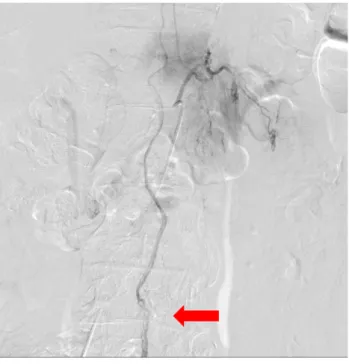
Figure 2. Spinal angiography in L5 arteriovenous fistula with feeding arteries from L2 spinal artery.
Figure 3. TceMEP formed after spinal decompression.
써 신경의 위치를 확인하여 주변 신경의 손상을 막을 수 있다. 본 검사는 진행하기 앞서 피하전극과 자극기를 준비하고 피하전극 을 기준전극으로 사용하여 중간볼기근(gluteus medius, GM) 에 전극을 삽입하였다. DNS는 반복적으로 자극을 주는 방식으 로 집도의가 필요시 수술하는 부위 주변에 자극기를 대고 자극 을 줌으로써 검사가 이루어진다. 만약 신경을 자극하게 될 경우 해당 근육이 반응을 하며 중저음의 큰소리와 함께 근전도 (electromyography, EMG)가 관찰된다(Figure 1A). 그러나 신경이 아닌 근육 또는 혈관에 자극을 줄 경우 자극에 의한 인공 산물만 관찰되고 소리가 나지 않는다(Figure 1B). 본 증례에서 는 4.7 Hz, 6~7 V 사이로 자극을 설정해 검사를 진행하였다. 만 약 근전도 파형이 관찰될 경우 집도의에게 알리고 수술이 원활 하게 진행될 수 있게 하였다. 운동등급의 평가는 신경외과의사 의 진찰로 Oxford motor grade scale에 따라 분류하였다 (Table 1).
2. 마취
TceMEP와 DNS 모두 마취의 영향을 받기 때문에 검사 중 마 취로 인해 발생하는 파형의 왜곡을 막기 위해 전신정맥마취 (total intravenous anesthesia, TIVA)로 유도하였고 마취의 심도가 깊거나 근이완제가 과다 투여되면 파형의 형성이 어렵기 때문에 사연속(train of four, TOF)자극은 적어도 3/4 이상의 근수축이 일어나도록 마취과와 협의하여 근이완제의 농도를 유 지하였다.
3. 증례
75세 남자환자 허리통증과 하지위약과 보행장애로 본원에 내원하였다. 하지 운동등급은 왼쪽은 4, 오른쪽은 3등급이었고, 환자가 느끼는 감각은 오른발이 왼발에 비해 70% 정도 느껴진 다고 하였다. 척수혈관조영술(spinal angiography)에서는 요 추 2번에서 기원한 척수동맥으로부터 요추 5번까지의 동맥혈관 이 정맥과 바로 연결되어 척수신경으로 혈액이 공급되지 않고
두꺼워지는 동정맥루가 관찰되었고(Figure 2), 요추 4, 5번의 협착증(stenosis)도 관찰되었다.
상기 병변에 대해 요추 5번, 천추 1번에서 후궁절제술 (laminectomy)과 동정맥루의 외과적 결찰, 감압술을 시행했 다. 수술 시작 후 TceMEP는 오른쪽 앞정강근과 양쪽 엄지벌림 근에서 파형이 약하게 관찰되었고, 나머지 하지근육에서는 파 형이 형성되지 않았고 SSEP 또한 파형 형성이 관찰되지 않았다.
경막을 절제해 척수를 감압 시킨 후 TceMEP에서 약하게 관찰 되었던 파형들이 정상적으로 형성이 되어 TceMEP의 기준점으 로 삼아 검사를 진행하였고(Figure 3), SSEP의 파형은 관찰되 지 않았으며 수술 종료시까지 파형은 유지되었다.
Figure 5. Change in TceMEP after first ligation. (A) About a minute after fistula ligation, the TceMEP waveform dis- appears from the left gastrocnemius muscle. (B) Waveform recovered immediately after ligation removal.
Figure 6. Change in TceMEP after second ligation. (A) After repositioning three minutes after fistula ligation, the TceMEP waveform 50% reduction from the right tibialis anterior muscle and disappeared in gastrocnemius muscle, adductor hallucis. (B) Waveform recovered four minutes after ligation removal.
Figure 4. Ligation of spinal dural arteriovenous fistula.
DNS를 이용해 동정맥루 주변을 자극하여 결찰 부위의 신경 을 확인하였다(Figure 1C). 그 후에 클립으로 fistula를 일시적 으로 결찰하였다(Figure 4). 첫 번째 결찰 후 약 1분 뒤 TceMEP 에서 좌측 비복근의 파형의 소실이 있었고 결찰을 풀고 즉시 파 형 회복되었다(Figure 5). 위치를 바꿔 두번째 결찰 후 3분 뒤 TceMEP에서 우측 앞정강근에서 50% 파형 감소, 비복근, 엄지 벌림근의 파형의 소실이 관찰되었고, 제거 후 4분 뒤 파형 회복 되었다(Figure 6). 세 번째 결찰 후 1분 30초 뒤 TceMEP에서 우측 앞정강근의 파형 소실이 관찰되었고 제거 후 5분 뒤 파형이 회복되었다(Figure 7). 결찰로 인한 TceMEP의 변화로 결찰을
진행하지 못하고 척수 감압만 진행하고 수술은 종료되었다.
INM에서는 수술 종료시까지 파형의 변화는 확인되지 않았다.
수술 후 환자의 하지 운동등급은 5/5였으며, 환자가 느끼는 증 상 또한 수술 전과 마찬가지로 보행장애가 있었지만 걷는 모습 이 반듯해지고, 발바닥 닿는 느낌을 느낄 수 있을 정도의 호전이 있었다. 환자는 퇴원 후에도 주기적으로 동정맥루 추적검사를 진행하고 있으며 재활을 병행하며 치료 중이다.
고 찰
척수 동정맥 기형은 척수의 공간 점유성 병변의 약 3.4%에서 11.5%를 차지하는 비교적 드문 질환으로 적절한 치료를 시행하 지 않은 경우에는 신경학적 마비증상이 진행되어 심각한 장애를 유발하는 질환이다. 그러므로 조기진단과 이에 따른 적절한 치 료가 강조되는데 치료당시의 환자의 신경학적 상태에 따라 예후 가 결정된다[5].
척수경막동정맥루는 수술로써 직접 동정맥루를 차단하거나 정제하는 방법, 혈관내 색전술로 차단하는 방법, 유출정맥을 결 찰하는 방법 등이 있다. 유출 정맥의 단순 결찰은 결찰 부위에서 역행적 혈전증을 유발하여 동정맥루가 막혀 절제 없이도 좋은 결과를 얻을 수 있는 단순한 수술방법이다. 본 증례에서는 사전
Figure 7. Change in TceMEP after third ligation. (A) A minute and a half after fistula ligation, the TceMEP waveform disappears from the right tibialis anterior muscle. (B) Waveform recovered five minutes after ligation removal.
에 집도의와 소통해 결찰 후 5분까지 여러 부위에서 TceMEP의 변화를 관찰하였다. 하지만 TceMEP의 감소 및 소실로 인해 결 찰을 진행하지 못한 채 수술은 종료하였다. 국내에서는 동정맥 루의 결찰술에서 BCR의 소실로 환자 대소변기능에 부작용이 생긴 사례가 보고되었으며[8], 척수경막동정맥루가 아닌 경막 내 연주주위 동정맥루(intradural perimedullary arterio- venous fistula)에서 동정맥루를 폐색하였으나 우측하지의 근 력약화가 있었던 사례가 보고되었지만 INM을 시행하지 않았던 수술이었다[5].
이에 본 증례는 외과적 결찰술로 인한 TceMEP 감소로 환자 의 신경학적 부작용을 예방한 사례라고 생각한다. TceMEP의 결찰술에서 결찰 후 TceMEP의 변화뿐만 아니라 복합 (multimodal) 신경계검사를 통해 환자의 수술 후 부작용을 최 소화하기 위해 다양한 검사의 추가적인 연구가 필요할 것으로 생각된다. 이에 검사를 진행하는 선생님들은 고착화되어 있는 검사 외에 환자의 상태와 신경과의사 및 집도의와의 소통으로 환자의 부작용을 최소화할 수 있게 효과적인 검사를 진행해야 하며 수술의 원활한 진행을 위한 검사기술의 숙지와 환자의 신 경학적 후유증을 최소화해야 한다고 생각하는 바이다.
요 약
척수경막동정맥루의 수술적기법은 동정맥루에 결찰을 통해 혈액공급을 원활하게 하여 신경학적 손상을 막는 수술법이다.
이에 INM 검사는 환자의 신경학적 증상에 따른 수술 후의 부작 용을 최소화하기 위해 복합(multimodal)적인 신경계검사가 요 구된다. TceMEP는 환자의 피질척수로(corticospinal tract) 의 상태를 확인할 수 있는 검사이다.
척수경막동정맥루에서 결찰할 때마다 TceMEP를 분단위로 검사를 진행해 이상 유무를 확인해야 한다. 하지만 검사자가 수
술과정이나 검사에 대한 술기가 부족하거나 검사자와 집도의 간 에 원활하지 못한 의사소통으로 TceMEP의 잘못된 자극 시점은 수술진행에 방해가 되며 수술 후 환자에게 마비와 위약과 같은 부작용이 생길 수 있다. 척수경막동정맥루 결찰술에서 INM은 앞으로 더 많은 연구와 함께 추가적인 증례보고들이 필요할 것 이라고 생각하며 검사자들 또한 환자의 신경학적 손상을 최소화 하기 위해 노력해야 할 것이다.
Acknowledgements: None Conflict of interest: None
Author’s information (Position): Jang MH, M.T.; Lee IS, M.T; Lim SH, M.T.
REFERENCES
1. Chul HK, Young SP, Hong KS, Chul HS. Myelopathy due to spinal dural arteriovenous fistula: a case report. J Korean Soc Spine Surg. 2006;13:126-131. https://doi.org/10.4184/jkss.2006.13.2.126 2. Chung SK, Jeon SR, Chung CK, Kim HJ. Surgical treatment of ar- teriovenous malformation of the spinal cord. J Korean Neurosurg Soc. 1997;26:1592-1598.
3. Ghadirpour R, Nasi D, Iaccarino C, Romano A, Motti L, Farneti M, et al. Intraoperative neurophysiological monitoring in surgical treatment of spinal dural arteriovenous fistulas: technique and results. Asian J Neurosurg. 2018;13:595-606. https://doi.org/
10.4103/ajns.AJNS_209_16
4. Kim YS, Park YG, Yi HS, Park JY, Lee JW, Pyo SE, et al. Anterior choroidal artery infarction with no abnormalities of motor evoked potentials during cerebral aneurysm surgery -a case re- port-. J Korean Assoc Electrodiagn Med. 2017;19:76-82.
https://doi.org/10.18214/ikaem.2017.19.2.76
5. Sohn MJ, Park HC, Park HS, Kim JJ, Kim EY. Surgical treatment of intradural perimedullary arteriovenous fistula(type Ⅳ spinal cord arteriovenous malformation). J Korean Neurosurg Soc. 2002;31:
384-387.
6. Gopalakrishna KN, Menon P, Singh P, Pruthi N, Bharadwaj S.
Multimodal intraoperative monitoring during microsurgical treatment of spinal dural arteriovenous fistula. Turk J Anaesthesiol Reanim. 2020;48:423-426. https://doi.org/10.5152/
TJAR.2020.88942
7. Neuloh G, Schramm J. Monitoring of motor evoked potential compared with somatosensory evoked potentials and micro- vascular doppler ultrasonography in cerebral aneurysm surgery. J
Neurosurg 2004;100:389-399. https://doi.org/10.3171/jns.2004.
100.3.0389
8. Kim SY, Kim JS, Park KS. Utility of intraoperative bulbocaverno- sus reflex monitoring for post-operative bladder function in sur- gical correction of conus medullaris arteriovenous fistula: case report. J Intraoper Neurophysiol. 2020;2:98-102. https://doi.org/
10.33523/join.2020.2.2.98