Introduction
Angelica tenuissima Nakai (= Ligusticum tenuissimum Kitagawa) belongs to the family Umbelliferae and is distributed in certain areas in China and rocky slopes in the Korean peninsula (Ka et al., 2005). The plant grows to a height of 30-80 ㎝, has no hair throughout thereof and emits fragrance. The plant bears oval fruits, and the root thereof is used for medicinal purposes. The root gathered in the season of fall has been used as traditional medicines treating headache and remedies for women with gynecological diseases and anemia in Asia (Kim et al., 1997;
Han et al., 1998). Recently, interferon-mediated anti-viral, anti- osteoporosis, and anti-inflammatory activities of A. tenuissima have been identified (Lee et al., 2010; Ahn et al., 2015;
Weeratunga et al., 2016). Various types of compounds, including decursin, Z-ligustilide, ferulic acid, and 3-butylidene-4,5- dihydrophthalide have been identified in A. tenuissima (Ka et al., 2005). Although studies on the separation and identification of the components of A. tenuissima have been made, there are few relevant reports on its biochemical activities currently.
The melanin pigment is a polymer produced inside the melanosomes under the influence of tyrosinase, which converts L-tyrosine to dopaquinone during melanogenesis (Smit et al., 2009; Skoczynska et al., 2017). Numerous factors, such as ultraviolet light (UV), inflammation, and rubbing of the skin as
Inhibitory Effects of Ethanol Extract from Angelica tenuissima Root on Oxidative Stress and Melanogenesis
Hyun Jung Koo
1†, Sung Ryul Lee
2†, Yuna Park
3, Jin Woo Lee
3, Gyeongseop So
3, Sung Hyeok Kim
3, Chang Woo Ha
3, Sang Eun Lee
3, Jong Phil Bak
3, Su Ryeon Ham
4, Hyosun Lim
4, Youn Kyu Kim
4and Eun-Hwa Sohn
3*
1
Department of Medicinal and Industrial Crops, Korea National College of Agriculture and Fisheries, Jeonju 54874, Korea
2
Department of Convergence Biomedical Science, Cardiovascular and Metabolic Disease Center, College of Medicine, Inje University, Busan 47392, Korea
3
Department of Herbal Medicine Resource, Kangwon National University, Samcheok 25949, Korea
4
Korea Research Institute Bio Science Co., Ltd., Yeoju-si 12668, Korea
Abstract - Angelica tenuissima, also known as Ligusticum tenuissimum, is classified as a food-related plant and has been used as traditional medicines treating headache and anemia in Asia. However, its anti-melanogenic effect has not been reported in detail. When the extract of Angelica tenuissima (ATE) was prepared by the extraction with 70% EtOH at 80°C (final yield = 22%), the contents of decursin and Z-ligustilide in ATE were determined 0.06% and 8.43%, respectively.
Total flavonoid and phenolic content in mg ATE were 5.52±0.07 ㎍ quercetin equivalents and 237.27±13.24 ㎍ gallic acid equivalents, respectively. Antioxidant capacity of ATE determined by DPPH and ABTS assay was increased with a dose dependent manner up to 1000 ㎍/㎖. The amount of melanin synthesis followed by α-melanocyte stimulating hormone on B16F10 cells were significantly reduced in the presence of ATE (250 to 1000 ㎍/㎖, p<0.05). ATE (125 to 1000 ㎍/㎖, p<0.05) suppressed the tyrosinase activity but did not show any significant effect on α-glucosidase activity at the same condition. Taken together, ATE possesses tyrosinase inhibitory potential with significant antioxidant capacities. These effects of ATE might be involved in suppression of melanin synthesis, at least, in B16F10 cells. The anti-melanogenic potential of ATE will provide an insight into developing a new skin whitening product.
Key words – Angelica tenuissima Nakai, Anti-melanogenic, Anti-oxidant, Decursin, α-Glucosidase, Melanin, Tyrosinase, Z-Ligustilide
*Corresponding author. E-mail [email protected] Tel. +82-33-540-3322
†
These authors contributed equally to this work.
ⓒ 2018 by The Plant Resources Society of Korea
Original Research Article
well as abnormal α-melanocyte stimulating hormone (α-MSH) release, are involved in the progression of pigmentary disorders (Bastonini et al., 2016). Excessive production of cutaneous melanin may cause considerable problems of esthetic nature, especially in hyperpigmentary conditions, like melasma, age spots, postinflammatory hyperpigmentation, freckles or lentigines (Smit et al., 2009). The basic molecular mechanism of skin whitening is aimed at reduction of the melanin production (Smit et al., 2009). Development of preparations for bleaching hyperpigmented lesions or to safely achieve overall whitening is one of the challenges for cosmetic industry, especially in East Asia.
The skin is continuously exposed to reactive oxygen species (ROS) endogenous such as inflammation and exogenous sources including UV and environmental pollutants. Oxidative stress plays a crucial role in upregulated synthesis of melanin and thus accelerates skin pigmentation (Masaki, 2010). Moreover, topical application of antioxidants provide potential benefits for skin as reviewed elsewhere (Silva et al., 2017). Skin ageing, such as a wrinkle formation and abnormal pigmentation, is also associated with skin damage caused by endogenous reactive oxygen species (ROS) (Masaki, 2010). Hence, the suppression of oxidative stress in the skin will be favorable in reduction and/or prevention of atypical pigmentation. Therefore, in addition to single agent which aims at inhibiting tyrosinase, the use of complex mixtures that target different mechanism like anti-oxidant and anti- inflammatory effects have been undergoing (Smit et al., 2009;
Bin et al., 2016).
Although A. tenuissima has been used widely in treating headache, diarrhea, epilepsy and rheumatic arthralgias (Kim et al., 1997; Han et al., 1998), its anti-melanogenic effect has not been reported in detail. Hence, in the present study, anti- oxidative potential of 70% ethanol extract of A. tenuissima (ATE) was determined by DPPH and ABTS scavenging activities, and total reducing potential power assay which is one of important factors led to excessive melanin production in the skin. Moreover, the anti-melanogenic potential of ATE on B16F10 cells was investigated through its inhibitory activity on the melanin production which is highly associated with tyrosinase and/or α-glucosidase activity.
Materials and Methods
Chemicals
2,2-diphenyl-1-picryl-hydrazyl-hydrate (DPPH), β-phycoerythrin, mushroom tyrosinase, and α-melanocyte stimulating hormone ( α-MSH), decursin, Z-ligustilide, dimethylsulfoxide (DMSO), L-3,4-dihydroxyphenylalanine (L-DOPA), and deionized distilled water were purchased from Sigma (St Louis, MO, USA).
Dulbecco's modified eagle medium (DMEM) and fetal bovine serum (FBS) were obtained from Gibco (NY, USA). Unless indicated otherwise, all other chemicals were obtained from Sigma.
Preparation of a 70% ethanol extract of A. tenuissima root (ATE)
A. tenuissima Nakai roots were purchased from Jaechun oriental medicine market and identified by Dr. S.C. Kang of Kyung Hee University. Voucher specimens have been deposited at the herbarium of the School of Medicine, Kyung Hee University. Dried roots of A. tenuissima were chopped into small pieces and extracted in 70% ethanol at 80℃ for 4 h. The extract was subsequently filtered to remove any particulates, and was concentrated under vacuum at 50℃ to be 53 brix of solution. Then, the extract was lyophilized to obtain a powder and stored at -20℃ for further experiments. The yield of ATE was 22% (weight/weight) from a dried root of A. tenuissima Nakai.
HPLC Analysis
HPLC analysis was performed using an Waters 600 HPLC system with a PDA detector and a reversed-phase analytical column (Capcell Pak C18 (2), 3 ㎛, 4.6 × 150 ㎜) at a flow rate 0.8 ㎖/min. HPLC grade acetonitrile was purchased from Samchon Chemical, Korea, and used without further purification. All solvents had formic acid (FA) as acid buffer (0.1% v/v). Each lyophilized sample was dissolved in ethanol fully with sonication for 30 min. Injection volume for the analysis was 15 ㎕ of the 1
㎎/㎖ concentration sample. For the decursin and Z-ligustilide analysis, methanol and water (1:9, v/v), acetonitrile and water (9:1, v/v) (both with 0.1% FA) were used for the gradient solvent condition. Chromatograph was monitored at UV 310
㎚. Each of the analysis was repeated for three times.
Determination of total phenolic and total flavonoid content in ATE
Total phenolic content was determined according to the method of Folin-Ciocalteu (Sasidharan et al., 2010). The extract was mixed with Folin-Ciocalteu’s (50 g/100 ㎖), phenol reagent and then, Sodium carbonate (2 g/100 ㎖) was added, and the final volume was made up to 5 ㎖ with deionized water. The mixture was allowed to stand at room temperature for 30 min and the absorbance was measured at 750 ㎚ by UV/VIS spectro- photometer. The concentration of total phenolic content of ATE was calculated from the standard curve of gallic acid and expressed as ㎍ gallic acid equivalent (GAE)/㎎. Total flavonoid content was measured using the aluminum chloride colorimetric method (Chang et al., 2002; Marinova et al., 2005). The extract was mixed with aluminum chloride hexahydrate (10 g/100 ㎖) and then, 95% EtOH and 1M Potassium acetate was added, and the final volume was made up to 5 ㎖ with deionized water.
After mixing, the solution was incubated for 40 min at room temperature. The absorbance of the reaction mixtures was measured at 415 ㎚ by UV/VIS spectrophotometer. The con- centration of total flavonoid content in the sample was calculated from the standard curve and expressed as ㎍ quercetin equivalent (QE)/ ㎎ of ATE. All the experiments were carried out, at a least, in triplicate.
Effect of ATE on DPPH radical scavenging activity Different concentrations of ATE were adjusted at 100 ㎕ with reaction mixture and then reacted with 100 ㎕ of 0.4 mM DPPH (2,2-diphenyl-1-picrylhydrazyl) solution in 99% EtOH.
After vigorous shaking, reaction mixtures were allowed to reach a steady state at room temperature for 30 min. Decolorization of DPPH was determined by measuring the absorbance at 540 ㎚ with using a Molecular Devices microplate reader. IC
50con- centration required to inhibit DPPH radical formation by 50%
was calculated from the graph after plotting inhibition percentage against extract concentration.
Effect of ATE on ABTS radical scavenging activity The 2,2’-azino-bis (3-ethylbenzthiazoline-6-sulfonic acid (ABTS) antioxidant assay was generated by adding 7.4 mM ABTS to 2.4 mM potassium persulfate and the mixture was left at room temperature for overnight in dark. The ABTS radical
cation solution was diluted with distilled water to obtain an absorbance of approximately 1.1 ± 0.02 at 734 ㎚. 400 ㎕ of ATE at various concentrations (0 to 1000 ㎍/㎖) was added to 800 ㎕ of ABTS reagent. Reaction was conducted at room temperature for 10 min in dark, and then absorbance was measured at 734 ㎚ with using UV/VIS spectrophotometer (Shimadzu, Japan). IC
50concentration required to inhibit ABST radical formation by 50% was calculated from the graph after plotting inhibition percentage against extract concentration.
Determination of the reducing power of ATE
Different concentrations of ATE were incubated with potassium ferricyanide (1 g/100 ㎖) and 0.2 M sodium phosphate buffer (pH 6.6) at 50℃ for 20 min the reaction was terminated by adding TCA solution (10 g/100 ㎖), centrifugated at 3000 g for 10 min and the supernatant was mixed with ferric chloride (0.1 g/100 ㎖). Reaction was conducted at room temperature for 10 min. The absorbance measured at 700 ㎚. The extract concent- ration providing absorbance of 0.5 (EC
50) was calculated from the graph of absorbance at 700 ㎚ against extract concentration.
Cell culture
B16F10 (mouse skin melanoma) cells were obtained from Korean cell line bank (Seoul, Korea) and were maintained in DMEM supplemented with 2 mM L-glutamine, 100 IU/㎖
penicillin, 100 ㎍/㎖ streptomycin, and 10% heat-inactivated FBS. Cells were maintained in a humidified atmosphere of 5%
CO
2at 37℃.
Cell viability
Cell viability was measured by quantitative colorimetric assay with a 3-(4,5-dimethylthiazol-2-yl)-2,5-diphenyltetrazolium bromide (MTT) as previously reported (Koo et al., 2017).
Exponentially growing B16F10 cells were seeded at 1×10
4cells/well in 96-well tissue culture plates and treated with
different dose of ATE (0-2000 ㎍/㎖) for 24 and 48 h. After
incubation of MTT (150 ㎍/㎖) for 4 h, the formazan crystals were
dissolved in dimethyl sulfoxide (DMSO), and then measured
the optical density at 540 ㎚ using a Molecular Devices micro-
plate reader (Menlo Park, CA). Cytotoxicity was expressed as a
percent of untreated control cells.
Effect of ATE on the production of melanin in B16F10 cells
B16F10 cells were treated with α-MSH (100 nM) in the presence or absence of ATE (0 to 1000 ㎍/㎖) for 48 h. The cells were washed with PBS and lysed with lysis buffer con- taining 50 mM sodium phosphate (pH 6.8) buffer, 1% Triton X-100, and 0.1 mM PMSF. After collecting the cell supernatant by centrifugation, the pellets were dissolved in 1 N NaOH for 1 h at 60 ℃. The absorbance was measured at 405 ㎚ with a microplate reader.
Tyrosinase activity assay
B16F10 cells were treated with α-MSH (100 nM) in the presence of absence of different concentrations of ATE (0 to 1000 ㎍/㎖) for 48 h. The culture medium was then removed, and the cell was washed with PBS and then lysed with lysis buffer containing 50 mM sodium phosphate (pH 6.8) buffer, 1% Triton X-100 and 0.1 mM PMSF. After collecting cell supernatant with centrifugation, tyrosinase activity was determined by addition of reaction mixture (40 ㎕ of 100 mM sodium phosphate buffer (pH 6.8) and 160 ㎕ of 10 mM L-DOPA) in the presence of cell lysate (40 ㎍) for 1 h. Then absorbance was measured at 490 ㎚ with microplate reader.
Effect of ATE on the in vitro inhibitory activity of tyrosinase Tyrosinase inhibitory activity was measured by a colorimetric method (Lee et al., 2017). Briefly, 40 ㎕ of mushroom tyrosinase (110 units/ ㎖) was added to 100 ㎕ of reaction mixture 175 mM sodium phosphate buffer (pH 6.8) and added 10 mM L-DOPA 40 ㎕ in the presence or absence of sample. Ascorbic acid (50
㎍/㎖) was used as a positive control. The reaction was conducted at 25℃ for 5 min and the absorbance was measured at 490 ㎚.
Tyrosinase inhibitory activity (%) was calculated using the following equation: [1-(Sample with enzyme - Sample without enzyme)/(Blank with enzyme - Sample without enzyme)] × 100.
Effect of ATE on the in vitro activity of α-glucosidase activity
The inhibition of α-glucosidase activity was determined using the modified published method (Kim et al., 2000). Briefly, α- glucosidase (Saccharomyces cerevisiae, Sigma-Aldrich, USA)
was dissolved in 100 mM of Sodium phosphate buffer (pH 6.8) containing 200 ㎎ of bovine serum albumin (Merck, German).
The reaction mixture consisting 10 ㎕ of sample was premixed with 90 ㎕ α-glucosidase (1 unit/㎖). After preincubating at 37℃
for 15 min, 100 ㎕ of 1 mM p-nitrophenyl α-D-glucopyranoside (Sigma-Aldrich, Switzerland) and incubated at 37℃ for 5 min.
α-Glucosidase activity was determined spectrophotometrically at 405 ㎚ on spectrophotometer. Quercetin (10 ㎍/㎖) was used as positive control of α-glucosidase inhibitor.
Statistical Analysis
Each experiment was repeated three or four times, and the results of a representative experiment are shown. The results are expressed as means ± SEM and were analyzed using one- Way ANOVA followed by Turkey’s method (Systat Software Inc., San Jose, CA, USA). A statistical probability of p<0.05 was considered significant.
Results
Determination of content of decursin and Z-ligustilide in ATE
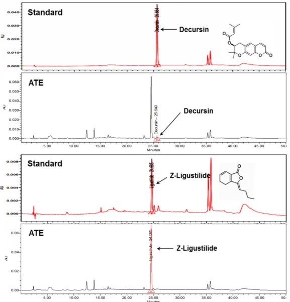
Decursin and Z-ligustilide are reliable marker compounds for the authenticity of ATE (Nam et al., 2014). When the content of decursin and Z-ligustilide were determined in ATE by HPLC method, the retention times of decursin and Z-ligustilide were approximately 25.66 min and 24.68 min, respectively (Fig. 1).
ATE contained 0.60 ㎍/㎎ (0.06%) of decursin and 84.34 ㎍/㎎
(8.43%) of Z-ligustilide (Fig. 1).
The content of total phenolic and total flavonoid
Total phenolic and total flavonoid content were expressed as a quercetin equivalents (QE) and gallic acid equivalents (GAE), respectively. ATE (1 ㎎) contained flavonoid as a 5.52 ㎍/QE and phenolic as a 237.27 ㎍/GAE (Table 1).
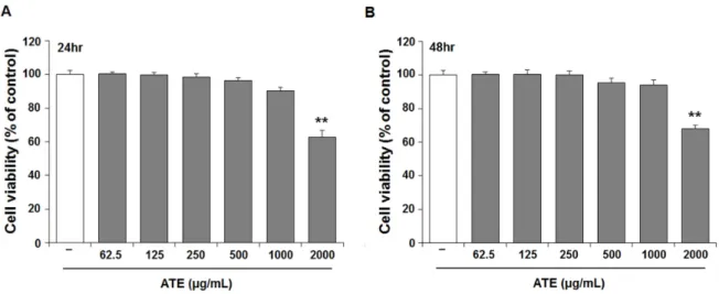
Effect of ATE on the cell viability
MTT assay was conducted at both 24 and at 48 h to determine the cytotoxicity of ATE in B16F10 cells. ATE up to 1000 ㎍/㎖ did not show significant cell death at both 24 (Fig.
2A) and 48 h (Fig. 2B). However, ATE at the concentration of
2000 ㎍/㎖ displayed approximately 40% reduction of cell
viability at both 24 and 48 h compared to untreated control.
Hence, subsequent experiments were conducted at below 1000
㎍/㎖ of ATE.
Antioxidant capacity of ATE
The radical scavenging activities of ATE were determined
by DPPH and ABTS assays. As shown Fig. 3A and B, DPPH and ABTS scavenging activity were increased as the concentration of ATE increased. The effective concentration for 50% scavenging activity (IC
50) was determined by the regression equation, and the IC
50values (㎎/㎖) of DPPH and ABTS radical scavenging activity were 2.78 and 0.76, respectively. The reducing power Fig. 1. High-performance liquid chromatography (HPLC)/photodiode array (PDA) detector chromatograms of the Angelica tenuissima root extract (ATE) and standards for decursin and Z-ligustilide.
Table 1. Total phenolic and total flavonoid content of A. tenuissima extract Total phenolic ( ㎍ GAE
z/ ㎎ dried extract)
Total flavonoid ( ㎍ QE
y/ ㎎ dried extract) Ethanol extract of A. tenuissima root (ATE) 237.27 ± 13.24
x5.52 ± 0.07
xz