—61—
INTRODUCTION
The Miho spine loach Cobitis choii (Cypriniformes;
Cobitidae), a synonym of Iksookimia choii, is one of the critically threatened freshwater species in the Korean peninsula. This endemic species has also been designated as a Natural Monument (No. 454; Cultural Properties Administration, Republic of Korea) (Kim et al., 2008;
Song et al., 2008). However during the last decades, they have lost, at least in part, their natural habitats pos- sibly caused by anthropogenic and industrial activities (Son and Byeon, 2005). Considering they often occupy a narrow range of habitats in natural water body, careful efforts for the conservation of this species are urgently needed. Moreover, recent molecular studies have claimed the currently resident C. choii populations have a very
low level of genetic diversity irrespective of geographic locations of their habitats (Lee et al., 2008; Bang et al., 2009). Conservation projects for C. choii are underway with respect to the stock assessment, artificial propaga- tions and ex situ and in situ restorations. Exploitation of functional genes relevant to stress/toxicity responses and host defense could be fundamental for better understand- ing its stress physiology, consequently providing useful means for effective designation of the conservation plan.
Metallothionein (MT), a low molecular weight (6~7 kDa) and cysteine-rich protein, is a metal-binding house- keeper, which plays central role in the homeostatic reg- ulation of metals (Klaassen et al., 2009). This protein is responsible for not only the reservation of essential metals but also the detoxification of excess metals. Owing to its high inducibility upon exogenous administration of various heavy metal ions, the MT-based expression assay has been one of versatile ways to notice the risks associ- ated with metal pollution in aquatic environments (Bour-
Gene Structure and Altered mRNA Expression of Metallothionein in Response to Metal Exposure and Thermal Stress in Miho Spine
Loach Cobitis choii (Cobitidae; Cypriniformes)
By Sang Yoon Lee and Yoon Kwon Nam*
Department of Marine Bio-Materials & Aquaculture, Pukyong National University, Busan 608-737, Korea
ABSTRACT Gene and promoter structures of metallothionein (MT ) from Miho spine loach (Cobitis choii; Cypriniformes) were characterized, and the transcriptional responses to experimental exposures to heavy metals and heat stress were examined. The C. choii metallothionein displayed well-conserved features of teleostean metallothioneins at gDNA, mRNA and amino acid levels. Bioinformatic analysis predicted that the C. choii MT regulatory region potentially possessed various motifs or elements targeted by various transcription factors associated with metal-coordinating regulation (e.g., metal transcription factor-1), immune responses (e.g., nuclear factor kappa B), and thermal modulations (e.g., heat shock factor). Acute heavy-metal exposures to 0.5 or 1.0μμM of cadmium (Cd), copper (Cu), man- ganese (Mn), nickel (Ni) or zinc (Zn) showed that MT transcription was significantly stimulated by Cd (9.6-fold relative to non-exposed control) and Cu (10.4-fold), only moderately by Mn (2.4-fold), but hardly by Ni and Zn. Elevation of water temperature from 25��C to 31��C caused a rapid modulation of MT mRNAs toward upregulation to 9.5-fold; however, afterward the elevated mRNA level slightly decreased during further incubation at 31�C for 6 h. Results from this study suggest that MT-based expression assay could be a useful basis for better understanding the metal- and/or heat-caused stresses in this endangered fish species.
Key words : Cobitis choii, gene structure, heat stress, heavy metals, metallothionein, mRNA expression
*Corresponding author: Yoon Kwon Nam Tel: 82-51-629-5918, Fax: 82-51-629-5908, E-mail: [email protected]
Accepted : March 28, 2011
http://www.fishkorea.or.kr
dineaud et al., 2006; Cho et al., 2009c). It has not been yet clarified whether or not the pollutants particularly including heavy metals have played certain roles in the loss of Cobitis choii populations in natural streams. How- ever, the functional evaluation of this multivalent detox- ifying protein could be helpful for build-up of basic know- ledge on the toxicant-related stress physiology of this endangered species, since MTs have also been proven to be related with many signaling pathways involved in aging, antioxidant defense and immune response (Guo et al., 2009; Fu et al., 2010; Swindell, 2011). Further- more the information on the MT modulation in response to metal toxicants could be a useful basis for the post-moni- toring plan (i.e., evaluation of general stress or health status of re-introduced stocks) along with the examina- tion of the possible changes of water quality in the reci- pient site.
The objective of this study is to isolate and characterize the genetic determinant of C. choii metallothionein at both mRNA and genomic levels in order to provide a basis for further development of MT-based biomarker.
For this, we characterized the full-length cDNA and genomic gene sequences of the C. choii MT, highlighted the structural characteristics of the 5′-flanking upstream regulatory region and examined the transcriptional alter- ations of MT in response to acute metal exposures and heat stress.
MATERIALS AND METHODS
1. Molecular cloning of cDNA and genomic gene for C. choii MT
The specimens (Cobitis choii) used in this study were laboratory-bred individuals produced using the captive
broodfish, which had been sampled from the Baekgok stream under the authorized permission. To identify the MT cDNA sequence, expressed sequence tag (EST) data- base constructed with liver, intestine, ovary and whole body of C. choii was surveyed (unpublished data). From the database search, an EST clone showing the signifi- cant homology with previously known teleost metallo- thioneins was selected and further sequenced at both 5′- and 3′-directions. A continuous fragment of the MT cDNA from 5′-untranslated region (UTR) to 3′-UTR was isolat- ed by RT-PCR using oligonucleotide primers ICMT-1F and ICMT-1R. Information on the oligonucleotide primers used in this study was provided in Table 1. RT-PCR was carried out with the liver cDNA that had been used for the construction of the cDNA library. Amplified frag- ment was cloned into pGEM®-T easy vector (Promega, Madison, WI, USA), and insert sequence was confirmed at both directions. Based on the cDNA sequence, geno- mic MT sequence was PCR-isolated. Preparation of C.
choii genomic DNA from the whole blood was carried out using the conventional proteinase K/SDS method and an aliquot (500 ng) of the purified genomic DNA template was subjected to a PCR reaction using the Ex- pand High Fidelity PCR System (Roche Applied Science, Mannheim, Germany). The primer pair was the same as described above. PCR product was gel-purified, TA clon- ed and six-randomly-chosen recombinant clones were sequenced in order to obtain the representative sequence.
All the raw sequence data was edited using the Sequen- cher®4.9 (Gene Codes, Ann Arbor, MI, USA). Genomic organization (exon/intron organization) was compared to other teleost MT genes. Nucleotide composition analy- sis of each intron was done using the GENE BOY tool (http://www.dnai.org/geneboy/index.html). Homology search against GenBank was performed using BLASTx
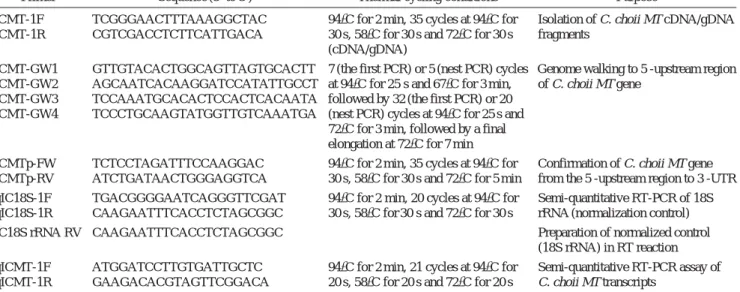
Table 1. Sequences of oligonucleotide primers and thermal cycling conditions used in this study
Primer Sequence (5′ to 3′) Thermal cycling conditions Purpose
ICMT-1F TCGGGAACTTTAAAGGCTAC 94�C for 2 min, 35 cycles at 94�C for Isolation of C. choii MT cDNA/gDNA ICMT-1R CGTCGACCTCTTCATTGACA 30 s, 58�C for 30 s and 72�C for 30 s fragments
(cDNA/gDNA)
ICMT-GW1 GTTGTACACTGGCAGTTAGTGCACTT 7 (the first PCR) or 5 (nest PCR) cycles Genome walking to 5′-upstream region ICMT-GW2 AGCAATCACAAGGATCCATATTGCCT at 94�C for 25 s and 67�C for 3 min, of C. choii MT gene
ICMT-GW3 TCCAAATGCACACTCCACTCACAATA followed by 32 (the first PCR) or 20 ICMT-GW4 TCCCTGCAAGTATGGTTGTCAAATGA (nest PCR) cycles at 94�C for 25 s and
72�C for 3 min, followed by a final elongation at 72�C for 7 min
ICMTp-FW TCTCCTAGATTTCCAAGGAC 94�C for 2 min, 35 cycles at 94�C for Confirmation of C. choii MT gene ICMTp-RV ATCTGATAACTGGGAGGTCA 30 s, 58�C for 30 s and 72�C for 5 min from the 5′-upstream region to 3′-UTR qIC18S-1F TGACGGGGAATCAGGGTTCGAT 94�C for 2 min, 20 cycles at 94�C for Semi-quantitative RT-PCR of 18S qIC18S-1R CAAGAATTTCACCTCTAGCGGC 30 s, 58�C for 30 s and 72�C for 30 s rRNA (normalization control)
IC18S rRNA RV CAAGAATTTCACCTCTAGCGGC - Preparation of normalized control
(18S rRNA) in RT reaction qICMT-1F ATGGATCCTTGTGATTGCTC 94�C for 2 min, 21 cycles at 94�C for Semi-quantitative RT-PCR assay of qICMT-1R GAAGACACGTAGTTCGGACA 20 s, 58�C for 20 s and 72�C for 20 s C. choii MT transcripts
or BLASTp option. Amino acid sequence identities bet- ween C. choii MT and other MTs were compared using the GeneDoc software (http://www.psc.edu/biomed/gene- doc).
2. Characterization of 5′-upstream region by genome walking
The 5′-flanking region of the C. choii MT was cloned in order to characterize the putative transcription factor binding motifs. Genome walking libraries were construct- ed using the GenomeWalkerTM Universal Kit (BD Bio- sciences Clontech, Mountain View, CA, USA). PCR amplifications were carried out using oligonucleotide primers, ICMT-GW1 to ICMT-GW4. PCR products were TA cloned and insert DNA was sequenced. Each frag- ment sequence was assembled into a contig to generate a contiguous sequence. From the contig sequence, a genomic DNA fragment from the 5′-upstream region to 3′-UTR was PCR-amplified again by using the primers ICMTp-FW and ICMTp-RV, and then the sequence was confirmed. Putative transcription factor binding motifs was predicted using the Transcription Element Search System (TESS; http://www.cbil.upenn.edu/cgi-bin/tess/
tess?RQ==WELCOME) and Transcriptional Factor Search (TFSEARCH; http://www.cbrc.jp/research/db/TFSEA RCH.html).
3. Experimental exposures to heavy metals and thermal stress
To examine the transcriptional response of MT to heavy metal exposure, acute immersion exposures of C. choii fry to various heavy metals were performed. Eight C.
choii fry (average body weight==1.1±0.2 g) were allocat- ed into one of two replicate tanks (10 L) filled with 8 L of well-aerated tap water (5.0μm-filtered) containing 0.5 or 1.0μM of cadmium (Cd), copper (Cu), manganese (Mn), nickel (Ni) or zinc (Zn). A non-exposed control group was also prepared identically except the adminis- tration of the heavy metals. During exposure, no feed was provided. Water temperature was adjusted to 22±
0.5�C throughout exposure period. At 24 h post exposure, whole body total RNA was extracted from the six-ran- domly-chosen fry from each tank (two replicate tanks per group).
In order to examine the modulation of MT gene in response to heat stress, a thermal treatment was perform- ed. Forty fry (same-sized as above) were allocated into one of two identical tanks (50 L) containing 40 L of tap water at 25�C, and incubated further for 24 h in order to make the fish to acclimate to the starting temperature.
Water temperature for one of the two tanks was gradually elevated with a rate of 1�C/h until 31�C. Eight randomly- chosen fry were sampled at 0 h (25�C; starting point), 3 h (reaching 28�C), 6 h (reaching 31�C) and 12 h (kept con-
stant at 31�C for 6 h after reaching 31�C) post thermal elevation, respectively. At the same time, a random sample of eight individuals was also obtained from the control group, which was maintained at the constant temperature (25�C). The temperature was controlled to be ranged within ±0.5�C and the level of dissolved oxygen was also kept from 5 to 6 ppm.
4. MT mRNA assay
Extraction of total RNA from whole body fry was car- ried out using TriPure reagent (Roche) and the total RNA extracted was purified again with RNeasy Mini Kit includ- ing DNase treatment step (Qiagen, Hilden, Germany).
In order to prepare an invariant normalization control, a partial segment of C. choii 18S rRNA was cloned by PCR isolation using the primers conserved in fish 18S rRNA (unpublished data). An aliquot (2μg) of the puri- fied total RNA was reverse transcribed into cDNA using the Omniscript Reverse Transcription Kit (Qiagen).
During the reverse transcription (RT), a reverse primer complementary to the C. choii 18S rRNA (IC18S rRNA RV; 0.1μM) was also included in the RT reaction. The cDNA pool synthesized was four-fold (for MT ) or eight- fold (for 18S rRNA) diluted and two μL of the diluted cDNA was used as the template for thermal cycling reac- tion. Primer pairs for C. choii MT (qICMT-1F/1R; ampli- con==240 bp) and 18S rRNA (qIC18S-1F/1R; amplicon== 569 bp) were included in the PCR reaction independent- ly. Six μL of amplified product was separated on a 1.5%
agarose gel, visualized by ethidium-bromide (EtBr) stain- ing and analyzed by the Quantity-OneTMimage analysis software implemented in the VersaDoc 4000 (Bio-Rad, Hercules, CA, USA). Triplicate assays were performed in an independent fashion. Based on the scanning densi- tometry, differences in the relative expression levels among groups were assessed by analysis of variance (ANOVA) followed by Duncan’s multiple range tests.
On the other hand, significant difference from the control levels was tested by Student’s t-test. All the statistics were performed using the SPSS software (ver. 10.1.3) and differences were considered to be significant when P⁄0.05.
RESULTS AND DISCUSSION
The C. choii MT cDNA was composed of 56 bp of 5′- UTR, 180 bp of single open reading frame (ORF) encod- ing 60 amino acids and 243 bp of 3′-UTR excluding the poly(A)++tail (Fig. 1; GenBank accession no. JF419523).
Two putative polyadenylation signals (AATATA and AATAAA) were found at 153 bp and 16 bp prior to the poly(A)++ tail, respectively, suggesting the potential processing of MT transcript variants with different 3′- UTR lengths (Cho et al., 2008). The C. choii MT poly-
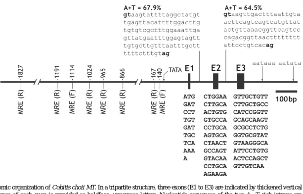
peptide consisted of twenty cysteine residues at conserv- ed positions when aligned with other representative ortho- logs from teleosts (alignment not shown). The richness of Cys residues in the Cys-X-Cys or Cys-Cys form at both β- and α-domains of MT proteins has already been reported widely in other vertebrate orthologs (Cho et al., 2005; Knapen et al., 2005; Gao et al., 2009). From the genomic PCR isolation, the C. choii MT was proven to possess a tripartite organization which is typical for most known vertebrate MTs (Fig. 2). The conserved exon/in-
tron splicing rule (GT-AG) was also found in each junc- tion site. Like many teleost MTs, C. choii MT showed a high proportion of adenine and thymine in its two introns (Chen et al., 2004; Lin et al., 2004; Cho et al., 2008).
Most teleosts are known to have at least two copies of MT genes in their genomes, although the chromosomal organization as well as chromosomal synteny between species has not been studied yet in most fish species so far (Knapen et al., 2005; Cho et al., 2009c). Unfortunate- ly, the classification of the C. choii MT into MT-I (or MT-
Fig. 1. Complementary DNA and deduced amino acid (single code) sequences of Cobitis choii metallothionein. Coding region is indicated by boldface uppercase letters while non-coding region by lowercase letters. Twenty conserved cysteine residues are bolded. Two putative polyadenylation signals are underlined.
Fig. 2. Genomic organization of Cobitis choii MT. In a tripartite structure, three exons (E1 to E3) are indicated by thickened vertical lines and the sequence of each exon is provided as boldface, uppercase letters. Nucleotide sequences of the two A++T rich introns are indicated by lowercase letters in which gt-ag splicing sites are bolded. Consensus TATA box and two putative polyadenylation signals are indicated arrows. In the 5′-flanking region, relative positions of eight metal responsive elements (MREs) from the translation initiation site (ATG) are indicated in a forward (F) or reverse (R) orientation. For complete nucleotide sequence of the C. choii MT gene and its promoter, refer to GenBank (accession no. JF419523).
A) or MT-II (or MT-B) has been difficult currently due to the limited information on MT orthologs from Cobitidae.
Moreover, the present C. choii MT showed a higher amino acid sequence identity (100%) with MT from the stone loach (Barbatula barbatula; GenBank no. CAA4 2036) belonging to different family (Balitoridae) than MT isoforms (98%) from the mud loach (Misgurnus mizolepis: GenBank nos. ACH90423 and ACH90424) belonging to the same family (Cobitidae). Gene duplica- tion of MT isoforms in fish genomes has been suggested to occur frequently in a taxon-specific manner (Cho et al., 2009c), indicating that further exploitation of MT paralog(s) from the C. choii genome should be crucial for more comprehensive comparison between the MT isoforms from Cobitidae.
Genome walking to the 5′-direction of C. choii MT
recovered a 4,090 bp of upstream sequence from the tran- slation initiation codon (ATG) (GenBank no. JF419523) and essential sites or motifs targeted by various transcrip- tion factors were predicted (Table 2; Fig. 2). Firstly, eight copies of metal responsive elements (MREs), which are known to be essential for the binding of metal transcrip- tion factor-1 (MTF-1), were found in both proximal and distal parts of the regulatory region. MTF-1 is the key factor to regulate both basal and induced expression of the genes to code the metal-coordinating proteins such as MT and superoxide dismutase (SOD) (Laity and And- rews, 2007; Cho et al., 2009a; Ferencz and Hermesz, 2009). The presence pattern of MRE copies in the C.
choii MT regulatory region was similar with those found in other fish MT promoters in terms of the clustering of multiple MREs in proximal and/or distal regions rather than random or even distribution (Ren et al., 2006; He et al., 2007; Cho et al., 2008). Other transcription factors identified in the C. choii MT promoter included GATA factor, activator protein1 (AP-1) and hepatocyte nuclear factor (HNF), which are known to be often found in most fish MT promoters, although their specific roles in the modulation of fish MT have not been clearly clarified yet (Yan and Chan, 2004; Cheung et al., 2005; Cho et al., 2008). In addition, several noteworthy transcription factors predicted in the C. choii MT promoter were the factors that have been known to be closely involved in the immune responses of vertebrates. They included the nuclear factor kappa B (NF-κB/c-Rel), CAAT-enhancer binding protein (C/EBP) and upstream stimulatory factor (USF), which all have been usually found in the antimi- crobial or antioxidant genes, suggesting the proposed employment of MTs in host defense mechanism (Atif et al., 2006; Thirumoorthy et al., 2007; Wang et al., 2009).
Finally, the present C. choii MT promoter represented two binding sites for heat shock factor (HSF), suggesting the possible modulation of the MT gene under thermally stressed conditions (Cho et al., 2009c).
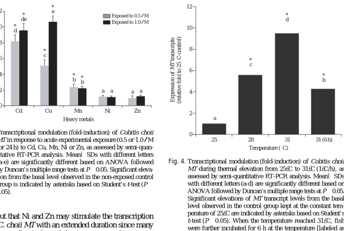
As expected, the C. choii MT transcripts were actively induced in response to the acute heavy metal exposures, although the inducibility was highly variable depending upon metal inducers (i.e., Cd, Cu, Mn, Ni and Zn) (Fig.
3). Of the five metals tested, most potent metals were Cd (8.2- and 9.6-fold induction of MT mRNA, respective- ly for 0.5 and 1.0μM-treated groups when compared to basal level of non-exposed control) and Cu (5.1- and 10.4- fold), while fish exposed to Mn showed only the mode- rate activation of MT at both 0.5 and 1.0μM concentra- tions (2.4- and 2.2-fold, respectively) (P⁄0.05). On the other hand, significant alteration was detected in neither Ni nor Zn irrespective of the metal concentrations (P¤
0.05). Differential responses of MT expression to differ- ent metal inducers have been commonly observed in a number of other fish species (Hermesz et al., 2001; Woo et al., 2006; Lee et al., 2010). However, it could not be
Table 2. Putative transcription factor binding sites predicted in the 5′-regulatory region of Cobitis choii MT
Transcription Consensus Positionb Sequence factor sequencea
TFIID TATAAA -116 TATAAA
STAT TTCNNNGAA -3345 TTCAGTGAA
-3036 TTCTCTGAA
USF CANNTG -540 CACGTG
HNF-3 TRTTKRYTY -3934 AAACAAATA*
-3773 AAACAAATA*
-2794 TGTTTGTTT -2146 GAGTAAATA*
-1779 TATTTATTT -330 AAACAAATA*
AP-1 TGASTMA -3514 TGACTCA
-850 TGACACA
C/EBP TTDNGNAA -2889 TTTCAAAA*
-2097 TTATGCAA
GATA WGATAR -1546 TGATAG
-1305 CTATCC*
MTF-1 TGCRCNC -1827 GTGTGCA*
-1191 GTGTGCA*
-1114 TGCACCC -1024 GAGTGCA*
-965 GAGTGCA*
-866 GTGTGCA*
-167 GTGTGCA*
-140 TGCACTC
AhR CACGCW -1425 TGCGTG*
-947 CACGCA
NF-κB/c-Rel GGGRNNYYCC -400 GGGACCTTCC
NF-AT WGGAAAA -4038 AGGAAAA
-1061 TGGAAAA
GR-h TGTTCT -2915 AGAACA*
-2763 TGTTCT
-987 TGTTCT
HSF GAAKKTTC -3027 GAATTTTC
-37 GAACCTTC*
aN==any base; R==A or G; Y==C or T; W==A or T; K==G or T; S==C or G;
D==A or G or T; M==A or C
bUpstream from the translation start (ATG) site
*Reverse orientation to the consensus sequence
ruled out that Ni and Zn may stimulate the transcription of the C. choii MT with an extended duration since many previous studies indicated the importance of treatment duration as well as the dose strength (Cho et al., 2008;
Rhee et al., 2009). In addition, the possibility that tissue- specific modulation of the MT transcripts might be mask- ed under the present assay condition because we perform- ed the analysis with the total RNA samples from whole fry rather than independent adult tissues, which should be addressed in future experiment. Nevertheless, result from this study could support, at least in part, the recent claim on the need of revision of the classic dogma for the MT induction, in the sense of that Zn is not always an inducer for activating the MT transcription (Bi et al., 2006; Bourdineaud et al., 2006; Cho et al., 2009c).
Metal toxicity and modulation of MT in response to a given heavy metal exposure have also been known to vary among fish species, and the response patterns are generally in relation with the taxonomic position of the species (Kalman et al., 2010).
Thermal stress could alter significantly the mRNA expression of C. choii MT (Fig. 4). Basal expression level observed at the starting temperature (25�C) was sharply elevated up to 5.6-fold at 28�C and then more increased to 9.5-fold at 31�C. This highest level observ- ed at 31�C was slightly decreased down when the fish were kept further at the constant 31�C for 6 h, although the level remained still significantly higher than that observed in the 25�C-control (4.3-fold relative to non- treated control). The modulation of the C. choii MT by thermal treatment was well in congruent with our finding
the presence of binding motifs for HSFs in its regulatory region. Regulatory mechanism of fish MT gene in res- ponse to acute thermal change has been little explored.
However, external temperature is one of the primary abiotic factors to affect the regulation of various physio- logical pathways in poikilothermal animals, and heat stress has been known to often cause the formation of reactive oxygen species (ROS) in those animals (Buckley et al., 2006; Cho et al., 2006; Cho et al., 2009b). Alth- ough we haven’t yet examined the expression pattern of antioxidant enzyme genes together with MT gene, the temperature-dependent upregulation of MT may be in accordance with its potential involvement in the antioxi- dant pathway to relieve the prooxidant stress arisen from the thermal elevation. On the other hand, the decline of MT transcript levels after reaching peak at 31�C could be explained, in general, by the stabilized or acclimated process after the initial shock-phase, indicating future experiments are needed to examine if the MT mRNA levels could return to the basal level or not. It is widely agreed that metallothionein is a useful early warning indicator of the risks associated with heavy metal-mediat- ed toxicity (Rodríguez-Ortega et al., 2009; Falfushynska et al., 2010). However, it has been claimed that the tran- scription (both basal and induced expressions) of MT genes could be greatly modulated by various biotic (e.g.,
Expression of MT transcripts (relative fold to control) 12
10
8
6
4
2
0
Heavy metals
Cd Cu Mn Ni Zn
*d de*
*c
*e
*b * b
a a a a
Exposed to 0.5μM Exposed to 1.0μM
Expression of MT transcripts (relative fold to 25。C-control) 12
10
8
6
4
2
0
25 28 31 31 (6 h)
Temperature (。C) a
*c
*d
*b
Fig. 3. Transcriptional modulation (fold-induction) of Cobitis choii MT in response to acute experimental exposure (0.5 or 1.0μM for 24 h) to Cd, Cu, Mn, Ni or Zn, as assessed by semi-quan- titative RT-PCR analysis. Mean±SDs with different letters (a-e) are significantly different based on ANOVA followed by Duncan’s multiple range tests at P⁄0.05. Significant eleva- tion from the basal level observed in the non-exposed control group is indicated by asterisks based on Student’s t-test (P⁄
0.05).
Fig. 4. Transcriptional modulation (fold-induction) of Cobitis choii MT during thermal elevation from 25�C to 31�C (1�C/h), as assessed by semi-quantitative RT-PCR analysis. Mean±SDs with different letters (a-d) are significantly different based on ANOVA followed by Duncan’s multiple range tests at P⁄0.05.
Significant elevations of MT transcript levels from the basal level observed in the control group kept at the constant tem- perature of 25�C are indicated by asterisks based on Student’s t-test (P⁄0.05). When the temperature reached 31�C, fish were further incubated for 6 h at the temperature [labeled as
‘31(6 h)’].
age, sex and health status etc.) and abiotic (e.g., tempera- ture, salinity, and seasons, etc.) factors (Ringwood et al., 1999; Amiard et al., 2006; Dragun et al., 2009; Lee et al., 2010). As evidenced in this study, thermal condition should be carefully considered for all the MT-based expression assays in this species.
In summary, gene and promoter structures of the C.
choii were determined and transcriptional modulations of C. choii MT in response to experimental exposures with different heavy metals and heat stress were examin- ed. Result of this study proposes that MT could be a potential indicator to address the various cellular stresses in this endangered fish species, however also suggests that careful and extensive analyses should be followed in order to employ the MT as an environment-realistic biomarker to aid the conservation of this species. In par- ticular, the assessment of MT modulation with more realistic doses of the heavy metals, which are expected potentially to be present in candidate recipient sites for the restoration of this species (i.e. the sites for re-intro- duction of the fishes), would be a valuable subject in future study. However for this assessment also, it is recom- mended that seasonal variation of MT gene expression caused by the change of water temperature should be taken together into account.
ACKNOWLEDGEMENTS
We thank Dr. In-Chul Bang, Soonchunhyang Univer- sity for his kind providing the experimental fish speci- mens as well as for his critical comments on this study.
This study was supported by a fund from the Korea- Ukraine international cooperative research project (#2010- 00091) from National Research Foundation.
REFERENCES
Amiard, J.C., C. Amiard-Triquet, S. Barka, J. Pellerin and P.S. Rainbow. 2006. Metallothioneins in aquatic inver- tebrates: their role in metal detoxification and their use as biomarkers. Aquat. Toxicol., 76: 160-202.
Atif, F., M. Kaur, S. Yousuf and S. Raisuddin. 2006. In vitro free radical scavenging activity of hepatic metalloth- ionein induced in an Indian freshwater fish, Channa punctatta Bloch. Chem. Biol. Interact., 162: 172-180.
Bang, I.C., W.J. Kim and I.R. Lee. 2009. Characterization of polymorphic microsatellite loci in the endangered Miho spine loach (Iksookimia choii) and cross-species amplification within the Cobitidae family. Mol. Ecol.
Resour., 9: 281-284.
Bi, Y., G.X. Lin, L. Millecchia and Q. Ma. 2006. Superin- duction of metallothionein I by inhibition of protein
synthesis: role of a labile repressor in MTF-I mediated gene transcriptioin. J. Biochem. Mol. Toxicol., 20:
57-68.
Bourdineaud, J.P., M. Baudrimont, P. Gonzalez and J.L.
Moreau. 2006. Challenging the model for induction of metallothionein gene expression. Biochimie, 88:
1787-1792.
Buckley, B.A., A.Y. Gracey and G.N. Somero. 2006. The cellular response to heat stress in the goby Gillichthys mirabilis: a cDNA microarray and protein-level analy- sis. J. Exp. Bio., 209: 2660-2677.
Chen, W.Y., J.A.C. John, C.-H. Lin, H.-F. Lin, S.-C. Wu, C.-H. Lin and C.-Y. Chang. 2004. Expression of metallothionein gene during embryonic and early larval development in zebrafish. Aquat. Toxicol., 69:
215-227.
Cheung, A.P.L., V.K.L. Lam and K.M. Chan. 2005. Tilapia metallothionein genes: PCR-cloning and gene expres- sion studies. Biochim. Biophys. Acta., 1731: 191- 201.
Cho, Y.S., B.N. Choi, E.M. Ha, K.H. Kim, S.K. Kim, D.S.
Kim and Y.K. Nam. 2005. Shark (Scyliorhinus tora- zame) metallothionein: cDNA cloning, genomic sequ- ence, and expression analysis. Mar. Biotechnol., 7:
350-362.
Cho, Y.S., I.C. Bang, I.R. Lee and Y.K. Nam. 2009a. Hepa- tic expression of Cu/Zn-superoxide dismutase tran- scripts in response to acute metal exposure and heat stress in Hemibarbus mylodon (Teleostei: Cypriniform- es). Fish. Aqua. Sci., 12: 179-184.
Cho, Y.S., S.Y. Lee, C.H. Noh, Y.K. Nam and D.S. Kim.
2006. Survey of genes responsive to long-term heat stress using a cDNA microarray analysis in mud loach (Misgurnus mizolepis) liver. Kor. J. Ichthyol., 18:
65-77.
Cho, Y.S., S.Y. Lee, K.H. Kim, S.K. Kim, D.S. Kim and Y.K. Nam. 2009b. Gene structure and differential modulation of multiple rockbream (Oplegnathus fasciatus) hepcidin isoforms resulting from different biological stimulations. Dev. Comp. Immunol., 33:
46-58.
Cho, Y.S., S.Y. Lee, K.-Y. Kim, I.C. Bang, D.S. Kim and Y.K. Nam. 2008. Gene structure and expression of metallothionein during metal exposures in Hemibar- bus mylodon, Ecotoxicol. Environ. Saf., 71: 125-137.
Cho, Y.S., S.Y. Lee, K.-Y. Kim and Y.K. Nam. 2009c. Two metallothionein genes from mud loach Misgurnus mizolepis (Teleostei; Cypriniformes): Gene structure, genomic organization, and mRNA expression analy- sis. Comp. Biochem. Physiol. B Biochem. Mol. Biol., 153: 317-326.
Dragun, Z., M. Podrug and B. Raspor. 2009. Combined use
of bioindicators and passive samplers for the assess- ment of the river water contamination with metals.
Arch. Environ. Contam. Toxicol., 57: 211-220.
Falfushynska, H.I., L.L. Gnatyshyna, C.V. Priydun, O.B.
Stoliar and Y.K. Nam. 2010. Variability of responses in the crucian carp Carassius carassius from two Ukrainian ponds determined by multi-marker appro- ach. Ecotoxicol. Environ. Saf., 73: 1896-1906.
Ferencz, Á. and E. Hermesz. 2009. Identification of a splice variant of the metal-responsive transcription factor MTF-1 in common carp. Comp. Biochem. Physiol.
C Toxicol. Pharmacol., 150: 113-117.
Fu, Z., J. Guo, L. Jing, R. Li, T. Zhang and S. Peng. 2010.
Enhanced toxicity and ROS generation by doxorubi- cin in primary cultures of cardiomyocytes from neo- natal metallothionein-I/II null mice. Toxicol. In Vitro, 24: 1584-1591.
Gao, D., G.T. Wang, X.T. Chen and P. Nie. 2009. Metallo- thionein-2 gene from the mandarin fish Siniperca chuatsi: cDNA cloning, tissue expression, and imm- unohistochemical localization. Comp. Biochem.
Physiol. C Toxicol. Pharmacol., 149: 18-25.
Guo, R., H. Ma, F. Gao, L. Zhong and J. Ren. 2009. Metallo- thionein alleviates oxidative stress-induced endoplas- mic reticulum stress and myocardial dysfunction. J.
Mol. Cell. Cardiol., 47: 228-237.
He, P., M. Xu and H. Ren. 2007. Cloning and functional characterization of 5′-upstream region of metallothi- onein-I gene from crucian carp (Carassius cuvieri).
Int. J. Biochem. Cell Biol., 39: 832-841.
Hermesz, E., M. Abraham and J. Nemcsok. 2001. Tissue- specific expression of two metallothionein genes in common carp during cadmium exposure and tempera- ture shock. Comp. Biochem. Physiol. C Toxicol.
Pharmacol., 128: 457-465.
Kalman, J., I. Riba, T. Angel DelValls and J. Blasco. 2010.
Comparative toxicity of cadmium in the commercial fish species Sparus aurata and Solea senegalensis.
Ecotoxicol. Environ. Saf., 73: 306-311.
Kim, K.-Y., S.Y. Lee, Y.S. Cho, I.C. Bang, D.S. Kim and Y.K. Nam. 2008. Characterization and phylogeny of two beta-cytoskeletal actins from Hemibarbus mylo- don (Cyprinidae, Cypriniformes), a threatened fish species in Korea. DNA Seq., 19: 87-97.
Klaassen, C.D., J. Liu and B.A. Diwan. 2009. Metallothion- ein protection of cadmium toxicity. Toxicol. Appl.
Pharmacol., 238: 215-220.
Knapen, D., E.S. Redeker, I. Inácio, W. De Coen, E. Verh- eyen and R. Blust. 2005. New metallothionein mRNAs in Gobio gobio reveal at least three gene duplication events in cyprinid metallothionein evolution. Comp.
Biochem. Physiol. C Pharmacol. Toxicol. Endocrinol.,
140: 347-355.
Laity, J.H. and G.K. Andrews. 2007. Understanding the mechanisms of zinc-sensing by metal-response ele- ment binding transcription factor-1 (MTF-1). Arch.
Biochem. Biophys., 463: 201-210.
Lee, S.Y., O. Stoliar and Y.K. Nam. 2010. Transcriptional alteration of two metallothionein isoforms in mud loach (Misgurnus mizolepis) fry during acute heavy metal exposure. Fish. Aqua. Sci., 13: 112-117.
Lee, Y.A., Y.E. Yun, Y.K. Nam and I.C. Bang. 2008. Gene- tic diversity of an endangered fish Iksookimia choii (Cypriniformes), from Korea as assessed by amplified fragment length polymorphism. Kor. J. Limnol., 41:
98-103.
Lin, C.H., J.A.C. John, L.W. Ou, J.C. Chen, C.H. Lin and C.Y. Chang. 2004. Cloning and characterization of metallothionein gene in ayu Plecoglossus altivelis.
Aquat. Toxicol., 66: 111-124.
Ren, H., M. Xu, P. He, N. Muto, N. Itoh, K. Tanaka, J. Xing and M. Chu. 2006. Cloning of crucian carp (Carassius cuvieri) metallothionein-II gene and characterization of its gene promoter region. Biochem. Biophys. Res.
Commun., 342: 1297-1304.
Rhee, J.S., S. Raisuddin, D.S. Hwang, K.W. Lee, I.C. Kim and J.S. Lee. 2009. Differential expression of metal- lothionein (MT) gene by trace metals and endocrine- disrupting chemicals in the hermaphroditic mangrove killifish, Kryptolebias marmoratus. Ecotoxicol. Envi- ron. Saf., 72: 206-212.
Ringwood, A.H., M.J. Hameedi, R.F. Lee, M. Brower, E.C.
Peters, G.I. Scott, S.N. Luoma and R.T. Di Giulio.
1999. Bivalve biomarker workshop: overview and discussion group summaries. Biomarkers, 4: 391-399.
Rodríguez-Ortega, M.J., A. Rodríguez-Ariza, J.L. Gómez- Ariza, A. Muñoz-Serrano and J. López-Barea. 2009.
Multivariate discriminant analysis distinguishes metal- from non metal-related biomarker responses in the clam Chamaelea gallina. Mar. Pollut. Bull., 58: 64- 71.
Son, Y.-M. and H.-K. Byeon. 2005. The ichthyofauna and dynamics of the fish community in Miho stream, Korea. Kor. J. Ichthyol., 17: 271-278 (in Korean).
Song, H.Y., W.J. Kim, W.O. Lee and I.C. Bang. 2008. Mor- phological development of egg and larvae of Ikso- okimia choii (Cobitidae). Korean J. Limnol., 41: 104- 110.
Swindell, W.R. 2011. Metallothionein and the biology of aging. Ageing Res. Rev., 10: 132-145.
Thirumoorthy, N., K.T.M. Kumar, A.S. Sundar, L. Panayap- pan and M. Chatterjee. 2007. Metallothionein: an overview, World J. Gastroenterol., 13: 993-996.
Wang, L., L. Song, D. Ni, H. Zhang and W. Liu. 2009.
—69— Alteration of metallothionein mRNA in bay scallop Argopecten irradians under cadmium exposure and bacteria challenge. Comp. Biochem. Physiol. C Phar- macol. Toxicol. Endocrinol., 149: 50-57.
Woo, S., S. Yum, J.H. Jung, W.J. Shim, C.H. Lee and T.K.
Lee. 2006. Heavy metal-induced differential gene
expression of metallothionein in Javanese medaka, Oryzias javanicus. Mar. Biotechnol., 8: 654-662.
Yan, C.H. and K.M. Chan. 2004. Cloning of zebrafish met- allothionein gene and characterization of its gene promoter region in HepG2 cell line. Biochim. Bio- phys. Acta., 1679: 47-58.
미호종개 metallothionein 유전자의 구조 및 중금속 노출과 고온 자극에 대한 MT mRNA의 발현 특징 분석
이상윤∙남윤권
부경대학교 해양바이오신소재학과
요 약 :멸종위기 어류 미호종개(Cobitis choii)로부터 중금속해독 단백질(metallothionein) 유전자를 분리, 클로 닝하고 중금속 및 고온 스트레스에 대한 전사 발현 특징을 분석하였다. 미호종개 metallothionein는 gDNA, mRNA 및 아미노산 서열 모두에서 경골 어류 MT들의 구조적 특징을 잘 보전하고 있었으며, 생물정보분석을 통 해 미호종개 MT 유전자 5′-upstream 영역은 중금속 조절, 면역 반응 및 온도 반응에 관여하는 다양한 전사 조절 인자들의 부착 위치들을 포함하는 것으로 관찰되었다. 카드뮴(Cd), 구리(Cu), 니켈(Ni), 망간(Mn) 및 아연(Zn)을 이용한 침지 노출 실험(0.5 및 1.0μM; 24시간)에서 미호종개 MT mRNA 발현은 구리 및 카드뮴 처리군에서 가 장 많이 유도되었고(1.0μM Cu 처리군에서 최대 10배), 망간 처리군에서는 비교적 적은 양의 MT 발현이 유도된 반면(2배), 아연 및 니켈 노출 군에서는 유의적인 MT 발현의 증감이 관찰되지 않았다. 또한 미호종개 MT 전사 발현은 고온 자극(25�C로부터 31�C까지 증가)에도 민감하게 반응하는 것으로 나타나, 31�C 도달시점에서 25�C 초기 MT mRNA 발현 수준보다 9배 높은 mRNA 발현이 관찰되었다. 본 연구 결과는 MT 기반의 유전자 발현 분 석을 이용함으로써, 향후 멸종위기 어류 미호종개의 스트레스 반응 연구에 유용한 기초 자료를 제공할 수 있다 고 기대된다.
찾아보기 낱말 :미호종개, metallothionein 유전자, 프로모터 구조, 중금속, 고온 자극