전통식품 유래 유산균의 해조류 발효 및 Probiotic 특성
김진학․박나영․이신호 대구가톨릭대학교 식품공학과
Seaweed Fermentation and Probiotic Properties of Lactic Acid Bacteria Isolated from Korean Traditional Foods
Jin-Hak Kim, La-Young Park, and Shin-Ho Lee
Department of Food Science & Technology, Catholic University of Daegu
ABSTRACT Lactic acid bacteria showing alginate-degrading and cellulolytic activity were isolated and identified as a starter for seaweed fermentation. A total of 331 strains of lactic acid bacteria isolated from various Korean traditional foods, such as Kimchi, Jeotgal, and Makgeolli, were examined alginate-degrading and cellulolytic activity by the plate assay method. Six strains showed strong alginate-degrading and cellulolytic activity among the isolated 331 strains.
Among these six strains, four strains (strain No. 162, 164, 192, and 196) showed probiotic properties (antimicrobial activity, tolerance to simulated gastric juice, artificial bile acid, and NaCl). No. 192 strain (Gram-positive cocci, catalase negative, and homofermentative) showed the best probiotic properties among selected strains and was identified as Enterococcus faecium by 16S rRNA sequencing. Strain No. 192 (E. faecium) showed the best growth and antioxidative activity during seaweed (sea mustard and sea tangle) fermentation for 72 h at 37°C among the four selected strains.
Key words: seaweed, alginate lyase, cellulase, lactic acid bacteria, fermentation characteristics
Received 30 May 2016; Accepted 2 August 2016
Corresponding author: Shin-Ho Lee, Department of Food Science
& Technology, Catholic University of Daegu, Gyeongsan, Gyeong- buk 38430, Korea
E-mail: [email protected], Phone: +82-53-850-3217
서 론
해조류는 양이온과 결합력이 높은 카르복실기와 황산기 가 결합된 다당류를 많이 함유하고 있어 유해 중금속을 배출 하는 효과가 있는 것으로 보고되고 있다(1). 또한, 정장작용 과 변비를 개선하는 효과(2), 항산화 효과(3) 및 당내성 증진 효과(4) 등의 생리활성이 있다고 알려져 있다. 녹조류와 홍 조류보다 높은 생리활성을 나타내고 있는 갈조류(5)에는 alginic acid, fucoidan 및 laminaran 등의 다당류가 함유되 어 있어 혈중 콜레스테롤 저하 효과(6), 항암 효과(7), 항혈 액응고 효과(8) 및 혈압 강하 효과(9) 등이 있는 것으로 보고 되었으며, 특히 갈조류의 주요 다당류 성분인 alginic acid는 장내 유해 미생물을 억제시키고
Bifidobacterium
과Lacto- bacillus
의 증식을 촉진한다(10). 이러한 기능성을 함유한 갈조류 즉 미역과 다시마는 대부분 건조 등 단순 가공형태로 유통되어 그 활용성이 매우 낮은 실정이므로, 발효기법을 이용한 기능성 소재화를 통해 식품에 응용할 수 있다면 다양 한 형태의 기능성 제품이 개발될 수 있을 것이다. 해조류의 가공에서 가장 문제가 되는 단단한 조체와 세포벽 충진 물질인 세포 간 유용성분을 추출하기 어려운 가공 문제(11)를 안고 있다.
최근 건강 지향적 소비성향으로 기능성 식품시장에서 probiotic 식품이 주목받고 있고 전 세계적으로 기능성 식품 시장의 60~70%를 차지하고 있으며, 그 종류도 매우 다양하 게 출시되고 있다(12). Probiotics는 숙주의 건강에 이로운 영향을 미치는 살아있는 미생물로 주로 인간의 장 건강에 유익한 영향을 미치며, 소화관의 안정성 유지, 설사 예방이 나 증상의 완화, 콜레스테롤 수치의 감소, 고혈압 완화, 면역 체계의 개선 등의 효과가 알려져 있다(13-15). 흔히 사용되 는 probiotics는
Lactobacillus
,Bifidobacterium
등이고Lactococcus
,Streptococcus
,Enterococcus
속의 유산균 이나 몇 종의 yeast도 활용되고 있다(13,14). 대부분 산업적 으로 이용되고 있는 probiotics는 인간의 장관이나 유제품 등에서 분리한 동물유래 유산균이었으나, 최근Pediococcus
,Leuconostoc
속 등 식물유래 유산균이 probiotic 기능이 있다고 밝혀지고 있다(16). 본 연구는 위에서 언급한 다양한 기능성을 갖춘 미역과 다시마를 이용한 probiotic 식품을 개발하고자 우리나라 전통식품에서 probiotic 유산균을 분 리하여 미역과 다시마에서 발효가능성을 검토하였다.재료 및 방법
균주의 분리
균주의 분리원은 대구 경북지역 각기 다른 가정에서 수집 한 김치, 대구 근교 대형 마트에서 구입한 시판 젓갈, 대구지 역의 시판 막걸리를 사용하였으며, 각 샘플을 직접 또는 적 정 희석하여 0.02% sodium azide가 포함된 MRS Agar (Difco Co., Detroit, MI, USA)에 도말하고, 37°C에서 24시 간 배양 후 나타난 특징적인 colony를 취하여 순수 분리하 였다. 순수 분리한 균주는 MRS 사면배지에 접종하여 24시 간 배양한 후 4°C에 보관하면서 사용하였다.
Cellulose와 alginate 분해 균주 선발
순수 분리한 유산균을 24시간 배양한 후 분리 유산균의 cellulose 분해능은 CMC 배지(CMC 10 g/L, peptone 5 g/L, agar 15 g/L), alginate 분해능은 alginate 배지(CMC 10 g/L, peptone 5 g/L, agar 15 g/L)에 점적하여 37°C에 서 2~3일간 배양하였다(17,18). 배양된 균주는 0.1% Con- go red 염색을 통한 plate assay를 거쳐 cellulase와 algi- nate lyase 활성 균주를 선발하였다.
선발균주의 성장검사(측정)
Plate assay를 통하여 분해환이 큰 균주를 선별하고 modifyed-MRS broth[CMC or alginate 10 g, peptone 10 g, beef extract 10 g, yeast extract 10 g, Tween 60 1 g, K2HP4 2 g, MgSO4・7H2O 0.2 g, MnSO4・4H2O 0.05 g, CH3COONa 5 g, (NH4)3C6H5O7 2 g/L]에 접종한 후 24 시간 동안 배양하여 600 nm에서 흡광도를 측정하여 성장을 비교하였다.
항균 활성 측정
선발된 균주에 대한 항균 활성은
Listeria monocyto- genes
ATCC 19115,Staphylococcus aureus
ATCC 2937,Bacillus cereus
ATCC 14581,Pseudomonas flu- orescens
ATCC 21541,Salmonella enteritidis
KCTC 12400,Vibrio parahaemolyticus
ATCC 17802에 대해 paper disc method로 생육 억제환 생성 여부를 비교하여 측정하였다.인공위액 및 인공담즙액에 대한 내성
인공위액 내성은 Kobayashi 등(19)의 방법에 따라 1 N HCl을 사용하여 pH 2.5로 조정한 nutrient broth(Difco Co.)에 pepsin 1%를 첨가하여 조제한 인공 위액에 선발 균 주을 접종하여 37°C에서 3시간 배양한 다음, 0.1% peptone 수로 적정 희석하여 MRS agar(Difco Co.)에 접종하고 37
°C에서 24시간 배양한 후 나타난 colony 수를 계측하여 생 균수를 측정하였다. 분리균주의 인공담즙액 내성은 Lee와 No(20)의 방법으로 MRS broth(Difco Co.)에 pancreatin (Kanto Chemical, Tokyo, Japan) 1%를 첨가하여 멸균시 킨 다음 멸균된 10% Oxgall(Difco Co.) 용액 1%를 첨가한 배지를 사용하였으며, 인공위액에서 3시간 배양한 후 원심
분리 하여 상등액을 제거한 균체를 접종하고 37°C에서 24 시간 배양한 다음 생균수를 측정하였다.
NaCl 내성
NaCl 내성은 0.2% potassium phosphate(Duksan Pure Chemicals Co., Ansan, Korea) 용액에 NaCl을 0, 2, 4, 8, 16, 32%(w/v)의 농도로 첨가한 후 멸균하여 MRS broth (Difco Co.)에서 24시간 배양한 공시균주를 접종하고 37°C 에서 4시간 배양한 다음 상기와 같은 방법으로 생균수를 측 정하였다.
균주의 동정
최종 선별된 균주를 Gram 염색, spore 염색, catalase test, API 50 CHL kit(bioMérieux Co., Marcy L'Etoile, France)을 이용하여 간이 동정하였다. 효소 활성과 항균 활 성이 뛰어난 균주를 동정하기 위해 Zhang 등(21)의 27F (5'-AGAGTTTGATCCTGGCTCAG-3')와 1429R(5'-GG TTACCTTGTTACGACTT-3') primer를 이용하여 RNA 를 증폭한 후 16S rRNA 염기서열 분석을 수행하여(Solgent Co., Daejeon, Korea) 확인하였다.
미역 및 다시마를 이용한 유산 발효
건조 미역(sea mustard) 및 다시마(sea tangle) 분말(40 mesh)을 1%가 되도록 증류수에 넣은 다음 121°C에서 15 분간 멸균하여 식힌 후 alginate와 cellulose 분해능이 있고 probiotics의 기능을 나타내는 4균주를 각각 0.2% 농도로 접종한 후 37°C에서 72시간 배양하면서 발효특성 및 항산 화 활성을 비교 측정하였다.
pH 및 생균수 측정
발효 중 일정 시간별로 시료를 취하여 pH와 생균수를 측 정하였다. pH는 pH meter(ORION 410A, Orion Research Inc., Boston, MA, USA)를 이용하여 측정하였고, 생균수는 발효 중 일정 시간별로 시료를 무균적으로 취한 후 0.1%
peptone 수로 적정 희석하여 MRS agar에 접종하고 37°C 에서 24시간 배양한 후 나타나는 colony 수로 나타내었다.
환원당 함량 측정
환원당 함량은 dinitrosalicylic acid(DNS)에 의한 비색 법(22)으로 측정하였다. 각각의 추출물 1 mL에 DNS 시약 3 mL를 가하여 잘 교반한 후 끓는 물에서 5분간 반응시키고 냉각시켜 발색된 용액을 550 nm에서 흡광도를 측정하여 glucose를 표준물질로 한 표준곡선에 의하여 산출하였다.
총폴리페놀 함량 측정
총폴리페놀 함량은 Folin-Denis 법(23)에 따라 측정하였 다. 즉 시료 1 mL에 0.2 N Folin-Ciocalteu's phenol re- agent(Sigma-Aldrich Co., St. Louis, MO, USA) 1 mL를
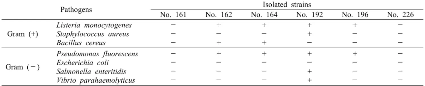
Table 2. Antimicrobial activity of selected lactic acid bacteria against various pathogens
Pathogens Isolated strains
No. 161 No. 162 No. 164 No. 192 No. 196 No. 226 Gram (+)
Listeria monocytogenes
Staphylococcus aureus Bacillus cereus
-
-
-
+
- +
+
- +
+ +
-
+
-
-
-
-
-
Gram (-)
Pseudomonas fluorescens Escherichia coli Salmonella enteritidis Vibrio parahaemolyticus
-
-
-
-
+
-
-
-
+
-
-
-
+
- + +
+
-
-
-
-
-
-
- +: positive, -: negative.
Table 1. Growth of isolated lactic acid bacteria in alginate and
CMC medium after 24 h at 37°CStrains Cell growth (OD at 600 nm) Alginate medium CMC medium No. 161
No. 162 No. 164 No. 192 No. 196 No. 226
1.058±0.040d 0.915±0.003c 0.854±0.029b 1.160±0.042e 1.088±0.001d 0.752±0.006a
1.215±0.004d 1.021±0.010b 1.054±0.030b 1.407±0.004e 1.175±0.017c 0.962±0.004a Value are means±standard deviations of triplicate determinations.
a-eMeans within each column with no common superscripts are significantly different (P<0.05).
가하여 실온에서 3분간 반응시킨 후, 7.5% Na2CO3(Duksan Pure Chemicals Co.) 1 mL를 가한 다음 암소에서 1시간 동안 방치한 후 765 nm에서 흡광도를 측정하여 gallic acid (Sigma-Aldrich Co.)를 표준물질로 한 표준곡선에 의하여 산출하였다.
ABTS 라디칼 소거 활성
ABTS[2,2'-azino-bis(3-ethylbenzothiazoline-6- sulfonic acid) diammonium salt; A9941, Sigma-Aldrich Co.] 라디칼 소거능은 ABTS radical cation decoloriza- tion assay(24)를 이용하여 측정하였다. 7.4 mM의 ABTS와 2.6 mM potassium persulfate(Duksan Pure Chemicals Co.)를 혼합하여 실온・암소에서 24시간 동안 방치하여 라 디칼을 형성시킨 후 실험 직전에 ABTS 용액을 732 nm에서 흡광도가 0.700±0.030이 되도록 phosphate-buffered saline(pH 7.4)으로 희석하여 사용하였다. 발효액 50 μL에 ABTS 용액 950 μL를 첨가하여 암소에서 10분간 반응시킨 후 732 nm에서 흡광도를 측정하여 계산식 ABTS radical scavenging activity (%)=100-[(OD of sample/ OD of control)×100]에 의하여 산출하였다.
통계처리
모든 실험은 3회 반복으로 시행하였으며, 유의성 검증은 SPSS(Statistical Package for Social Sciences, SPSS Inc., Chicago, IL, USA) software package(version 19.0) 를 이용,
P
<0.05 수준으로 Duncan's multiple range test에 의하여 검증하였다.결과 및 고찰
Alginate 및 cellulose 분해 균주의 선발
갈조류 세포벽 구성다당류인 알긴산은 D-mannuronic acid와 L-guluronic acid가 여러 가지 비율로 β-1,4 결합한 polyuronide(25)로 다양한 생리활성을 나타낸다. 김치, 젓 갈, 막걸리 등 한국 전통식품으로부터 분리한 유산균 331 균주 중 alginate lyase와 cellulase 활성을 동시에 나타내 는 균주는 31개 균주였으며, 이중 비교적 활성이 우수한 6균 주에 대해 modified-MRS 배지를 이용하여 성장을 확인한
결과(No. 161, 162, 164, 192, 196) 5균주 모두 성장이 양 호하였으며, No. 192 균주가 alginate-MRS 배지에서 1.160 (OD600), CMC-MRS 배지에서 1.407(OD600)로 가장 우수하 였다(Table 1). 선발된 6균주는 해조 구성 다당인 cellulose 와 alginate를 이용하여 성장할 수 있으므로 미역과 다시마 를 이용한 발효에 스타터로 사용 가능할 것으로 판단되며, probiotic 기능을 검토하고자 항균 활성 및 생리적 특성을 측정하였다.
항균 활성
분리 유산균의 probiotic 특성 유무를 관찰하기 위하여 No. 161, 162, 164, 192, 196, 226 균주의 그람 양성균인
L. monocytogenes
,S. aureus
,B. cereus
와 그람 음성균인P. fluorescens
,E. coli
O157:H7,S. enteritidis
,V. para- haemolyticus
에 대한 항균 활성을 측정한 결과는 Table 2 에서 보는 바와 같다. No. 161과 No. 226 균주는 공시 병원 성 균에 대한 성장억제 활성을 나타내지 않았으나, No. 162, 164, 192, 196은 일부 그람 양성 병원성균과 그람 음성 병 원성균에 대해 뚜렷한 항균 활성을 나타내었다. No. 192는 선발 공시균주 중 가장 넓은 항균 spectrum을 나타내었다.인공위액 및 인공담즙액에 대한 내성
구강을 통하여 섭취되는 균은 강산성의 위산과 담즙산에 서 살아남아 위액과 담즙을 분비하는 췌장 및 십이지장을 통과하여 최종 목적 부위인 장에 도달하여야 정장의 효과를 발휘하게 된다(26,27). Alginate와 cellulose 분해능이 있
Table 4. Survival of selected lactic acid bacteria in MRS broth containing different concentrations of NaCl after incubation for
4 h at 37°C (log No. CFU/mL)Incubation
time (h) NaCl (%) Isolated strains
No. 162 No. 164 No. 192 No. 196
0 0 8.84±0.01 8.60±0.10 8.67±0.10 8.68±0.03
4
0 2 4 8 16 32
7.84±0.04dC 8.21±0.03eD 7.85±0.07dC 7.50±0.03cC 6.89±0.04bC 6.62±0.01aC
7.00±0.40fA 6.68±0.05eA 6.55±0.07dA 5.64±0.07cA 4.77±0.01bA 4.02±0.06aA
7.95±0.04dD 7.86±0.03cC 7.75±0.01bC 7.69±0.14abD 7.59±0.08aD 7.60±0.14aD
7.69±0.03eB 7.56±0.09eB 7.01±0.05dB 6.88±0.04cB 5.39±0.10bB 4.63±0.03aB Value are means±standard deviations of triplicate determinations.
Means with different superscripts within each column (a-f) and row (A-D) indicate significant differences (P<0.05).
Table 5. Identification of selected lactic acid bacteria by 16S rRNA gene sequencing
Isolated strains Source Gram staining Shape Catalase test Identification (similarity, %) No. 162
No. 164 No. 192 No. 196
Kimchi Kimchi Kimchi Kimchi
+ + + +
Cocci Rod Cocci
Rod
-
-
-
-
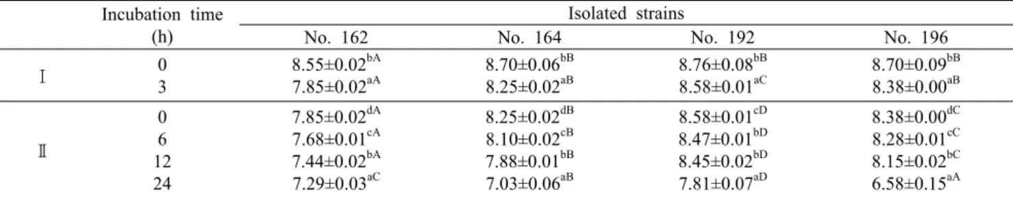
Pediococcus pentosaceus (99%) Lactobacillus plantarum (100%) Enterococcus faecium (100%) Lactobacillus plantarum (100%) Table 3. Survival of selected lactic acid bacteria in artificial gastric juice and bile acid at 37°C (log No. CFU/mL)
Incubation time (h)
Isolated strains
No. 162 No. 164 No. 192 No. 196
Ⅰ 0
3
8.55±0.02bA 7.85±0.02aA
8.70±0.06bB 8.25±0.02aB
8.76±0.08bB 8.58±0.01aC
8.70±0.09bB 8.38±0.00aB
Ⅱ
0 6 12 24
7.85±0.02dA 7.68±0.01cA 7.44±0.02bA 7.29±0.03aC
8.25±0.02dB 8.10±0.02cB 7.88±0.01bB 7.03±0.06aB
8.58±0.01cD 8.47±0.01bD 8.45±0.02bD 7.81±0.07aD
8.38±0.00dC 8.28±0.01cC 8.15±0.02bC 6.58±0.15aA
Ⅰ: Survival cell in artificial gastric juice after 3 h at 37°C.
Ⅱ: Survival cell in artificial bile acid for 24 h at 37°C treated with artificial gastric juice for 3 h at 37°C.
Value are means±standard deviations of triplicate determinations.
Means with different superscripts within each column (a-d) and row (A-D) indicate significant differences (P<0.05).
고 성장이 우수하며, 그람양성균과 그람음성균에 대한 항균 활성을 나타낸 No. 162, 164, 192, 196 균주에 대해 인공위 액과 인공담즙산염 내성을 측정한 결과(Table 3) 선발 균주 모두 인공위액에서 3시간 경과 후 7.85~8.58 log CFU/mL 범위를 나타내었으며, 인공 담즙에서 24시간 경과 후 6.58~
7.81 log CFU/mL를 나타내어 생존율이 높았으며, 특히 No. 192 균주는 인공위액에서 3시간 경과 후 8.58 log CFU/
mL, 인공 담즙액에서 24시간 경과 후 7.81 log CFU/mL를 각각 나타내어 미역 및 다시마 발효용 스타터로 사용 가능할 것으로 판단되었다.
NaCl 내성
과다한 소금(NaCl) 섭취는 고혈압을 일으키는 중요한 위 험인자로 지적되고 있으며(28), 고염섭취에 노출된 현대인 의 식생활 환경에서 생균제로서의 기능을 발휘하기 위해서 는 고농도의 염에 대한 내성이 있어야 한다. No. 162, 164, 192, 196 균주에 대해 NaCl 내성을 측정한 결과(Table 4) No. 192 균주는 32% NaCl을 포함한 용액에서도 배양 4시
간 후 7.60 log CFU/mL의 우수한 생존율을 나타내어 고염 섭취에 노출된 현대인들의 장내 환경에서도 생존이 가능할 것으로 판단되었다.
균주의 동정
16S rRNA 염기서열 분석을 통한 미생물 동정 결과 (Table 5) No. 162, 192는 Gram positive, cocci, catalase negative였으며, 각각
Pediococcus pentosaceus
(No. 162),Enterococcus faecium
(No. 192)으로 동정되었다. No.164와 196은 Gram positive, rod, catalase negative로
Lactobacillus plantarum
으로 동정되었다.선발 유산균의 미역과 다시마 기질에서의 발효특성
선발 유산균을 해조류 발효 시 스타터로 사용하기 위해서 는 해조류가 첨가된 조성물에서 성장할 수 있어야 한다. 미 역 분말과 증류수 혼합물(SME), 다시마 분말과 증류수 혼합 물(STE)을 이용하여 선발 유산균 4균주를 각각 0.2% 접종 하여 37°C에서 72시간 배양하면서 pH(Fig. 1)와 유산균수4 5 6 7 8
0 12 24 36 48 60 72
Fermentation time (h)
pH .
162 164 192 196
SME
4
5 6 7 8
0 12 24 36 48 60 72
Fermentation time (h)
pH .
162 164 192 196
STE
Fig. 1. Changes in pH of sea mustard and sea tangle during fermentation for 72 h at 37°C with different isolated starters. SME:
sea mustard, STE: sea tangle.
4 5 6 7 8 9 10
0 12 24 36 48 60 72
Fermentation time (h)
Viable cell count (log CFU/mL) .
162 164 192 196
SME
4
5 6 7 8 9 10
0 12 24 36 48 60 72
Fermentation time (h)
Viable cell count (log CFU/mL) .
162 164 192 196
STE
Fig. 2. Changes in viable cell count of sea mustard and sea tangle during fermentation for 72 h at 37°C with different isolated
starters. SME: sea mustard, STE: sea tangle.0.0 0.1 0.2 0.3 0.4 0.5 0.6
0 12 24 36 48 60 72
Fermentation time (h)
Reducing sugar (mg/mL) . 162 164 192 196SME
0.0
0.1 0.2 0.3 0.4 0.5 0.6
0 12 24 36 48 60 72
Fermentation time (h)
Reducing sugar (mg/mL) . 162 164 192 196STE
Fig. 3. Changes in reducing sugar of sea mustard and sea tangle during fermentation for 72 h at 37°C with different isolated
starters. SME: sea mustard, STE: sea tangle.의 변화(Fig. 2), 환원당(Fig. 3)을 비교 검토하였다. 모든 공시균주는 미역과 다시마에서 배양 시간이 경과할수록 pH 는 서서히 감소하였으며, 미역의 경우 No. 192 균주로 발효 한 실험구에서 24시간 이후 pH 5.5 범위를 나타내었다. 발 효균주와 해조류에 종류에 따라 pH 감소 정도는 달랐으나 모두 미역과 다시마 조성물에서 pH가 감소하는 것으로 보아 유산균의 성장에 기인한 것으로 판단되었다.
미역과 다시마 기질로 한 배양액에서 선발 유산균의 성장 은 미역에서 발효 전 6 log CFU/mL에서 배양이 진행됨에
따라 증가하였다가 서서히 감소하는 경향을 나타내었다. 미 역을 기질로 한 배양액(SME)에서는 배양 72시간 동안 No.
192(
E
.faecium
)를 제외한 공시균주는 배양 초기의 6~7 log CFU/mL의 범위를 나타내었으나, No. 192 균주는 배양 초기부터 서서히 성장하여 배양 48시간째에 8 log CFU/mL 를 나타내어 스타터로 활용이 가능할 것으로 판단되었다.다시마를 기질로 한 배양액(STE)에서 No. 162(
P
.pento-
saceus
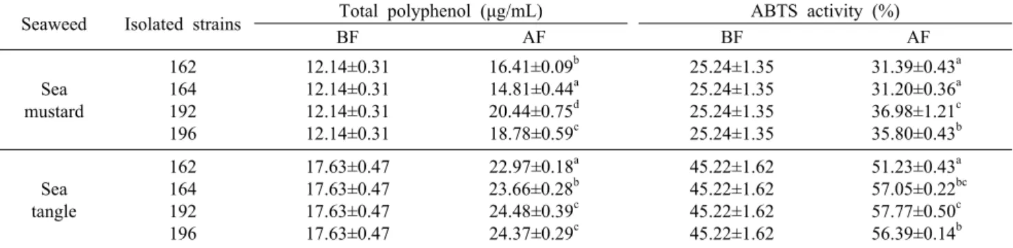
)는 배양 초기부터 서서히 감소하여 배양 60시간 이 후 6 log CFU/mL 이하를 나타내어 다시마를 기질로 한 배Table 6. Changes in total polyphenol contents and ABTS radical scavenging activity of sea mustard and sea tangle during fermentation
for 72 h at 37°C with different isolated startersSeaweed Isolated strains Total polyphenol (μg/mL) ABTS activity (%)
BF AF BF AF
Sea mustard
162 164 192 196
12.14±0.31 12.14±0.31 12.14±0.31 12.14±0.31
16.41±0.09b 14.81±0.44a 20.44±0.75d 18.78±0.59c
25.24±1.35 25.24±1.35 25.24±1.35 25.24±1.35
31.39±0.43a 31.20±0.36a 36.98±1.21c 35.80±0.43b
Sea tangle
162 164 192 196
17.63±0.47 17.63±0.47 17.63±0.47 17.63±0.47
22.97±0.18a 23.66±0.28b 24.48±0.39c 24.37±0.29c
45.22±1.62 45.22±1.62 45.22±1.62 45.22±1.62
51.23±0.43a 57.05±0.22bc 57.77±0.50c 56.39±0.14b BF: before fermentation, AF: after fermentation.
Value are means±standard deviations of triplicate determinations.
a-dMeans within each column with no common superscripts are significantly different (P<0.05).
양액에서는 성장이 불가능한 것으로 판단되었다. 반면 No.
164(
L
.plantarum
), 192(E
.faecium
), 196(L
.plantarum
) 균주는 서서히 성장하여 배양 24시간 이후 7 log CFU/mL 이상의 생균수를 나타내어 다시마를 기질로 한 배양액에서 스타터로 사용이 가능할 것으로 판단되었다. 또한, 발효과정 중 환원당 함량을 측정한 결과 미역과 다시마는 초기 약 0.12 mg/mL에서 발효기간 경과에 따른 모든 실험구에서 환원당 함량이 증가하여 선발균주들은 미역과 다시마의 탄 소원을 이용하여 성장하는 것으로 판단되었다. Gupta 등 (29)은 갈조류 발효 24시간 이후L
.plantarum
의 생균수가 감소하며, 이는 초기 당류의 양이 유산균의 성장에 중요하기 때문이라고 보고하였다. 본 연구에서도 이와 유사하게 미역 과 다시마 단독 배양액에서 유산균의 왕성한 성장은 관찰되 지 않았으나, 발효 3일 동안 초기 균수 이상의 성장 및 생존 을 나타내어 분리 균주는 미역과 다시마를 이용한 유산 발효 물 제조에 스타터로 사용이 가능할 것으로 판단되었다.총폴리페놀 함량 및 ABTS 라디칼 소거능의 변화
Table 6은 선발 유산균을 이용한 미역과 다시마 발효 후 총폴리페놀 함량과 항산화 활성을 비교한 결과이다. 총폴리 페놀 함량은 모든 공시균주를 접종한 기질에서 발효 전보다 발효 후에 증가하였다. SME의 경우 초기 12.14 μg/mL에서 발효 후 14.81~20.44 μg/mL로 증가하였다. No. 192는 20.44 μg/mL를 나타내어 발효 후 약 8.30 μg/mL 증가하였 다. STE에서도 발효과정 중 초기 17.63 μg/mL에서 22.97
~24.48 μg/mL로 증가하였으며, No. 192 균주를 이용한 경 우 가장 많이 증가하여 발효 후 24.48 μg/mL로 약 6.85 μg/mL 증가하였다. ABTS 라디칼 소거능은 모든 공시균주 로 발효한 후 SME와 STE에서 모두 증가하였다. SME의 경우 초기 25.24%에서 31.20~36.98%의 범위로 증가하였 고, STE의 경우 초기 45.22%에서 51.23~57.77%로 증가 하였다. Kim 등(30)은 해조류 추출물의 총페놀 함량과 free radical 소거능이 정의 상관관계를 보인다고 보고하여 본 실험 결과와 비슷하였다.
이상의 결과로 미루어 보아 분리 유산균(
L
.plantarum
및E
.faecium
)은 병원성균에 대한 항균력, 위액과 담즙에 대한 내성 등의 특성뿐만 아니라 특히 미역과 다시마를 발효 시킬 수 있는 특성을 보유하고 있어 이를 이용한 기능성 식 품 및 유산균제제의 소재로 활용이 가능할 것으로 판단된다.미역 및 다시마 발효기질에 분리 유산균의 성장을 증진할 수 있는 탄소원, 질소원, 무기염류를 보충한 최적의 발효조 건에서는 더욱 왕성한 성장과 미역 및 다시마의 분해가 증가 하여 생리활성이 증가할 수 있을 것으로 판단되므로 생균제 로서 기능을 가진 이들 유산균의 대량생산을 위한 최적 배지 조성에 관한 연구가 보다 심도 있게 진행되어야 할 것이다.
요 약
해조류의 발효가 가능하고 probiotic 특성이 우수한 유산균 을 분리 선발한 후 이들의 미역과 다시마 발효능을 검토하였 다. 미역 및 다시마 발효가 가능한 균주를 김치 젓갈, 된장으 로부터 331 균주를 순수 분리하여 해조류 구성 다당(algi- nate, cellulose) 분해능, 균의 생육, 항균 활성 등을 비교 검토한 결과 4균주(stain No. 162, 164, 192, 196)가 우수 하였다. 선발 균주 모두 인공위액, 인공담즙액, NaCl에 높은 생존율을 나타내었고 이들 4균주 중 No. 192가 가장 우수하 였으며
Enterococcus faecium
으로 동정되었다. 미역과 다 시마를 이용하여 선발 유산균을 배양한 결과 No. 192 균주 가 발효특성이 가장 양호하였으며, No. 162, 164, 196 균주 도 양호하였다. 선발 유산균을 이용한 미역과 다시마에서 성장이 가능하였으며 발효 후 미역과 다시마 발효물의 항산 화 활성이 증진되었다.REFERENCES
1. Kim YY, Lee KW, Kim GB, Cho YJ. 2000. Studies on phys- iochemical and biological properties of depolymerized algi- nate from sea tangle, Laminaria japonicus by heating hy- drolysis; 3. Excretion effects of cholesterol, glucose and cad-
mium (Cd) in rats. J Korean Fish Soc 33: 393-398.
2. Lee HA, Lee SS, Shin HK. 1994. Effect of dietary fiber source on the composition of intestinal microflora in rats.
Korean J Nutr 27: 988-995.
3. Huang HL, Wang BG. 2004. Antioxidant capacity and lip- ophilic content of seaweeds collected from the Qingdao coastline. J Agric Food Chem 52: 4993-4997.
4. Cho YJ, Bang MA. 2004. Effects of dietary seaweeds on blood glucose, lipid and glutathione enzymes in streptozoto- cin-induced diabetic rats. J Korean Soc Food Sci Nutr 33:
987-994.
5. Jung BM, Ahn CB, Kang SJ, Park JH, Chung DH. 2001.
Effects of Hijikia fusiforme extracts on lipid metabolism and liver antioxidative enzyme activities in triton-induced hyper- lipidemic rats. J Korean Soc Food Sci Nutr 30: 1184-1189.
6. Cho KJ, Lee YS, Ryu BH. 1990. Antitumor effect and im- munology activity of seaweeds toward sarcoma-180. Bull
Korean Fish Soc 23: 345-352.
7. Yoon JA, Yu KW, Jun WJ, Cho HY, Son YS, Yang HC.
2000. Screening of anticoagulant activity in the extracts of edible seaweeds and optimization of extraction condition.
J Korean Soc Food Sci Nutr 29: 1098-1106.
8. Girard JP, Marion C, Liutkus M, Boucard M, Rechencq E, Vidal JP, Rossi JC. 1988. Hypotensive constituents of ma- rine algae; 1. Pharmacological studies of laminine. Planta
Med 54: 193-196.
9. Cha SH, Ahn GN, Heo SJ, Kim KN, Lee KW, Song CB, Cho SK, Jeon YJ. 2006. Screening of extract from marine green and brown algae in Jeju for potential marine angio- tensin-Ⅰ converting enzyme (ACE) inhibitory activity. J
Korean Soc Food Sci Nutr 35: 307-214.
10. Kim YY, Cho YJ. 2001. Studies on physicochemical and biological properties of depolymerized alginate from sea tangle, Laminaria japonicus by thermal decomposition; 6.
Effects of depolymerized alginate on fecal microflora in rats. J Korean Fish Soc 34: 77-83.
11. Kim JH, Kim YH, Kim SK, Kim BW, Nam SW. 2011.
Properties and industrial applications of seaweed poly- saccharides-degrading enzymes from the marine microor- ganisms. Korean J Microbiol Biotechnol 39: 189-199.
12. Mohammadi R, Sohrabvandi S, Mortazavian AM. 2012. The starter culture characteristics of probiotic microorganisms in fermented milks. Eng Life Sci 12: 399-409.
13. Ouwehand AC, Salminen S, Isolauri E. 2002. Probiotics:
an overview of beneficial effects. Antonie Van Leeuwenhoek 82: 279-289.
14. Leahy SC, Higgins DG, Fitzgerald GF, van Sinderen D.
2005. Getting better with bifidobacteria. J Appl Microbiol 98: 1303-1315.
15. de Vrese M, Schrezenmeir J. 2008. Probiotics, prebiotics, and synbiotics. In Food Biotechnology. Stahl U, Donalies
UEB, Nevoigt E, eds. Springer-Verlag, Berlin Heidelberg, Germany. Vol 111, p 1-66.
16. Jonganurakkun B, Wang Q, Xu SH, Tada Y, Minamida K, Yasokawa D, Sugi M, Hara H, Asano K. 2008. Pediococcus
pentosaceus NB-17 for probiotic use. J Biosci Bioeng 106:
69-73.
17. Kim CH, Lee SH. 2011. Isolation of Bacillus subtilis CK-2 hydrolysing various organic materials. J Life Sci 21: 1716- 1720.
18. Lee JH, Lee EY. 2003. Isolation of alginate-degrading ma- rine bacteria and characterization of alginase. J Life Sci 13:
718-722.
19. Kobayashi Y, Toyama K, Terashima T. 1974. Tolerance of a multiple antibiotic resistant strain, Lactobacillus casei PSR 3002, to artificial digestive fluids. Nihon Saikingaku
Zasshi 29: 691-697.
20. Lee SH, No MJ. 1997. Viability in artificial gastric and bile juice and antimicrobial activity of some lactic acid bacteria isolated from Kimchi. Korean J Appl Microbiol Biotechnol 25: 617-622.
21. Zhang Y, Xu J, Yuan Z, Xu H, Yu Q. 2010. Artificial neural network-genetic algorithm based optimization for the im- mobilization of cellulase on the smart polymer Eudragit L- 100. Bioresour Technol 101: 3153-3158.
22. Miller GL. 1959. Use of dinitrosalicylic acid reagent for determination of reducing sugar. Anal Chem 31: 426-428.
23. Folin O, Denis W. 1912. On phosphotungstic-phosphomo- lybdic compounds as color reagents. J Biol Chem 12: 239- 249.
24. Re R, Pellegrini N, Proteggente A, Pannala A, Yang M, Rice-Evans C. 1999. Antioxidant activity applying an im- proved ABTS radial cation decolorization assay. Free Radic
Biol Med 26: 1231-1237.
25. Fisher FG, Dorfel H. 1955. The polyuronic acids of brown algae. Hoppe Seylers Z Physiol Chem 302: 186-203.
26. Saarela M, Mogensen G, Fondén R, Mättö J, Mattila- Sandholm T. 2000. Probiotic bacteria: safety, functional and technological properties. J Biotechnol 84: 197-215.
27. Gilliland SE. 1979. Beneficial interrelationships between certain microorganisms and humans: Candidate microorgan- isms for use as dietary adjuncts. J Food Prot 42: 164-167.
28. MacGregor GA. 1997. Salt-more adverse effects. Am J Hyp-
ertens 10: 37S-41S.
29. Gupta S, Abu-Ghannam N, Scannell AGM. 2011. Growth and kinetics of Lactobacillus plantarum in the fermentation of edible Irish brown seaweeds. Food Bioprod Process 89:
346-355.
30. Kim BM, Jun JY, Park YB, Jeong IH. 2006. Antioxidative activity of methanolic extracts from seaweeds. J Korean Soc