153
유용 박테리오신을 생산하는 유산균의 분리와 동정
홍성욱1․배효주1․장진희1․김소영1․최은영1․박범영1․정건섭2․오미화1*
1농촌진흥청 국립축산과학원, 2연세대학교 생명과학기술학부
Isolation and Identification of Bacteriocin-Producing Lactic Acid Bacteria
Sung Wook Hong1, Hyo Ju Bae1, Jin Hee Chang1, So-Young Kim1, Eun-Young Choi1, Beom Young Park1, Kun Sub Chung2 and Mi-Hwa Oh1*
1
National Institute of Animal Science, RDA, Suwon 441-706, Korea
2
Division of Biological Science and Technology, Yonsei University, Wonju 220-710, Korea
Abstract
Lactic acid bacteria are microorganisms that are closely associated with human and/or animal environments, and are categorized as generally recognized as safe (GRAS) organisms due to their ubiquitous appearance in foods and their contribution to the healthy microflora of mucosal surfaces. This study was performed to isolate and identify lactic acid bacteria with antagonistic effects against food-borne pathogens. A total of 3,000 acid-producing bacteria were isolated from infant feces, cattle feces, goat feces, dog feces, pig feces, vaginal tracts, vegetables, fruits, Kimchi, Jeotgal, fermented sausages, raw milk, cheese, yogurt, Cheonggukjang, Meju, and Makgeolli cultured on MRS agar with 0.05% bromocresol purple. For the isolation of bacteriocin-producing bacteria, the diameter of the clear zone was measured on MRS agar plates. Twenty-six isolates exhibited strong antibacterial activity against indicator strains such as Listeria monocytogenes, Escherichia coli O157:H7, and Salmonella enterica serovar Enteritidis. Lactic acid bacteria were identified as Enterococcus faecalis, Enterococcus
faecium, Enterococcus hirae, Lactobacillus acidophilus, Lactobacillus amylovorus, Lactobacillus curvatus, Lactobacillus plantarum, and Pediococcus acidilactici by 16S rDNA gene sequence analysis. The results of this study suggest that the
isolates could be used as potential probiotic starters for functional food applications.Keywords: Bacteriocin, lactic acid bacteria, food-borne pathogen
* Corresponding author: Mi-Hwa Oh, National Institute of Animal Science, RDA, Suwon 441-350, Korea. Tel: +82-31-290-1689, Fax: +82-31-290- 1697, E-mail: [email protected]
서 론
식중독(food-poisoning)은 경구를 통한 병원체의 장관내 감염에 의하여 구토, 설사와 같은 증상과 장내 균총의 변화 를 나타나게 되는데, 이러한 증상의 원인이 되는 대표적인 병원균으로는 Listeria monocytogenes, E. coli O157:H7,
Salmonella enterica serovar Enteritidis
등이 있다(Faber et al., 1989). 식중독 사례는 전 세계적으로 다수 보고되며, 면역기능이 저하된 환자, 노인 및 어린이 등에서 균혈증, 수막 염, 수막뇌염, 폐렴, 요로감염증, 복막염, 안감염증 등을 발 병시키고 있으며, 특히 외국에서는 식중독으로 사망하는 사고가 적지 않은 것으로 보고되어 있다(CDC, 2013). 세균 성 식중독은 과거에는 주로 5~9월의 여름철에 주로 발생하 였지만, 최근에는 계절에 상관없이 연중 발생하고 있다. 따 라서 식중독의 위험에 항상 대비해야 하며, 날씨가 따뜻해 지면 세균이 번식하기 좋은 환경이 되므로 더욱 주의해야 한다. 2002년 이전에는 주로 살모넬라균, 황색포도상구균, 장염비브리오균에 의한 식중독이 주로 발생하였으나, 식생 활의 서구화와 학교급식 등 집단급식의 증가로 그 동안 발
생빈도가 낮았던 클로스트리디움 퍼프리젠스, 바실러스 세 레우스, 장출혈성 대장균, 리스테리아, Norovirus 등 서구형 식중독균 및 신종 병원균에 의한 식중독이 증가하고 있는 추세이다(Scallan et al., 2011). 최근 들어 식품산업의 발달 과 위생관리기술의 향상에도 불구하고, 우리나라에서는 매 년 식중독 발생 사례가 증가하고 있는 추세에 있으며, 특히 대형화, 집단화되는 경향을 보이고 있어 사회적 또는 경제 적인 손실면에서 큰 문제가 되고 있다. 또한 웰빙의 영향으 로 비가열 처리한 제품 또는 날로 섭취하는 식품이 증가함 에 따라 식중독이 점차 증가할 것으로 예상한다.
식품의 부패 및 변질을 방지하여 식품의 안전성 확보를 위한 식품 보존제는 다양한 식음료 제품에서 지속적으로 사 용되고 있다. 현재 산업적으로 사용되는 대부분의 보존제는 화학적인 공정에 의해 합성된 제품으로 발암 및 돌연변이 유발과 같은 문제점이 발생할 가능성이 있는 것으로 알려지 면서 최근 합성 보존제에 대한 안전성 문제가 심각하게 인 식되고 있다. 더욱이 식생활에 있어서 소비자들의 건강 지 향적 성향의 증가로 안전성에 대한 관심이 고조됨에 따라 합성 보존제에 대한 기피 현상이 일어나고 있다. 이러한 합 성보존제의 문제점이 대두되면서 독성이 낮은 천연항균성 물질의 개발은 매우 필요한 실정이다(Tagg et al., 1976).
유산균들은 요거트, 치즈, 버터 등의 유제품이나 발효 소 세지 같은 육제품 뿐만 아니라, 우리나라 전통발효 식품인 김치, 젓갈, 장류 등에 많이 존재하며, 사람이나 동물의 장 이나 질내에 정상 미생물군으로 존재하고 있다. 또한, 유산 균과 같은 식품유래 미생물들은 오랜 기간 동안 인류가 발 효식품과 함께 섭취한 GRAS(generally regarded as safe) 미 생물로서 안전성이 증명되었다. 미생물 유래의 항균물질인 bacteriocin은 천연의 antimicrobial polypeptide로서, 기존의 항생제가 2차 대사산물인데 반하여 자신의 유전자로부터 직접 생합성 되는 것이 특징이다(Cleveland et al., 2001). 따 라서 생물공학적 응용이 용이하고, 그 결과 산업현장의 소 요에 보다 다양하게 반응할 수 있을 뿐만 아니라, 분자가 단백질로 이루어져 있으므로 인체에 경구로 섭취되었을 때 즉시 소화기관의 단백질 가수분해 효소에 의해서 쉽게 분 해되어 인체에 무독성이고 잔류성이 적을 뿐만 아니라, 종 류에 따라서는 항균범위가 넓은 것도 있으며, 내열성의 것 도 있어서 식품 제조 시 새로운 생물학적 천연 보존제로서 그 효용이 크다(Tagg et al., 1976). 유산균이 생산하는 박테 리오신 중에서 가장 잘 알려진 nisin은 유제품을 포함한 다 양한 식품의 천연보존제로 사용되어 제품의 저장유통기한 을 향상시킬 뿐 아니라, 식중독 미생물의 증식을 억제하는 것으로 알려져 전 세계적으로 각종 가공식품들의 보존제로 활용되고 있다(Ralph et al., 1995). 이러한 관점에서 유산균
은 장내에서 정상 미생물군으로 존재하고 있기 때문에, 독 성이 비교적 적다고 판단된다. 유산균들이 생산하는 항균 물질은 식품 등의 새로운 생물학적 보존제로서 효용이 크 며, 식품 산업이나 의약품 산업에의 응용도 기대될 것이다.
따라서 본 연구는 다양한 식품 또는 분변으로부터 식중 독을 유발하는 유해성 세균을 저해하는 유산균들을 분리 및 탐색하고, 이를 유전학적인 방법을 사용하여 미생물을 동정하였다.
재료 및 방법
1. 미생물 분리시료
유아 분변, 우분, 염소 분변, 개 분변, 돼지 분변, vaginal tract 채취물, 채소류, 부패한 과일류, 김치, 젓갈, 발효 소시 지, 원유, 치즈, 발효유, 청국장, 메주, 막걸리 등 다양한 시 료로부터 미생물을 분리하였다.
2. 미생물의 분리
분리시료는 각각 10 g 또는 10 mL씩 취하여 멸균 생리 식염수에 1:9 비율로 혼합한 후, homogenizer(Stomacher 400, Seward, England)를 사용하여 10분 동안 균질화한 후, 현탁 액을 제조하였다. 이 현탁액을 멸균 생리식염수로 십진희석 한 후, 0.05%(w/v) bromocresol purple(BCP)이 첨가된 MRS agar(Difco, Detroit, MI, USA) plate에 100μL씩 도말하여 37℃에서 48시간 동안 배양하였다. 배양한 후, 미생물 집락 의 형태에 따라 서로 다른 미생물을 순수 분리하였다.
3. 사용균주와 배양조건
본 연구에 사용한 유산균주는 American Type Culture Collec- tion(ATCC), Korean Collection for Type Culture(KCTC), Korean Culture Center of Microorganisms(KCCM)으로부터 분양받은 Lactobacillus, Lactococcus, Leuconostoc, Streptococcus,
Weissella
표준균주로 MRS broth(Difco, Detroit, MI, USA) 를 사용하여 37℃에서 48시간 동안 배양하였다. 항균활성 실험을 위한 피검균주로는 식중독을 유발하는 병원성 세균 인 Listeria monocytogenes ATCC15313, Escherichia coli O157:H7 ATCC43894, Salmonella enterica serovar Enteritidis CCARM 8200을 American Type Culture Collection(ATCC)과 항생제 내성 균주은행(CCARM)으로부터 분양을 받아 사용하였다.
피검균주들은 tryptic soy broth(TSB; Difco, Detroit, MI)를 사용하여 37℃에서 24시간 동안 배양하였다.
4. 항균활성 측정
분리미생물은 MRS 액체배지에 1%(v/v) 접종한 후, 37℃
에서 48시간 동안 배양하였다. 항균활성 측정은 paper disc 방법으로 각각의 피검균주(108 CFU/mL)를 0.5%(v/v) 접종한 nutrient agar plate 상에 직경 8 mm의 paper disc(Advantec Toyo Roshi Kaisha, Ltd., Japan)를 올려 놓은 후, 원심분리 (8,000×g/15 min)하여 취한 배양상등액 50μL를 적가한 다 음, 37℃에서 18시간 동안 배양하여 생성된 생육 저지환(clear zone)의 직경(mm)을 측정하였다(Tagg and Mcgiven, 1971).
또한, well diffusion 방법은 MRS 액체배지에서 배양(37℃, 48시간)한 후, 원심분리(8,000×g/15 min)하여 배양상등액을 취하고 pH 7.0으로 조정한 다음, 여과제균(0.45μm, Whatman, Springfield Mill, England)을 하였다. Corkborer를 사용하여 well(직경 8 mm)을 제조한 nutrient agar plate에 여과제균한 배양상등액 150μL를 적가한 다음, 37℃에서 18시간 동안 배양하여 생성된 생육 저지환(clear zone)의 직경(mm)을 측 정하였다.
5. 미생물의 동정
분리 선발한 미생물의 동정은 Gram 염색법과 현미경 관 찰을 통해 형태학적 특성을 관찰하였다. 보다 더 정확한 동 정을 위하여 16S ribosomal DNA gene sequencing 분석을 통하여 동정하였다. 선발미생물의 chromosomal DNA는 DNeasy tissue kit(Qiagen, Valecia, CA, USA)를 사용하여 DNA를 추 출하였다. 16S rDNA gene 증폭을 위하여 일반적으로 사용 하는 27F(5'-AGAGTTTGATCATGGCTCAG-3')와 1492R(5'- GGATACCTTGTTACGACT T-3') primer를 사용하였다. PCR 반응시 0.4 mM dNTP, 0.5 units Taq polymerase, 4 mM Mg2+이 함유된 Takara Perfect Premix(Takara, Japan) 10μL 에 DNA template(20μg/mL) 1μL, 1.0μM forward primer 와 1.0μM reverse primer를 각각 1μL씩 넣고, 나머지는 증 류수를 첨가하여 총 부피가 20μL가 되도록 제조하였다.
PCR 증폭은 94℃에서 1분간 denaturation, 51℃에서 1분간 annealing, 72℃에서 1분 30초 동안 extension을 30 cycles 실 시하였고, 72℃에서 5분간 최종 extension을 실시하는 조건에 서 PCR(MinicyclerTM, MJ research Inc., Waltham, MA, U.S.A) 로 증폭하였다. 증폭된 약 1,400 bp의 fragment를 T vector (Invitrogen, Carlsbad, CA, U.S.A)에 결합시킨 후 형질 전환하 였다. T vector sequencing primer를 이용하여 염기서열을 분석하 고, BLAST search(http://www.ncbi.nlm.nih.gov) program을 이 용하여 GENBANK(NCBI, Bethesda, MD, U.S.A)의 ribosomal DNA gene sequence와 비교하여 동정하였다.
결과 및 고찰
1. 항균활성 미생물의 분리
수집한 17 종류의 시료는 멸균 생리식염수로 단계 희석 하고, BCP 지시약이 첨가된 MRS agar plate에 도말하여 37℃
에서 48시간 동안 배양하였다. 산이 생성되어 노란색으로 변한 colony의 형태에 따라 유아 분변에서 150주, 소 분변 에서 120주, 염소 분변에서 87주, 개 분변에서 142주, 돼지 분변에서 130주, vaginal tract 채취물에서 76주, 채소류에서 210주, 부패한 과일류에서 202주, 김치에서 610주, 젓갈에서 165주, 발효 소시지에서 86주, 원유에서 330주, 치즈에서 148 주, 발효유에서 310주, 청국장에서 110주, 메주에서 58주, 그리고 막걸리에서 46주를 각각 분리하였다(Table 1).
분리한 미생물 2,965주와 분양받은 표준균주 35주를 MRS broth에서 각각 배양한 후, paper disc 방법을 통해 식중독균 인 L. monocytogenes, E. coli O157:H7, S. enterica serovar Enteritidis에 대해 항균활성을 측정한 결과, 유아 분변 유래 분리균주에서 21주, 소 분변 유래 분리균주에서 18주, 염소 분변 유래 분리균주에서 12주, 개 분변 유래 분리균주에서 15주, 돼지 분변 유래 분리균주에서 11주, vaginal tract 채취 물 유래 분리균주에서 5주, 부패한 과일류 유래 분리균주에
Table 1. Isolation and selection of lactic acid bacteria with antagonistic effect against food-borne pathogenic strains
No. Origins Isolated
LAB
1)LAB with antagonistic
effect
Selection rate (%)
1 Infant feces 150 21 14.0
2 Cattle feces 120 18 15.0
3 Goat feces 87 12 13.8
4 Dog feces 142 15 10.5
5 Pig feces 130 11 8.5
6 Vaginal tract 76 5 6.6
7 Vegetable 210 -
2)0.0
8 Fruit 202 12 5.9
9 Kimchi 595 71 11.9
10 Jeotgal 165 11 6.6
11 Fermented sausage 86 5 5.8
12 Raw milk 330 38 11.5
13 Cheese 148 - 0.0
14 Yogurt 310 30 9.7
15 Cheonggukjang 110 5 4.5
16 Meju 58 - 0.0
17 Makgeolli 46 - 0.0
18 Type culture 35 1 2.9
Total 3,000 207
1)
LAB, lactic acid bacteria
2)
-, no inhibition
서 12주, 김치 유래 분리균주에서 71주, 젓갈 유래 분리균주 에서 11주, 발효 소시지 유래 분리균주에서 5주, 원유 유래 분리균주에서 38주, 발효유 유래 분리균주에서 30주, 청국 장 유래 분리균주에서 5주, 표준균주에서는 1주를 항균활성 보유 미생물로 선발하였다(Table 1). 다양한 분리시료들 중 에서 분변(8.5~15.0%), 김치(11.9%), 원유(11.5%) 시료로부터 분리한 미생물에서 항균활성 효과가 더 많은 것으로 확인되 었다. 그러나, 부패한 채소류, 치즈, 메주, 그리고 막걸리로 부터 분리한 미생물에서는 항균활성을 확인할 수 없었다.
최근까지 많은 연구를 통해 항균활성이 우수한 미생물들
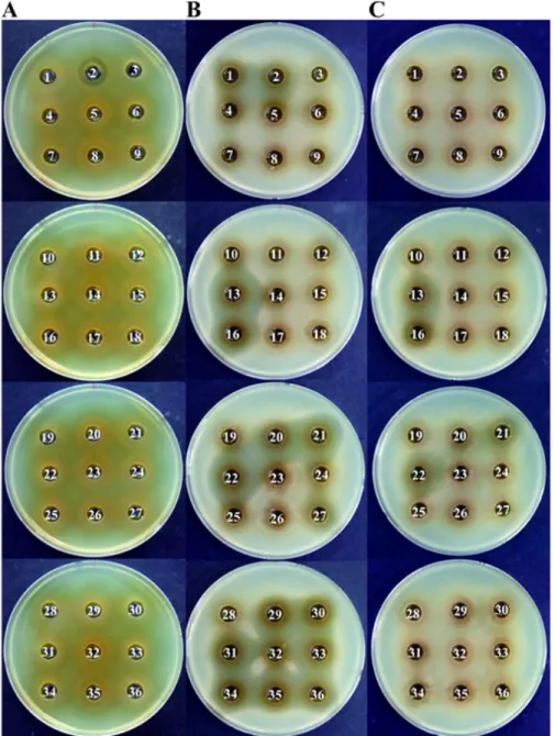
Fig. 1. Antibacterial activity of isolates against food-borne pathogenic strains. A, Listeria monocytogenes; B, E. coli O157:H7; C, Salmonella enterica serovar Enteritidis. Results from the 16S rDNA sequence analysis of the isolates are summarized in Table 2.
은 김치(Kim et al., 2009), 신생아 및 유아 분변(Kim et al., 2008; Lee et al., 2008; Kang et al., 2010), 송아지 분변(Bae
et al., 2006), Kefir(You et al., 2005)
등의 시료에서 분리한 유 산균으로 보고되어 있는데, 이는 본 연구결과와 유사하였다.2. 박테리오신 생산이 우수한 미생물 선발
유산균은 glucose 및 lactose를 탄소원으로 사용하여 최 종산물로 lactic acid, acetic acid, propionic acid, benzoic acid, mevalonic acid 등과 같이 유기산을 주로 생산하는데, 유산균이 생성한 유기산은 pH를 감소시켜 병원성 미생물
이나 식품위생 미생물의 생육을 억제하는 작용을 하며, 유 기산 자체가 항균력을 나타내기도 한다고 보고되어 있다 (McGroarty, 1993). 따라서, 항균물질인 bacteriocin을 생산 하는 미생물을 선발하기 위해서는 pH를 6.5~7.0으로 조정 한 후, 항균활성을 조사해야 한다. 분리미생물의 배양상등 액을 pH 7.0으로 조정하고 여과제균한 후, well-diffusion 방법을 사용하여 피검균주의 생육저지환 직경을 측정하여 박테리오신 생산 미생물 26주를 최종 선발하였다(Fig. 1).
선발한 유산균주들이 생산하는 박테리오신은 E. coli O157:
H7와 S. enterica serovar Enteritidis와 같은 그람음성 식중
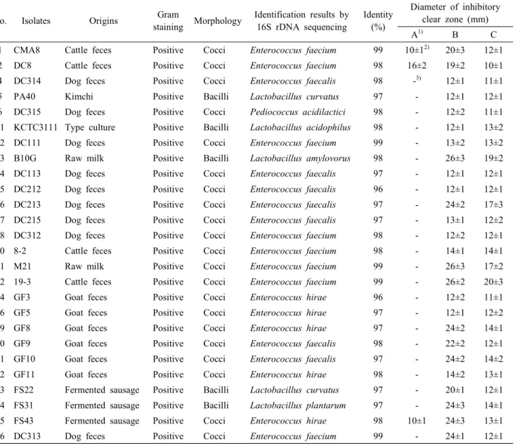
Table 2. Identification and antibacterial activity of bacteriocin-producing isolates
No. Isolates Origins Gram
staining Morphology Identification results by 16S rDNA sequencing
Identity (%)
Diameter of inhibitory clear zone (mm)
A
1)B C
1 CMA8 Cattle feces Positive Cocci Enterococcus faecium 99 10±1
2)20±3 12±1
2 DC8 Cattle feces Positive Cocci Enterococcus faecium 98 16±2 19±2 10±1
4 DC314 Dog feces Positive Cocci Enterococcus faecalis 98 -
3)12±1 11±1
5 PA40 Kimchi Positive Bacilli Lactobacillus curvatus 97 - 12±1 12±1
6 DC315 Dog feces Positive Cocci Pediococcus acidilactici 98 - 12±2 11±1
11 KCTC3111 Type culture Positive Bacilli Lactobacillus acidophilus 98 - 12±1 13±2
12 DC111 Dog feces Positive Cocci Enterococcus faecium 99 - 13±2 13±2
13 B10G Raw milk Positive Bacilli Lactobacillus amylovorus 98 - 26±3 19±2
14 DC113 Dog feces Positive Cocci Enterococcus faecalis 97 - 12±1 12±1
15 DC212 Dog feces Positive Cocci Enterococcus faecalis 96 - 12±1 12±1
16 DC213 Dog feces Positive Cocci Enterococcus faecalis 97 - 24±2 17±3
17 DC215 Dog feces Positive Cocci Enterococcus faecalis 97 - 13±1 12±2
18 DC312 Dog feces Positive Cocci Enterococcus faecium 98 - 12±2 12±1
20 8-2 Cattle feces Positive Cocci Enterococcus faecium 98 - 14±1 14±1
21 M21 Raw milk Positive Cocci Enterococcus faecium 99 - 26±3 17±2
22 19-3 Cattle feces Positive Cocci Enterococcus faecium 99 - 26±2 20±3
24 GF3 Goat feces Positive Cocci Enterococcus hirae 96 - 12±2 11±1
26 GF5 Goat feces Positive Cocci Enterococcus hirae 97 - 12±1 12±2
29 GF8 Goat feces Positive Cocci Enterococcus hirae 97 - 24±2 14±1
30 GF9 Goat feces Positive Cocci Enterococcus faecalis 98 - 22±2 12±1
31 GF10 Goat feces Positive Cocci Enterococcus faecalis 97 - 24±2 14±2
32 GF11 Goat feces Positive Cocci Enterococcus hirae 98 - 14±2 13±1
33 FS22 Fermented sausage Positive Bacilli Lactobacillus curvatus 97 - 20±1 12±1
34 FS31 Fermented sausage Positive Bacilli Lactobacillus plantarum 97 - 24±3 14±1
35 FS43 Fermented sausage Positive Cocci Enterococcus hirae 98 10±1 24±3 13±1
36 DC313 Dog feces Positive Cocci Enterococcus faecium 99 - 24±1 12±1
1)
A, Listeria monocytogenes; B, E. coli O157:H7; C, Salmonella enterica serovar Enteritidis
2)
-, no inhibition
3)
Numbers are mean±SD.
독 세균에 대해서 항균활성을 나타내었는데, 그 중에서 원 유 유래 유산균주 B10G(생육저지대 19±2 mm와 26±3 mm) 와 M21(생육저지대 17±2 mm와 26±3 mm), 소 분변 유래 유산균주 19-3(생육저지대 20±3 mm와 26±2 mm)이 가장 우수한 항균활성능을 나타내었다. 그람양성 식중독 세균인
L. monocytogenes
에 대한 항균활성은 소 분변 유래 유산균주 CMA8(생육저지대 10±1 mm)와 DC8(생육저지대 16±2 mm), 발효 소시지 유래 유산균주 FS43(생육저지대 10±1 mm)만 항균활성을 나타내었다(Table 2). 선발한 박테리오신 생산 미생물은 그람양성 식중독 균주보다 그람음성 식중독 균주
에 대해서 비교적 넓은 항균활성을 나타내었다.
3. 선발미생물의 동정
박테리오신 생산 미생물로 선발한 미생물의 동정은 그람 염색법과 현미경 관찰을 통해 형태학적 특성을 관찰한 결 과, 선발미생물 모두 그람양성의 구균과 간균으로 확인되었 다. 보다 더 정확한 동정을 위하여 16S rDNA gene sequencing 분석을 통하여 동정한 결과, Enterococcus faecalis(7주), E.
faecium(8
주), E. hirae(5주), Lactobacillus acidophilus(1주),L. amylovorus(1주), L. curvatus(2주), L. plantarum(1주), Pediococcus acidilactici(1
주)와 같이 8종의 미생물이 박테리 오신을 생산하는 것으로 확인되었다(Table 2). 그 중에서 분 변유래의 E. faecalis(7주)와 E. faecium(8주)은 박테리오신을 생산하는 미생물 중에서 58%를 차지하였는데, 본 연구를 통 해 사람 또는 가축 유래 장내미생물에서 항균활성이 우수한 미생물이 다수 존재할 것으로 사료되었다. 그람음성 식중독 세균에 대해서 항균활성이 우수한 유산균주인 B10G은 L.amylovorus(98%), M21과 19-3은 E. faecium(99%), 그리고 그람
양성 식중독 세균인 L. monocytogenes에 대한 항균활성이 우 수한 유산균주인 DC8은 E. faecium(98%)으로 동정하였다.Cintas 등(1997)은 Spanish dry-fermented sausage로부터 분리한 E. faecium P13이 생산한 Enterocin P 박테리오신은 L.
monocytogenes, Staphylococcus aureus, Clostridium perfringens, C. botulinum의 생육을 억제한다고 보고하였고, Line 등(2008)
은 닭의 맹장으로부터 분리한 E. durans, E. faecium, E. hirae 등이 생산한 박테리오신은 S. enterica serovar Enteritidis, S.enterica serovar Choleraesuis, S. enterica serovar Typhimurium, S. enterica serovar Gallinarum, E. coli O157:H7, Yersinia enterocolitica, Citrobacter freundii, Klebsiella pneumoniae, Shigella dysenteriae, Pseudomonas aeruginosa, Proteus mirabilis, Morganella morganii, Campylobacter jejuni, L. monocytogenes, S. aureus, S. epidermidis
등의 생육을 억제한다고 보고하였 다. 또한 Han 등(2007)은 돼지장으로 부터 분리한 L. acidophilus GP1B이 생산한 Acidocin 1B 박테리오신은 E. coli O157:H7,P. aeruginosa, Shigella sonnei, Yersinia enterocolitica
의 생 육을 억제한다고 보고하였고, Hernandez 등(2007)은 치즈로 부터 분리한 L. plantarum TF711이 생산한 Plantaricin TF711 박테리오신은 Bacillus cereus, S. aureus, Shigella sonnei, K.pneumoniae
의 생육을 억제한다고 보고하였다. 박테리오신생산 미생물의 주요 분리원과 동정 결과는 다른 연구들과 유사한 것으로 확인하였다.
요 약
유아 분변, 우분, 염소 분변, 개 분변, 돼지 분변, vaginal tract 채취물, 채소류, 부패한 과일류, 김치, 젓갈, 발효 소세 지, 원유, 치즈, 발효유, 청국장, 메주, 막걸리 등 다양한 시 료로부터 3,000주의 미생물을 분리하였다. 분리미생물은 L.
monocytogenes, E. coli O157:H7, S. enterica serovar Enteritidis
에 대해 항균활성을 측정하여 박테리오신 생산 미생물 26주 를 최종 선발하였다. 16S rDNA gene sequencing 분석을 통하 여 동정한 결과, Enterococcus faecalis(7주), E. faecium(8주),E. hirae(5
주), Lactobacillus acidophilus(1주), L. amylovorus (1주), L. curvatus(2주), L. plantarum(1주), Pediococcus acidi-lactici(1
주)와 같이 8종의 미생물이 박테리오신을 생산하는것으로 확인되었다. 미생물이 생산한 박테리오신은 대부분 이 단백질 또는 펩타이드성 물질이어서 인간 또는 동물의 소화효소에 의해 분해되므로 식품에서 안전한 천연보존제 로 사용할 수 있을 것으로 기대된다.
감사의 글
본 논문은 농촌진흥청 공동연구사업(과제번호: PJ009221) 의 지원에 의해 이루어진 것입니다.
참고문헌
1. Bae, I. H., Byun, J. R., Bae, G. S., Lee, S. S., Chang, M. B. and Yoon, Y. H. 2006. Inhibition activity against pathogenic organism of probiotic bacteria and characterization of inhibition activity of isolated bacteria from calf dejecta.
J. Anim. Sci. Technol. 48:907-920.
2. CDC. 2013. Incidence and trends of infection with path- ogens transmitted commonly through food - foodborne diseases active surveillance network, 10 U.S. sites, 1996- 2012. Morbidity and Mortality Weekly Report 62:283- 287.
3. Cintas, L. M., Casaus, P., Havarstein, L. S., Hernandez, P. E. and Nes, I. F. 1997. Biochemical and genetic cha- racterization of enterocin P, a novel sec-dependent bacte- riocin from Enterococcus faecium P13 with a broad anti- microbial spectrum. Appl. Environ. Microbiol. 63:4321- 4330.
4. Cleveland, J., Montville, T. J., Nes, I. F. and Chikindas, M. L. 2001. Bacteriocin: safe, natural antimicrobials for food preservation. Int. J. Food Microbiol. 71:1-20.
5. Faber, J. W., Sanders, G. W. and Johnston, M. A. 1989.
A survey of various foods for the presence of Listeria
species. J. Food Prot. 52:456-458.
6. Han, K. S., Kim, Y. H., Kim, S. H. and Oh, S. J. 2007.
Characterization and purification of acidocin 1B, a bac- teriocin produced by Lactobacillus acidophilus GP1B. J.
Microbiol. Biotechnol. 17:774-783.
7. Hernandez, D., Cardell, E. and Zarate, V. 2005. Anti- microbial activity of lactic acid bacteria isolated from Tenerife cheese: Initial characterization of plantaricin TF711, a bacteriocin-like substance produced by Lactobacillus
plantarum TF711. J. Appl. Microbiol. 99:77-84.
8. Kang, J. H., Kim, D. H., Lee, S. W., Kim, H. C., Cho, Y. U. and Gal, S. W. 2010. Biological probiotic properties of Lactobacillus rhamnosus GG-4 isolated from infant feces. J. Life Sci. 20:1882-1888.
9. Kim, E. A. and Yi, D. H. 2008. The probiotic charac- teristics of Lactobacillus acidophilus isolated from infant feces. J. Korean Soc. Appl. Biol. Chem. 51:93-101.
10. Kim, Y. J., Jang, S. J., Park, J. M., Kim, C. U. and Park, Y. S. 2009. Isolation of garlic resistant lactic acid bacteria for feed additives. Food Eng. Prog. 13:352-359.
11. Lee, S. H., Yang, E. H., Kwon, H. S., Kang, J. H. and Kang, B. H. 2008. Potential probiotic properties of Lactobacillus
johnsonii IDCC9203 isolated from infant feces. Korean
J. Microbiol. Biotechnol. 36:121-127.12. Line, J. E., Svetoch, E. A., Eruslanov, B. V., Perelygin, V. V., Mitsevich, E. V., Mitsevich, I. P., Levchuk, V.
P., Svetoch, O. E., Seal, B. S., Siragusa, G. R. and Stern, N. J. 2008. Isolation and purification of enterocin E-760 with broad antimicrobial activity against gram- positive and gram-negative bacteria. Antimicrob. Agents Chemother. 52:1094-1100.
13. McGroarty, J. A. 1993. Probiotic use of Lactobacilli in the human female urogenital tract. FEMS Immunol. Med.
Microbiol. 6:251-264.
14. Scallan, E., Hoekstra, R. M., Angulo, F. J., Tauxe, R.
V., Widdowson, M., Roy, S. L., Jones, J. L. and Griffin, P. M. 2011. Foodborne illness acquired in the united states-major pathogens. Emerg. Infect. Dis. 17:7-15.
15. Tagg, J. and Mcgiven, A. R. 1971. Assay system for bacteriocin. Appl. Environ. Microbiol. 21:943-948.
16. Tagg, J. R., Dajani, A. S. and Wannamaker, L. W. 1976.
Bacteriocins of gram-positive bacteria. Bacteriol. Rev.
40:722-756.
17. You, S. J., Cho, J. K., Hwang, S. G. and Heo, K. C. 2005.
Probiotic characteristics of Lactobacillus rhamnosus isolated from kefir. Korean J. Food Sci. Ani. Resour. 25:357- 364.
(Received: November 1, 2013 / Accepted: November 20, 2013)