원고 접수일 2012년 5월 31일, 원고 수정일 2012년 7월 16일, 게재 확정일 2012년 11월 27일
책임저자 이상한
(700-705) 대구시 중구 달구벌대로 2175, 경북대학교 치과병원 구강악안면외과 Tax: 053-426-5365, Tel: 053-600-7571, E-mail: [email protected]
RECEIVED May 31, 2012, REVISED July 16, 2012, ACCEPTED November 27, 2012
Correspondence to Sang-Han Lee
Department of Oral and Maxillofacial Surgery, Kyungpook National University Dental Hospital
2175, Dalgubeol-daero, Jung-gu, Daegu 700-705, Korea
Tax: 82-53-426-5365, Tel: 82-53-600-7571, E-mail: [email protected]
CC This is an open access article distributed under the terms of the Creative Commons Attribution Non-Commercial License (http://creativecommons.org/licenses/
by-nc/3.0) which permits unrestricted non-commercial use, distribution, and reproduction in any medium, provided the original work is properly cited.
하악골의 불연속 결손부 재건 시
비혈행화 장골이식술의 골흡수율에 관한 연구
최진욱ㆍ이충오ㆍ황희돈ㆍ김진욱ㆍ권대근ㆍ김진수ㆍ이상한
경북대학교 치의학전문대학원 구강악안면외과학교실
Abstract
Evaluation of Bone Resorption Rate after Nonvascularized Iliac Bone Graft for Mandibular Discontinuity Defect
Jin-Wook Choi, Chung-O Lee, Hee-Don Hwang, Jin-Wook Kim, Tae-Geon Kwon, Chin-Soo Kim, Sang-Han Lee Department of Oral and Maxillofacial Surgery, School of Dentistry, Kyungpook National University
Purpose: Mandible resection and discontinuity defect created lead to aesthetic and functional problems. The iliac crest bone graft exhibits relative ease for bone harvesting, possibility of two team approach, ability to close the wound primarily, large amount of corticocancellous bone and relatively few complications. Whereas the use of free vascularized flaps has donor site morbidity and worse-fitting bone contour, the use of nonvascularized iliac bone graft has advantages in the operation time and patients' recovery time. So, nonvascularized iliac bone graft could be an attractive option.
Methods: Twenty-one patients (M:F=1:1.1) underwent iliac crest bone harvesting for reconstruction of mandibular discontinuity defect (mean length : 61.6±17.8 mm), from May 2005 to October 2011 at the Department of Oral and Maxillofacial Surgery in Kyungpook National University. The average age was 44.1±16.4 years and the mean follow up periods was 28.2±22.7 months. Bone resorption rate, according to age, sex, primary lesion, location and distance of defect, type of fixation plate, time of graft and pre-operative radiation therapy, were measured in each patient.
Results: The mean bone resorption rate was 16.1±9.0%. Bone resorption rate was significantly increased in mandibular defect that is over 6 cm in size (
P
=0.015,P
<0.05) and the cases treated pre-operative radiation therapy (P
=0.017,P
<0.05).All was successfully fixed and maintained for the long-term follow-up. There were a few donor site complications and almost all patients were shown favorable outcome without severe bone resorption in this study.
Conclusion: The nonvascularized iliac bone graft seems to be a reasonably reliable treatment option for reconstruction of mandibular discontinuity defects.
Key words: Mandibular discontinuity defect, Nonvascularized iliac bone graft, Bone resorption rate
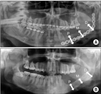
Fig. 1. Methods of measurement. (A) Immediate panoramic view
after nonvascularized iliac bone graft. (B) Panoramic view of 2 years 5 months later surgery. P is proximal osteotomy line 1 cm distally, M is center of proximal and distal osteotomy line and D is distal osteotomy line 1 cm proximally. P, proximal portion;M, midline portion; D, distal portion.
서 론
대부분의 광범위한 하악골 결손부는 종양절제술, 외상 및 감염 등에 의해 이차적으로 발생하며, 특히 연속성이 단절된 하악골 결손은 다양한 기능적, 심미적인 문제들을 야기한다[1]. 악안면 영역의 골결손부 재건 시 다양한 방법의 골이식술이 사용될 수 있으며, 이 중 자가골은 항원-항체 반응을 일으키지 않고 치유가 양호하며 이식골편의 골아세포에 의한 골형성력을 기대할 수 있는 골이식재이다[2].
이러한 자가골의 공여부로는 하악골, 두개골, 경골, 늑골 및 장골 등이 있으며, 장골은 풍부한 골채취량, 접근의 용이성 및 낮은 공여부 합병증 등을 고려할 때 가장 널리 사용되는 공여부이다 [3]. 장골이식술은 혈행화 이식술 또는 비혈행화 이식술의 방법으 로 시행될 수 있으며, 혈행화 장골이식술은 하악골의 연속성이 단절된 골결손부에서 비혈행화 장골이식술에 비하여 높은 성공률 을 보인다는 보고가 있다. 특히, 혈행화 장골이식술은 장골이식 전 방사선치료를 시행하였거나 골이식과 함께 연조직의 이식이 필요한 경우 또는 이식골편의 길이가 긴 경우에 적극 추천되는 술식이다[4].
그러나 비혈행화 장골이식술은 혈행화 장골이식술에 비해 수술 시간과 술 후 환자의 회복기간을 줄여주며, 이식골편의 조작 용이 성, 심미성 및 낮은 공여부 합병증 등의 장점을 가지기 때문에 비교적 단순하고 상대적으로 적은 골결손부의 경우에는 여전히 널리 사용되는 하악골 재건 술식이다[5].
이에 본 연구에서는 하악골의 불연속적인 결손부위에 비혈행화 장골이식술의 효용성을 확인하고자 나이, 성별, 원발병소의 종류, 이식부위, 이식골편의 길이, 사용된 고정용 plate의 종류, 즉시 재건술 및 술 전 방사선치료 시행 유무에 따라 이식골편의 흡수량을 조사하였다. 그리고 각각의 요인들에 따른 흡수율의 차이를 확인하 고, 각각의 요인들과 흡수율과의 상관관계를 통계적으로 조사함으 로써 향후 치료계획 수립에 도움을 주고자 한다.
연구방법
1. 연구대상
본 연구는 2005년 5월부터 2011년 10월까지 경북대학교 치과 병원 구강악안면외과에서 악성 또는 양성종양으로 인한 하악골절 제술(mandibular resection) 후 불연속적인 결손부의 재건을 위하여 비혈행화 장골이식을 시행한 21명의 환자를 대상으로 하였 다. 남녀의 성비는 1:1.1이었으며, 평균나이 44.1±16.4세(최소 16세, 최대 73세)였다. 평균 경과관찰 기간은 28.2±22.7개월(최 소 3개월, 최대 76개월)이며, 평균 결손부위의 길이는 61.6±17.8 mm (최소 32 mm, 최대 97 mm)였다.
2. 연구방법
환자의 의무기록지를 통하여 나이, 성별, 경과관찰 기간, 원발병 소의 종류, 술 전 방사선치료 시행 유무를 조사하였고, 장골이식을 시행한 전후의 파노라마 방사선사진(Orthopantomograph
ⓇOP100D, Instrumentarium Imaging, Tuusula, Finland)을 통 하여 이식부위, 이식골편의 길이와 높이, 사용된 고정용 plate의 종류에 대해 각각 조사하였다.
장골이식술 직후(T1)와 최종 경과관찰시점(T2)에서의 파노라 마 방사선사진상에서 이식골편의 폭경과 높이를 측정하여 변화량 을 측정하였으며, 결과의 정확도를 높이기 위해 이식골편의 높이 변화량은 Fig. 1에서와 같이 근심부(proximal portion)는 근심 골절단선(osteotomy line)에서 1 cm, 중심부(midline)는 이식골 편의 중점, 원심부(distal portion)는 원심 골절단선에서 1 cm 떨어진 곳에서 각각 측정하여 평균치를 사용하였다. 또한, 파노라 마 방사선사진의 확대 및 왜곡을 보상하기 위하여 고정용 plate의 크기를 측정하고 이를 근거로 비율에 맞추어 골편의 크기를 측정하 였고, 술 후 plate를 제거한 환자에서는 120%의 파노라마 방사선 사진의 일반적인 확대율을 적용하였다.
측정된 이식골의 변화량을 근거로 나이, 성별, 원발병소의 종류,
이식부위, 이식골편의 길이, 사용된 고정용 plate의 종류, 즉시
재건술 및 술 전 방사선치료 시행 유무에 따른 이식골 흡수량과의
상관관계를 측정하였다.
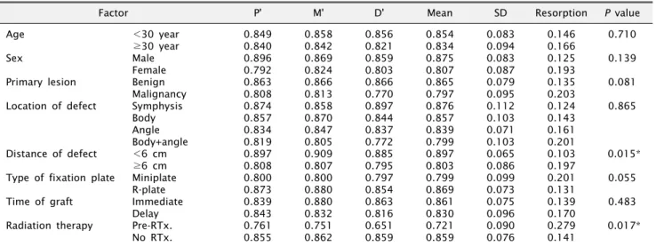
Table 1. Classification of patients treated for mandibular
discontinuity with nonvascularized iliac bone graftAge Male Female Total
<30 year
≥30 year Total
3 7 10
2 9 11
5 16 21
Table 2. Result of bone resorption after nonvascularized iliac bone graft
Factor P' M' D' Mean SD Resorption
P value
Age Sex
Primary lesion Location of defect
Distance of defect Type of fixation plate Time of graft Radiation therapy
<30 year
≥30 year Male Female Benign Malignancy Symphysis Body Angle Body+angle
<6 cm
≥6 cm Miniplate R-plate Immediate Delay Pre-RTx.
No RTx.
0.849 0.840 0.896 0.792 0.863 0.808 0.874 0.857 0.834 0.819 0.897 0.808 0.800 0.873 0.839 0.843 0.761 0.855
0.858 0.842 0.869 0.824 0.866 0.813 0.858 0.870 0.847 0.805 0.909 0.807 0.800 0.880 0.880 0.832 0.751 0.862
0.856 0.821 0.859 0.803 0.866 0.770 0.897 0.844 0.837 0.772 0.885 0.795 0.797 0.854 0.863 0.816 0.651 0.859
0.854 0.834 0.875 0.807 0.865 0.797 0.876 0.857 0.839 0.799 0.897 0.803 0.799 0.869 0.861 0.830 0.721 0.859
0.083 0.094 0.083 0.087 0.079 0.095 0.112 0.103 0.071 0.103 0.065 0.086 0.099 0.073 0.075 0.096 0.090 0.076
0.146 0.166 0.125 0.193 0.135 0.203 0.124 0.143 0.161 0.201 0.103 0.197 0.201 0.131 0.139 0.170 0.279 0.141
0.710 0.139 0.081 0.865
0.015*
0.055 0.483 0.017*
Resorption: (∆T1=T2)/T1.
P', proximal; M', midline; D', distal bone height of T2/T1; SD, standard deviation; R-plate, reconstruction plate; Pre-RTx., previous radiation therapy; No Rtx., no previous radiation therapy.
*P <0.05.
Fig. 2. Diagram for bone resorption with age (P =0.710).
3. 통계분석
각 연구항목에 대한 통계분석은 SPSS version 18.0 (SPSS Inc., Chicago, IL, USA)을 이용하였으며, 비모수적인 방법을 사용하여 분석하였다. 경과관찰 기간과 이식골편의 흡수량 사이의 상관분석(correlation analysis)를 시행하였으며, 각각의 요인에 따른 이식골편의 흡수량에 대한 통계학적 유의성을 평가하기 위해 Mann-Whitney U 검정을 사용하였고, 골이식 부위에 따른 통계학 적 유의성 평가는 Kruskal-Wallis 검정을 사용하였다.
결 과
본 연구에서는 종양절제 후 하악골의 연속성이 단절된 환자에서 장골이식술을 시행하였으며, 골이식을 시행한 21명 모두에서 이식 된 골편이 수여부에서 생착을 이루었으며, 심미적으로나 기능적으 로 양호한 결과를 나타내었다. 모든 환자에서 피질해면골편을 이식하였으며, 술 후 영구적인 보행장애나 공여부의 감각장애는 나타나지 않았다.
평균 경과관찰 기간 28.2±22.7개월(최소 3개월, 최대 76개월) 동안의 평균 흡수율은 16.1±9%였으며, 근심부, 중심부, 원심부
각각의 측정 위치에 따른 골흡수량은 모든 항목에서 큰 차이를 보이지 않았다. 또한, 본 연구에서 경과관찰 기간과 이식골편의 흡수량과의 상관분석(correlation analysis)을 시행한 결과는 상 관계수가 0.330으로 경과관찰 기간과 흡수량 사이에는 상관성을 보이지 않았다( P =0.144) (Table 1, 2).
남녀 성비는 1:1.1이고 성별에 따른 흡수율은 남자 12.5%이며, 여자 19.3%로 남자가 여자에 비하여 적은 흡수율을 보였으나 통계학적인 유의성은 보이지 않았다( P =0.139). 나이는 30세 미만 과 30세 이상으로 구분하였으며 그 비율은 1:3.2였으며, 30세 미만에서 이식골편의 흡수율은 14.6%였고 30세 이상에서의 흡수 율은 16.6%로 젊은 환자에서 낮은 흡수율을 보였지만 통계학적인 유의성은 없었다( P =0.710) (Fig. 2, 3).
원발병소가 악성종양인 환자는 8명, 양성종양은 13명이었으며
Fig. 3. Diagram for bone resorption with sex (P =0.139).
Fig. 4. Diagram for bone resorption with primary lesion (P =0.081).
Fig. 5. Diagram for bone resorption with location of defect (P=0.865).
Fig. 6. Diagram for bone resorption with distance of defect (P=0.015).
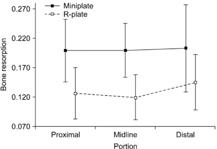
Fig. 7. Diagram for bone resorption with type of fixation plate
(P =0.055). R-plate, reconstruction plate.악성종양은 20.3%의 흡수율을 보인 반면, 양성종양은 13.5%의 흡수율을 나타내었으며, 통계학적인 유의성은 보이지 않았다 ( P =0.081) (Fig. 4).
이식부위에 따른 구분에서는 정중부 2명, 하악체부 7명, 하악 우각부 7명이었고, 하악체부와 우각부에 걸쳐 이식된 경우가 5명 이었다. 이 중 정중부에서 가장 낮은 흡수율(12.4%)을 보였고, 하악체부와 우각부에 걸쳐 이식된 경우가 가장 높은 흡수율 (20.1%)을 보였으며, 통계학적인 유의성은 없는 것으로 나타났다 ( P =0.865) (Fig. 5).
이식골편의 길이는 6 cm 미만인 경우와 6 cm 이상인 경우로 구분하였으며, 그 비율은 1:1.6이고 각각의 흡수율은 10.3%와 19.7%였다. 이식골편의 길이가 길수록 높은 흡수율을 나타내었으 며 통계학적으로 유의성 있는 상관관계를 보였다( P =0.015) (Fig.
6).
사용된 고정용 plate의 종류는 reconstruction plate와 miniplate 였으며, 그 비율은 1:1.3이었다. Reconstruction plate는 13.1%의 흡수율을 보였고, miniplate는 20.1%의 흡수율을 보였으며 통계학
적 유의성은 없는 것으로 나타났다(P=0.055) (Fig. 7).
하악골절제술을 시행하면서 장골이식술을 동시 시행한 즉시
재건술과 2차 지연 재건술의 비율은 1:2.5였고, 흡수율은 즉시
Fig. 8. Diagram for bone resorption with time of graft (P =0.483). Fig. 9. Diagram for bone resorption with pre-operative radiation
therapy (P =0.017). P, proximal; M, midline; D, distal; Pre-RTx, previous radiation therapy; No Rtx, no previous radiation therapy.재건술에서 13.5%이며 지연재건술에서 17.0%로 즉시 재건술을 시행한 경우에서 보다 낮은 흡수율을 보였으나 통계학적 유의성은 없었다( P =0.483) (Fig. 8).
술 전 방사선 치료를 시행한 경우는 3명이었고 평균 조사량은 56.7±5.8 Gy이며 방사선치료 후 장골이식술을 시행한 평균 기간 은 30±36.5개월이었다. 술 전 방사선치료를 시행한 경우에서 27.9%의 높은 흡수율을 보였으며 이는 통계학적으로 유의한 결과 를 나타내었다( P =0.017) (Fig. 9).
고 찰
종양, 감염 및 심한 외상으로 인한 하악골의 광범위한 결손부의 재건 시 혈행화 자가골, 비혈행화 자가골, 이종골 또는 합성이식재 를 사용하는데[5,6], 합성이식재를 사용하여 재건술을 시행한 경우 기능적으로나 심미적으로 장기적인 경과관찰에서 성공적이지 못 한 결과를 보인다[7]. 이에 반해, 자가골은 골형성 능력이 우수하기 때문에 많은 양의 골이식을 필요로 하는 경우에서 성공적인 생착률 을 나타내며[8-11], 자가골이식을 시행했을 경우 평균 흡수율은 5∼25% 정도이고[12], 대부분의 골흡수는 6개월 이내에 발생하며 18개월까지 골흡수가 지속되는 것으로 보고되고 있다.
자가골 중에서도 장골은 많은 양의 골채취가 가능하며 피질골과 해면골을 함께 채취할 수 있고 공여부의 창상을 1차봉합할 수 있으며 악골과 골질이 유사하기 때문에 block bone 이식 시에 널리 사용된다[13,14]. 장골이식술 시 이식골편의 생착성공에 대하 여 Tidstrom과 Keller[15]는 34증례의 유리 장골이식술을 시행한 결과 100%의 생착성공률을 보인다고 하였으며, Ogunlade 등[16]
은 37증례의 비혈행화 장골이식술을 시행하여 91.9%의 생착성공 률을 보고하고 있다. 또한, Pogrel 등[5]은 39증례의 혈행화 장골이 식술과 29증례의 비혈행화 장골이식술을 시행한 결과에서 각각 95%와 76%의 성공률을 보고하고 있다. 그러나 혈행화 장골이식술 은 보다 높은 성공률에도 불구하고 비혈행화 장골이식술을 시행한
경우보다 평균 수술시간은 3시간 길었으며, 평균 재원기간도 14일 정도 증가하는 것으로 보고되고 있다. 이처럼 혈행화 장골이식술에 서 높은 성공률을 보이고 있으나, 수술의 간편성과 환자의 회복기 간, 술 후 합병증 등을 고려할 때 비혈행화 장골이식술은 여전히 매력적인 하악골재건술 중의 하나이다.
본 연구에서는 나이와 성별에 따른 골흡수량과의 상관관계는 없었으며, 위치에 따른 골흡수량의 평가에서도 상관관계를 보이지 않았다. 비록, 통계적 유의성은 없지만 하악체부와 우각부에 걸쳐 이식된 경우에서 가장 높은 흡수율을 보였으며, 이는 결손부위의 길이가 길어짐에 따라 흡수율이 증가되었기 때문으로 생각되며, 하악 정중부보다 우각부에서 높은 골흡수율을 보인 것은 우각부의 저작근 부착으로 인한 지속적인 교합력의 작용 때문으로 생각한다.
원발병소의 종류에 따른 흡수율의 평가에서는 악성종양에서 높은 흡수율을 보였으며, 이는 악성종양의 경우 술 전 방사선요법 을 시행한 경우가 3증례 포함되어 있기 때문으로 생각한다.
Chen 등[4]의 연구에 따르면 이식골편의 길이가 이식술의 성공 률에 영향을 줄 수 있으며 이식골편의 길이가 6 cm 이상이면 실패율이 증가하며, 혈행화 장골이식의 적응증으로 고려될 수 있다. 따라서 본 연구에서는 이식골편의 길이를 6 cm 미만인 경우와 6 cm 이상인 경우로 구분하여 흡수율을 조사하였으며 이식골편의 길이와 이식골의 흡수량과의 관계는 통계학적인 유의 성을 보였다. 하지만 6 cm 이상에서 평균 골흡수율은 19.7%로 자가골이식술을 시행했을 때 발생하는 평균 흡수율과 거의 유사하 였으며, 13명의 모든 환자에서 안정적이고 지속적인 생착을 보이 면서 기능적, 심미적으로 양호한 결과를 보였다.
하악골의 재건술을 시행하는 시기에 관해서는 학자들마다 다른
견해가 있으며, 과거에는 종양절제술을 시행할 경우 종양의 재발
가능성 때문에 2차적으로 지연 수술하는 것이 효과적이라고 생각
하였다. Lawson 등[17]은 즉시 재건술을 시행한 경우의 성공률은
46%이며, 지연 재건술을 시행한 경우에는 90%의 성공률을 보고하
고 있으며, Komisar[18]는 즉시 재건술을 시행한 경우 저작이나 연하작용에 있어서 재건술을 시행하지 않은 환자와 비슷한 결과를 보이며, 입원기간과 술 후 회복기간이 길어지고 수술 과정이 복잡 해지면서 합병증의 가능성이 증가될 수 있다고 보고하고 있다.
본 연구에서는 즉시 재건술을 시행한 5명의 환자에서 술 후 합병증 은 없었으며, 지연 재건술을 시행한 경우에 비하여 낮은 흡수율을 보였으나 통계학적인 유의성은 없었다.
불연속적인 하악골의 재건술에서는 골결손부로 인한 골편의 이동이 발생하며, 일반적인 하악골절의 치료 시 시행하는 고정보다 견고한 내고정술이 필요하다[1]. 이를 위해 reconstruction plate 의 사용이 추천되고 있으며, 본 연구에서는 9명의 환자에서 re- construction plate를 사용하였으며 평균 흡수율은 13.1%로 min- iplate를 사용했을 때의 평균 흡수율인 20.1%보다 다소 낮은 흡수율을 보였으나 통계학적인 유의성은 없었다.
술 전 방사선치료를 시행한 경우의 골이식에 관하여 절개선의 치유지연으로 인한 합병증의 발생이 보고되었으며[15], 또 다른 연구에서는 방사선 치료가 선행된 환자에서는 이식술을 시행하기 전에 고압산소요법을 시행하는 것을 추천하고 있다[19]. 본 연구에 서는 술 전 방사선치료를 시행한 환자에서 27.9%의 높은 흡수율을 보였으며, 이는 이식골의 흡수량과 통계학적으로 유의성을 보였 다. 따라서 방사선치료를 시행한 환자에서 골이식술을 시행할 경우 술 전 고압산소요법을 시행하고 섬세한 외과적 술식을 진행하 며, 혈행화이식술에 대한 고려도 필요할 것으로 생각한다.
결 론
하악골의 불연속적인 결손부의 재건을 위하여 비혈행화 장골이 식술을 시행하였으며, 모든 환자에서 심각한 합병증을 보이지 않았으며 기능적, 심미적으로 양호한 결과를 보였다. 모든 증례에 서 이식술 시행 후 이식골편의 높이가 감소하는 골흡수가 일어났지 만 만족할만한 수준이었다. 여러 가지 요인에 따른 흡수율과의 상관관계에서 이식골편의 길이와 술 전 방사선치료의 시행 여부가 하악골 재건을 위한 장골이식술을 시행한 후 이식골편의 흡수율에 영향을 미치는 것으로 조사되었다. 따라서 하악골의 결손 부위가 6 cm 이상이거나 악성종양의 절제술 후 방사선요법을 시행한 환자에서는 좀 더 섬세한 외과적 술식이 요구되며, 경우에 따라서 는 혈행화 장골이식술을 고려해 볼 수 있을 것이다. 하지만 높은 흡수율을 감안하더라도 수술의 간편성과 수술 시간의 단축, 환자의 술 후 회복기간과 입원기간, 술 후 합병증 가능성 등을 고려해 볼 때 비혈행화 장골이식술은 여전히 예지성 있는 치료 방법이다.
References