소뇌-교각종양 수술시 수술 중 전기생리학적 신경감시에 따른 수술 후 기능적 결과
성균관대학교 의과대학 삼성서울병원 신경외과학교실, 신경과학교실*
이상구·박 관·박익성·서대원*·엄동옥*·남도현 이정일·김종수·홍승철·신형진·어 환·김종현
= Abstract =
Intraoperative Neurophysiologic Monitoring and Functional Outcome in Cerebellopontine Angle Tumor Surgery
Sang Koo Lee, M.D., Kwan Park, M.D., Ik Seong Park, M.D., Dae Won Seo, M.D.,* Dong Ok Uhm, Ph.D.,* Do-Hyun Nam, M.D.,
Jung-Il Lee, M.D., Jong Soo Kim, M.D., Seung Chyul Hong, M.D., Hyung Jin Shin, M.D., Whan Eoh, M.D., Jong Hyun Kim, M.D.
Departments of Neurosurgery and Neurology,* Samsung Medical Center, Sungkyunkwan University School of Medicine, Seoul, Korea
bjectives:Intraoperative neurophysiologic monitoring(INM) is a well known useful method to reduce intraoperative neurological complications during neurosurgical procedures. Furthermore, INM is required in most cerebello- pontine angle(CPA) surgery because cranial nerves or brain stem injuries can result in serious complications.
Object of this study is to the correlation between the changes of intraoperative monitoring modalities during cere- bellopontine angle tumor surgery and post-operative functional outcomes in auditory and facial functions.
Material and Methods::::Fifty-seven patients who underwent intraoperative neurophysiologic monitoring during CPA tumor surgery were retrospectively reviewed. Their lesions were as follows;vestibular schwannomas in 42, other cranial nerve schwannomas in seven, meningiomas in five and cysts in three cases. Pre- and postoperative audi- ologic examinations and facial nerve function tests were performed in all patients. Intraoperative neurophysiologic monitoring modalities includes brainstem auditory evoked potentials(BAEP) and facial electromyographies(EMG).
We compared the events of INM during CPA tumor surgeries with the outcomes of auditory and facial nerve fun- ctions.
Results::::The subjects who had abnormal changes during CPA tumor surgery were twenty cases with BAEP chan- ges and facial EMG changes in twenty one cases. The changes of intraoperative neurophysiologic monitoring did not always result in poor functional outcomes. However, most predictable intraoperative monitoring changes were wave Ⅲ-Ⅴ complex losses in BAEP and continuous neurotonic activities in facial EMG.
Conclusion::::These results indicate that intraoperative neurophysiologic monitoring in CPA tumor surgery usually provide predictive value for postoperative functional outcomes.
KEY WORDS:Intraoperative monitoring・Cerebellopontine angle tumor・Brain stem auditory evoked potentials・
Electromyography・Hearing・Facial nerve.
서 론
최근 첨단화된 의료기구와 미세수술기법의 발달로 신경외
과영역의 수술도 눈부신 발전을 이룩하여 왔다. 특히, 소뇌 교각부는 뇌간과 뇌신경이 밀집되어 있어 손상시 심각한 후 유장애를 초래할 수도 있으므로 현미경적 미세수술기법이
OOOO
꼭 필요한 부위이다17). 특히 이 부위의 종양은 정상적인 해 부학적 구조물을 변형시키고 뇌신경의 직접적인 관찰을 가 로막는 위치에 잘 발생하므로, 주요 뇌신경에 대한 기능적 감시는 수술도중 신경계의 손상을 최소화하며 수술후 합병 증의 발생을 줄이는데 크게 도움을 준다22).
안면신경에 대한 감시는 19세기부터 실험적으로 시도되어 오다, 1979년 Delgado등3)에 의해 처음 시도되었으며, 청각 뇌간반응(auditory brainstem response)은 1971년 Jewett 와 Williston8)에 의해 알려져 1978년 Levine11)에 의해 시 도된 이후 유용성에 대한 많은 보고가 있었다. 소뇌교각부 종 양의 수술시 안면신경 대한 감시가 보편적으로 시행되고 있 으며 수술직후 안면신경의 일시적 마비가 관찰되어도 수술시 신경손상이 없었고 종양제거 후 전기적 자극에 대해 반응이 있었다면 대부분 기능이 회복되는 것을 경험할 수 있다. 또한 청각뇌간반응을 동시에 이용하여 수술전의 청력을 보존시키 려는 노력이 시도되어지고 있다.
본 연구에서는 소뇌-교각 종양환자의 수술에 있어 수술 중 감시장치소견의 변화가 있었던 환자에 있어 수술전후 안 면 및 청신경의 기능적 변화와 수술후 기능적 악화를 예측할 수 있는 소견에는 어떤 것이 있는지에 대해 알아보고자 하 였다.
방 법
1. 대 상
1995년 1월부터 1998년 7월까지 본원에 입원하여 수술 중 신경계 집중감시(INM)를 시행한 소뇌교각종양 환자 57 명을 대상으로 의무기록 및 신경감시결과를 후향적으로 연구 하였다. 환자는 남자 20명, 여자 37명으로 여자가 많았으며, 중앙연령은 48세(19~68세)였다. 병리학적 소견상 전정신 경초종(vestibular schwannoma) 42예, 삼차신경초종(trig- eminal schwannoma) 3예, 경정맥공 신경초종(jugular for- amen schwannoma) 4예, 수막종(meningioma) 5예, 낭종 (cyst) 3예였다. 위치는 우측 29예, 좌측 28예로 비슷하였다.
수술적 방법으로는 유돌후방 후두하 두개골절제술(retro- mastoid suboccipital craniectomy) 38예, 방정중 후두하 두개골절제술(paramedian suboccipital craniectomy) 2예, 경미로 접근법(translabyrinthine approach) 10예, 측두하 접근법(subtemporal approach) 3예, 기타 4예[경추체 접근 법(transpetrosal approach) 2예, 추체후두 접근법(petroo- ccipital approach) 1예, 안와-관골 접근법(orbitozygo- matic approach) 1예]를 시행하였으며, 종양제거정도는 전 적출 44예, 아전적출 11예, 부분적출 2예였다.
2. 수술 전 검사
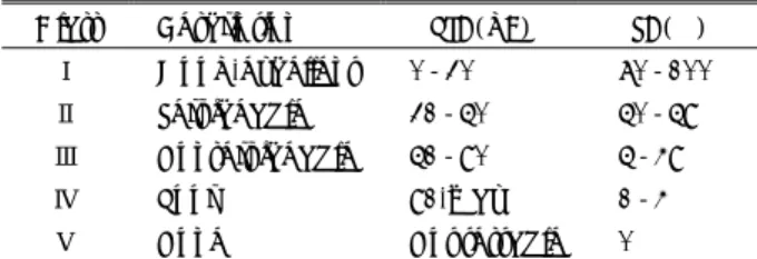
뇌신경을 포함한 수술 전 신경학적 검사를 면밀히 시행하 고 특히 안면신경기능은 House-Brackmann Grade에 따라 기능적 분류를 시행하였다7). 또한 청력검사, 안면근육 근전 도검사(facial muscle electromyogram;이후 fEMG), 청 각유발전위검사(brain stem auditory evoked potential;이 후 BAEP)를 시행하여 수술 전 기준검사소견(baseline stu- dy)을 확인하였다. 청력검사소견은 modified Gardner-Ro- bertson system에 따라 기능적 분류를 시행하였다(Table 1).
3. 수술 중 준비
기관삽관시 근육이완제를 사용하고 난 후 근전도를 시행하 기 때문에 정중신경을 4번 연속 자극하여 엄지손가락이 적 어도 2번 이상 움직일 수 있도록 유지하였으며, 흡입마취제 는 isoflurane(forane)을 이용하여 마취가 너무 깊지 않도록 minimal alveolar concentration(MAC)을 1.0이하로 유지하 였다. 만약 청각피질의 파형이 관찰되지 않으면 농도를 0.5 까지 낮추어 반응을 보면서 마취를 유지하였다. 수술 중 bar- biturates 계통의 약물은 사용하지 않고 주로 fentanyl을 사 용하였다.
4. 수술중 신경계감시
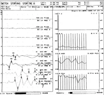
기계로는 8채널의 Viking Ⅳ®(Nicolet,Inc.)을 이용하여 4채널은 안면근육 근전도, 2채널은 청각유발전위, 2채널은 체 성유발전위(somatosensory evoked potential;이후 SEP) 를 동시에 기록하도록 하였고, 반복해서 기록한 파형을 그래 프로 그려서 변화나 이상을 연속적으로 관찰할 수 있도록 하 였다(Fig. 1).
안면근육 근전도는 안륜근(orbicularis oculi), 구륜근(or- bicularis oris), 저작근(massetor), 턱근(mentalis)에 각각 전극을 꽂고 소리와 함께 지속적으로 근전도반응(free run- ning EMG)을 관찰하다가 긴장성 파형(tonic activity)이 나 타나는지를 확인하였으며, 종양제거시 신경의 전기유발자극 을 시행하고 근전도상의 반응을 관찰하여 신경조직의 기능여 부를 확인한 후 종양제거후 신경조직을 다시 자극하여 여전
Table 1. Functional classification of modified Gardner and Robertson system
Class Description PTA(dB) SA(%)
Ⅰ Good-excellent 0-30 70-100
Ⅱ Serviceable 31-50 50-59
Ⅲ Nonserviceable 51-90 5-49
Ⅳ Poor 91-max 1-4
Ⅴ None Not testable 0
*PTA;pure tone audiogram, SA;speech discrimination score
*If PTA and SA do not qualify in same class, use the lower class
히 기능을 하는지 확인하였다. 청각유발전위는 tubal insert phone을 귀에 꽂고 자극을 주고 기록전극은 A1, A2 기준전 극하에 Cz에서 짧은 잠복기의 far field potential을 기록하였 다. 체성유발전위의 자극은 좌우측 정중신경(median nerve) 및 후경골신경(posterior tibial nerve)에서 시행하였으며, 기 록은 제5경추부위(C5)와 중심구 2cm 후방(Cz’)에서 FPz 기준기록전극하에 기록하였다. 접지전극은 어깨에 붙이고 기 타전극은 피하전극을 사용하였다(Table 2).
수술중 신경계감시에 대한 변화 및 이상소견은 기도삽관 후 기록한 청각유발전위의 기준자료와 수술중 추적관찰시 변 화를 비교하였으며, 이상소견은 제3파형(wave Ⅲ)과 제4~
5번 복합체(Ⅳ~Ⅴ complex)의 소실 또는 절대 잠복기의 변 화(0.2msec)로 정의하여 비정상 반응으로 판정하였다(Ta- ble 3). 수술시행도중 변화를 보이면 즉각 신경과의사 또는 신경과 전문요원에 의해 집도의에게 알려 주위를 환기시키고, 잠시 수술진행을 멈춘 후 반응이 회복되는지 여부를 확인하 였다.
5. 수술후 검사
수술시행 후 신경학적 검사와 청력검사를 시행하여 수술전 과 마찬가지로 안면신경 및 청각기능에 대한 기능적 분류를 시행하였고, 퇴원후의 외래기록을 검토하여 수술 전에 비해 기능이 호전 또는 악화되었는지 여부를 확인하였다. 수술 전 상태와 최종적으로 검사한 소견을 비교하여 호전, 보존, 일 시적 악화, 지속적 악화, 무변화로 분류하였다. 환자의 평균추 적기간은 11개월(1~38개월)이었다.
결 과
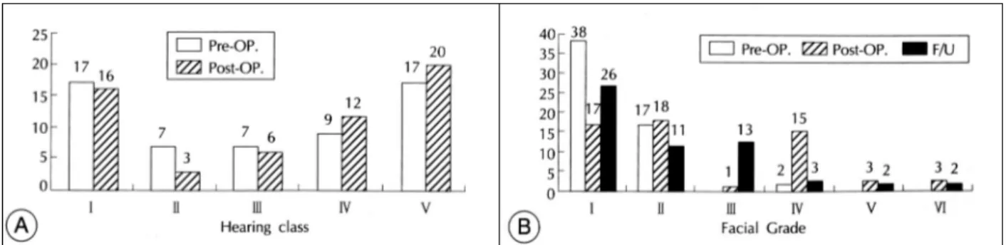
총 57명 환자의 수술 전 청력은 class Ⅰ 17예, class Ⅱ 7 예, class Ⅲ 7예, class Ⅳ 9예, class Ⅴ 17예였고, 수술 후 청력은 class Ⅰ 16예, class Ⅱ 3예, class Ⅲ 3예, class Ⅳ 12예, class Ⅴ 20예였다(Fig. 2-A). 수술 후 2예에서 청력 기능의 호전을 보였고 15예는 Class Ⅰ, Ⅱ이상의 술전 청력 을 그대로 보전하였다. 또한 2예는 수술직후 일시적 악화가 왔으나 회복되었으며 12예에서 술전에 비해 청력이 악화되 었고 술전부터 class Ⅳ, Ⅴ인 26명은 변화가 없었다. 수술 중 class Ⅲ이상의 31명의 환자에 대해 수술 중 청각유발전 위 감시를 시행하였으며 이중 20예에서 이상소견을 보였는 데 12예는 일시적으로 나타났다가 사라졌으며 8예에서는 수술이 끝날때까지 이상파형을 보였다.
양성검사소견중 각각의 청각유발전위 pattern별 청력의 변 화를 보면 일시적인 제1파형 소실(Wave Ⅰ loss) 소견을 보
Table 2. Intraoperative electrophysiologic neuromonitoring parameters during cerebellopontine angle tumor surgery
EMG BAEP SEP Device Flush tip electrode Tubal insertion phone Subdermal needle
Site Facial muscle Ear Median/post. tibial n.
Type Electrical Broad band click Electrical
Duration 0.2msec 100usec pulse 0.2msec Stimulation
parameters
Intensity Less than 4V 120dBpeSPL 23mA
+Electrode Muscle Ear lobe(A1, A2) FPz, C5s
-Electrode Subdermis Cz Cz’FPz
Time base 100ms 1ms 5-10ms
Recording parameters
Sensitivity 50μV 0.1-0.05μV 1-0.5μV
*EMG:Electromyography, BAEP:Brainstem auditory evoked potentials, SEP:Somatosensory evoked potentials, Cz’:2cm pos- terior to Cz electrode, C5s:Spinous process of 5th cervical vertebra
Fig. 1. Normal electrophysiologic neuromonitoring during cer- ebe-llopontine angle tumor surgery. Normal free running EMG on ipsilateral orbicularis oculi, oris, massetor and mentalis which were located on channel 1, 2, 3, 4, respectively. SEP of right median nerve stimulation on channel 5,6 and BAEP of right ear stimulation on channel 7, 8. The serial data of BAEP and SEP were plotted on right panel. Vertical bar indicates latency (msec) and continuous lines represent amplitude(uV)of each wave.
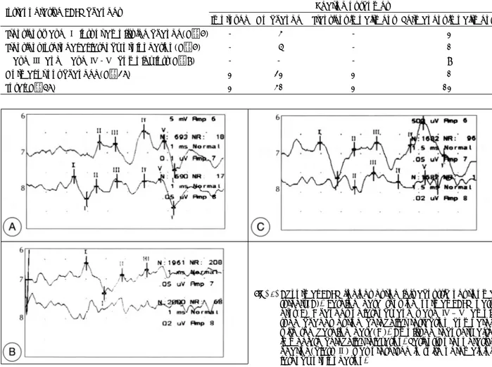
인 환자 6예 중 2예에서 청력이 악화되었으며, 최고파형간 잠복기(interpeak latency)의 일시적 연장 소견을 보인 6예 중 1예에서 청력이 악화되었다. 또한 wave Ⅲ 혹은 Ⅳ~Ⅴ complex 파형 소실이 수술 종료시까지 회복되지 않은 8예는 모두 수술 후 청력이 악화되었다(Table 4). 일시적 이상소견 을 보인 환자들은 수술 중 제Ⅳ~Ⅴ파형의 진폭감소와 잠복 기의 증가소견을 일시적으로 보였으나 견인을 완화시키고 기다린 후 파형의 회복을 보였으며 Fig. 3의 환자는 수술 전 class Ⅱ의 청력을 그대로 보존하였다. 따라서 본 조사결과 잠복기의 연장보다는 파형의 소실, 특히 제3~5파형의 소실 이 수술후 청력악화를 예견할 수 있는 검사소견이었다. 그외 에도 수술 중 감시소견상 변화가 없었지만 수술 후 청력악 화가 발생한 환자가 1예 있었다. 이미 기술한 기준을 적용한 BAEP 양성 검사소견과 청력의 지속적악화 환자를 대비해 볼 때 민감도(sensitivity)는 91.7%(11/12), 특이도(spe- cificity)는 80%(36/45)였고, 양성예측률(positive predi- ctive value)은 55%(11/20)였다(Table 4).
환자의 수술 전 안면신경기능은 House-Brackman grade
Ⅰ 38예, Ⅱ 17예, Ⅳ 2예였고, 수술 후 추적검사시 House- Brackman grade Ⅰ 26예, Ⅱ 11예, Ⅲ 13예, Ⅳ 3예, Ⅴ 2 예, Ⅵ 2예였다(Fig. 2-B). 수술중 근전도 감시결과 21명의 환자가 이상소견을 보였는데 이중 15명은 일시적인 소견 (transient)이었으나 6명은 수술이 종료될때까지(contin- uous) 이상소견이 지속되었다. 안면신경기능은 보존 26예 (45.6%), 일시적악화 25예(43.9%), 지속적악화 6예(10.
5%)였다(Table 5). 각각의 수술 중 안면근전도소견과 수
술 후 안면기능의 정도를 비교해보면, 근전도의 일시적 변화 를 나타냈던 15예 중(Fig. 4) 안면기능의 무변화 4예, 일시 적 악화 10예, 지속적악화 1예였고, 연속적 tonic activity를 보이고 trigger response가 수술종료시까지 없었던 환자 6 예 중 안면신경마비가 지속된 환자는 4예였다 . 수술중 변화 가 없었던 36예중 1예에서도 안면신경기능의 지속적악화가 관찰되었다. 따라서 수술중 근전도상 연속적 tonic activity와 trigger response의 소실이 수술후 실제 안면기능의 악화를 예측하는 중요한 소견이었다. 안면근전도 양성소견과 실제 안면기능의 지속적 악화를 대비해 보면 민감도는 83%(5/6), 특이도는 68.6%(35/51)였고, 양성예측률은 23.8%(5/21) 로 나타났다(Table 5).
고 찰
소뇌-교각부위의 종양수술에 있어 청력 및 안면신경의 기 능적 보존은 중요한 과제중 하나이다. 여기에는 종양의 수 술적 접근법, 종양의 크기, 신경유착정도, 종양표면에서의 청 신경형태, 수술 전 청력 및 안면신경기능의 유지여부 등이 중 요한 요소가 된다9)24). Colletti와 Fiorino등2)은 소뇌교각부 종양수술에서 내이도 근처의 종양제거시 가장 청력소실의 위 험성이 높다고 하였다. 특히 이 부위의 안면신경 및 청신경은 견인 또는 종양절제시 손상의 가능성이 높아 신경의 해부학 적 보존 뿐 만 아니라 기능적 보존을 위한 수술중감시장치가 도움이 된다26). 이러한 수술중감시장치의 사용은 신경학적 결손을 감소시키고, 변화시 수술자의 주의를 환기시켜 영구 적 신경손상의 가능성을 감소시킨다고 알려져 있다1)10)23). Kveton등9)은 수술중감시장치를 사용한 경우 유용한 청력 유지에 있어 그렇지 않은 경우보다 결과가 좋았으며(57% vs.
44%), Harner등6)은 특히 크기가 큰 종양에서 수술중감시 장치를 사용한 경우가 사용하지 않은 경우보다 안면신경의 해부학적 보존에 있어 결과가 좋았다고 보고했다(67% vs.
33%). 종양이 그리 크지않은 경우에도 수술중감시를 한 환
Table 3. Criteria for abnormal responses of intraoperative ele- ctro-physiologic neuromonitoring parameters
Intraoperative events
BAEP* Wave Ⅰ, Ⅲ, Ⅳ-Ⅴ complex loss Interpeak latency >0.2msec
EMG Free running EMG;neurotonic discharge Negative response in triggered EMG
*Baseline data were obtained after intubation
Fig. 2. Pre- and post-operative functional classification of auditory(A) and facial nerve function(B).
자에서 안면신경의 기능회복이 더 좋은 것으로 보고되는데 Niparko등16)은 2cm 이상의 청신경초종에 대해 경미로접근 법을 시행한 29명의 수술중 감시를 시행한 환자와 그렇지 않 은 75명의 환자를 1년 추적관찰하여, 감시를 한 경우 안면신 경의 기능회복이 더 좋았다고 보고하였다. Fisher 등5)도 4cm 이상의 청신경초종에서 신경감시를 시행하여 기능적, 해부학 적 보존을 하는 경우 수술 후 일시적 안면신경마비가 86%에 서 호전되었다고 하였다. Samii등21)은 1000례의 청신경초종 수술환자에서 93%의 높은 안면신경보존률을 발표하였고, 수 술 중 감시장치의 사용이후 점차적으로 보존률이 호전되었다 고 하였다. 즉 신경감시장치를 이용하여 수술 중 육안으로 구 분이 힘든 주요 뇌신경의 존재를 확인하고 이를 해부학적으 로 보존함으로써 수술후 기능적 보존을 기대할 수 있었던 것 이다. Erikson등4)은 안면신경이 해부학적으로 보존되어 있 으면 기능소실이 적어도 1년 이내에 대부분 자연회복된다고 보고하였다.
기존의 보고와 같이 뇌신경을 보존하여 신경학적 손상을
줄일 수 있는 수술중감시장치의 효용성을 평가하는데는 여러 가지 어려움이 있다. 그중 하나로는 검사상 이상소견과 실 제적 손상과의 보편타당한 진단기준이 모호하다는 것이다. 실 제로 본 연구결과에서와 같이 청각유발전위에서 제Ⅲ~Ⅴ파 형복합체 소실이 있었던 환자에서는 청력악화를 있지만 다른 지만 다른 이상소견의 경우에는 청력악화를 나타내거나 그 렇지 않은 경우도 있었고, 수술 중 변화가 없었던 환자에서도 청력악화가 1예가 있었으며, 안면근육 근전도에서도 기능적 결과는 일치하고 있지 않았다. 기능적 결과를 예견할 수 있는 소견에는 여러 가지 기준이 제시되어왔다. 체성유발전위는 유 발전위의 진폭이 기준치의 50% 감소, 잠복기가 1ms이상 증 가할 때 의미가 있는 것으로 알려져 있고 진폭감소보다는 오 히려 잠복기의 증가가 의미있는 것으로 알려져 있다5). 청각 유발전위에는 5개의 중요파형이 나타나는데 각각의 파형의 진폭감소 또는 잠복기증가는 와우신경, 청신경핵, 뇌간에서의 변화를 의미하며, 영구적 파형소실은 기능적 손실을 반영하는 것으로 알려져 있다. 제Ⅰ~Ⅴ파간 잠복기의 증가는 청신경의
Table 4. Hearing outcomes according to intraoperative BAEP patterns
Hearing outcomes Intraoperative BAEP changes
Improved No change Transient impairment Permanent impairment
Trensient wave Ⅰ loss or amplituide change(N=6) - 4 - 2
Trensient Interpeakal latency prolongation(N=6) - 5 - 1
Wave Ⅲ and Wave Ⅳ-Ⅴ complex loss(N=8) - - - 8
Normal or not changed(N=37) 2 32 2 1
Total(N=57) 2 41 2 12
Fig. 3. Abnormal BAEP findings during left acoustic neurinoma surgery(A). Baseline data showing normal BAEP patt- erns(B) Changes of latency and wave Ⅳ–Ⅴ ampli- tude chages during cerebellar retraction compared with the baseline data(C). Amplitude recovery after immediate cerebellar relaxation. Patient’s preoperative hearing(class Ⅱ) was preserved in spite of remaining latency prolongation.
견인 또는 압박으로 유발되고, 전반적인 파형의 갑작스런 소실은 internal auditory artery의 혈액공급 차단 또는 신 경절단에 의해 야기된다. 그중 제Ⅰ파형은 종양절제시 와우신 경으로부터 멀어지고 뇌간쪽으로 견인시 진폭감소 또는 소 실이 나타나며, 허혈성 변화에 가장 민감한 것으로 알려져 있 고, 8분 이내에 압박을 풀면 회복된다고 보고된 바 있다13). Matties 등13)은 청각유발전위의 파형변화를 Type B1에서 B5까지 5단계로 나누어 분석한 결과 제 Ⅲ 파형의 변화가 가 장 초기에 나타나는 민감한 소견이라고 하였고, 제Ⅰ및 Ⅲ파 형의 소실 후에 제Ⅴ파형의 소실이 뒤따른다고 하였으며, 위 험한 수술조작(드릴사용, 종양견인, 신경박리 등)시 제Ⅰ및
Ⅲ파형의 변화를 잘 감시해야 한다고 하였다. 또한 제Ⅲ파형 의 변화가 있었던 환자의 65%에서 청력소실을 보였다고 하
였다. 소뇌의 견인시 제Ⅴ파형의 변화가 나타나지만, 견인을 풀어주어 조정하면 청력의 영구적 소실을 초래하지는 않는다 고 하였다. Nadol등15)은 수술전 청각유발전위 소견에서 수 술후 청력을 예견할 수 있는 지표는 없으나, 수술 중 제Ⅴ파형 의 보존여부가 수술후 청력에 중요하다고 하였으며, Levine 등11)도 제Ⅴ파형의 잠복기와 진폭의 변화가 중요한 소견이 라고 하였다. Wadanabe 등에 의하면 제Ⅴ파형이 수술종료시 관찰된 환자 68명중 66명은 정상, 2명에서 청력소실, 종료 시 파형이 소실된 12명중 2명은 정상, 10명은 청력소실이 있 었으며, 제Ⅰ파형의 경우 종료시 파형이 소실된 13명중 3명 은 정상, 10명은 청력소실을 나타냈다24). 이러한 결과는 제
Ⅰ파형 또는 제Ⅴ파형을 감시하는 경우 어느정도 회복가능한 손상에 의해서도 파형의 소실이 있을 수 있다는 것을 나타내 며, 제Ⅴ파형이 좀더 정확하다는 것을 알 수 있다. 그러나 이 러한 소견이 있더라도 반드시 기능소실이 동반된다는 것을 의미하지는 않는다. 잠복기증가나 파형진폭의 감소가 청각유 발전위 monitoring에 있어 중요한 소견이지만 일부저자에 서는 파형의 완전소실에도 청력이 보존된 경우를 보고하고
있다15)20). Raudzen등19)은 청각유발전위파형의 잠복기 증가
가 있으면서 제Ⅰ파형 이후의 파형은 형태를 유지했던 7명의 환자에서 수술후 청력이 정상 또는 경미한 기능저하가 나타 났다고 하였다. Roberson등20)은 수술 중 청각유발전위파형 을 관찰할 수 없었던 6명의 환자에서 수술후 3~22개월 추 적기간동안 4명의 환자가 청각유발전위 파형이 나타나 청력 보존에 있어 청각유발전위 파형이 없는 것이 청력의 예후를 평가하는 지표가 될 수 없다고 하였다. 본 연구에서 수술후 오히려 청력이 호전된 환자가 1예 있었는데 가능한 기전을 설명한 문헌을 보면, 종양에 눌려 전도장애가 있다가 호전되 는 것과 와우(cochlear)나 청신경으로의 혈행호전으로 설명 하고 있다15). 일과성 전도성 청력장애는 장액성 중이염 또는 유양돌기내로의 액체유입으로 야기된다고 설명한 바 있다18).
이와 같이 수술 중 다양한 파형의 변화가 나타날 수 있고, 과연 어떤 것이 수술후 실제 손상을 반영할 수 있는지에 대 해서도 여러 주장이 제기되고 있으나, 본 연구에서는 청각유 발전위에 있어 잠복기의 증가보다는 진폭의 변화 특히, Ⅲ~
Table 5. Facial functional outcomes according to intraoperative EMG patterns
Facial outcomes Intraoperative EMG changes
No change Transient paresis Permanent paresis
Trensient neurotonic activity(N=15) 4 10 1
Continuous neurotonic activity or Negative response in triggered
EMG(N=6) - 2 4
No changes(N=36) 32 3 1
Total(N=57) 36 15 6
Fig. 4. EMG activities during Lt. acoustic neurinoma surgery(A).
Neurotonic activities were seen mainly on orbicularis oris and masseter for about 1 second, which was occ- ured by touching facial nerve(B). Facial nerve trig- ered EMG revealed excellent EMG activitie when trains of 5Hz, 4 volt, 0.2msec electrical stimuli were given.
Patient showed postoperative transient facial paralysis (grade Ⅰ → Ⅲ). But, functional recovery was obtai- ned 6 months after surgery.
AAA A
B BB B
Ⅴ파형복합체의 소실이 수술후 중요한 청력악화를 반영하였 고 Fig. 3의 예처럼 수술 중 Ⅲ~Ⅴ파형복합체의 변화가 나 타나자 즉시 소뇌견인을 중지하고 일시적으로 수술적 조작 을 중단하였다가 파형이 원래의 상태로 회복된 후 수술을 진 행함으로서 수술후 기능적 손상을 방지할 수 있었다.
그럼에도 불구하고 이러한 수술 중 감시장치를 이용하는데 는 몇 가지 문제점이 있는데 수술적 감시장치의 연속성을 유 지하기가 어려우며 감시환경의 다양성에 따른 부정확성이 존 재한다는 것이다. 장치기구에 따라 다르지만 청각유발전위의 파형평균화(averaging)는 대개 약 2분 정도의 시간이 소요 되어 실제 손상과 감시소견간의 시간적 간격이 발생한다9). 즉 적절한 되먹임이 빠른 시간내에 이루어져야 기능적 손상 을 줄일 수 있다는 것을 의미한다. 또한 청각유발전위는 유 발전위를 발생부위로 부터 멀리 떨어진 부위에서 검출하는 원거리법(far field technique)이므로 잡음에 의해 결과에 영향을 받을 수 있기 때문에 신호-잡음 비율을 향상시키기 위하여 필터를 쓰거나, 청각유발전위의 부정확성을 보완하 기 위해 electrocochleography를 병용하기도 한다14). 직접 적 안면신경 자극과 안면근육 근전도는 안면신경의 연속성을 확인하는데 유용하며 electrocochleography와 같은 직접적 청신경감시방법이 청각유발전위에 비해 수술후 청신경 기능 을 보존하는데 더 좋은 결과를 얻었다는 보고도 있다2). 열거 한 바와 같이 장치에 따라 수술 중 감시소견이 다양하게 나타 날 수 있으며, 실제 기능적 결과와는 일치하지 않을 소인이 존재한다는 것을 의미한다.
결 론
소뇌-교각종양 수술시 전기생리학적 신경감시장치를 이 용하여 수술 중 변화소견에 따라 수술후 기능적 결과를 예측 하는데 도움이 되었으며, 수술도중 감시소견 변화시 적절한 대처로 수술후 발생할 수 있는 합병증을 예방할 수 있었다.
수술 중 감시장치의 변화소견 중 기능적 결과에 영향을 미치 는 가장 중요한 소견은 청각유발전위에서 제Ⅲ~Ⅴ파형 복합 체의 소실이었고, 안면근육 근전도에서 연속적 tonic activity 와 trigger시 음성반응 이었다.
•논문접수일:1999년 9월 29일
•심사완료일:2000년 2월 29일
•책임저자:박 관
135-710 서울 강남구 일원동 50번지
성균관대학교 의과대학 삼성서울병원 신경외과학교실 전화:02) 3410-3496, 전송:02) 3410-0048 E-mail:kwanpark@smc.samsung.co.kr
References
1) Cheek JC:Posterior fossa intraoperative monitoring. J Clin Neurophysiol 10(4):412-424, 1993
2) Colleti V, Fiorino FG:Advances in monitoring of seventh and eight cranial nerve function during posterior fossa surg- ery. Am J Otol 19:503-512, 1998
3) Delgado TE, Buchheit WA, Rosenholtz HR, Chrissian S:In- traoperative monitoring of muscle evoked responses obtained by intracranial stimulation of the facial nerve;An accurate technique for nerve dissection. Neurosurgery 4:418-421, 1979 4) Erickson DL, Ausman JI, Chou SN:Prognosis of seventh ne-
rve palsy following removal of large acoustic tumors. J Neu- rosurg 47:31-34, 1977
5) Fischer RS, Raudzens P, Nunemacher M:Efficacy of intra- operative neurophysiological monitoring. J Clin Neurophysiol 12(1):97-109, 1995
6) Harner SG, Daube JR, Ebersold MJ, Beaty CW:Improved preservation of facial nerve function with use of electrical mo- nitoring during removal of acoustic neurinomas. Mayo Clin Proc 62:92-102, 1987
7) House JW, Brackmann DE:Facial nerve grading system. Ot- olaryngol Head Neck Surg 93:184, 1985
8) Jewett DL, Williston JS:Auditory evoked far fields averaged from the scalp of humans. Brain 94:681-696, 1971 9) Kveton JF:The efficacy of brainstem auditory evoked pot-
entials in acoustic tumor surgery. Laryngoscope 100:1171- 1173, 1990
10) Leonetti JP, Brackmann DE, Prass RL:Improved preserv- ation of facial nerve function in the infratemporal approach to the skull base. Otolaryngol Head Neck Surg 101:74-78, 1989 11) Levine RA, Montgomary WW, Ojemann RJ:Evoked poten- tials detection of hearing during acoustic neuroma surgery.
Neurosurgery 28:339, 1978
12) Matthies C, Samii M:Direct brainstem recording of auditory evoked potentials during vestibular schwannoma resection: nuclear BAEP recording. J Neurosurg 86:1057-1062, 1997 13) Matthies C, Samii M:Management of vestibular schwanno- mas(acoustic neuromas):The value of neurophysiology for evaluation and prediction of auditory function in 420 cases.
Neurosurgery 40(5):919-930, 1997
14) Mullatti N, Coakham HB, Maw AR, Butler SR, Morgan MH:
Intraoperative monitoring during surgery for acoustic neuro- ma:benefits of an extratympanic intrameatal electrode. J Neurol Neurosurg Psychiatry 66(5):591-599, 1999 15) Nadol JB, Chiong CM, Ojemann RG, McKenna MJ, Martuza
RL, Montgomery WW, et al:Preservation of hearing and fa- cial nerve function in resection of acoustic neuroma. Laryngo- scope 102:1153-1158, 1992
16) Niparko JK, Kileny PR, Kemink JL, Lee HM, Graham MD:
Neurophysiologic intraoperative monitoring:Ⅱ. Facial nerve function. Am J Otol 10:55-61, 1989
17) Pitts LH, Jackler RK:Treatment of acoustic neuromas. N Eng J Med 339(20):1471-1473, 1998
18) Radtke RA, Erwin CW, Wilkins RH:Intraoperative brains- tem auditory evoked potentials:significant decrease in posto- perative morbidity. Neurology 39:187-191, 1989
19) Raudzens PA, Shetter AG : Intraoperative monitoring of brainstem auditory evoked potentials. J Neurosurg 57:341- 348, 1982
20) Robertson JB, Jackson LE, McAuley JR:Acoustic neuroma surgery:absent auditory brainstem respone does not contra- indicate attempted hearing preservation. Laryngoscope 109 (6):904-910, 1999
21) Samii M, Matthies C:Management of 1000 vestibular schw-
annomas(acoustic neuromas):The facial nerve-preservation and restitution of function. Neurosurgery 40:684-695, 1997 22) Seo DW, Park K, An JY, Lee SK, Chung CS, Hong SB, et al:
Electrophysiologic neuromonitoring changes during tumor sur- gery in cerebellopontine angle. J Kor Neurol Ass 17(1):98- 105, 1999
23) Umezu H, Tadashi A, Shoichi T, Yojiro J:Early and late po- stoperative hearing preservation in patients with acoustic ne- uromas. Neurosurgery 39(2):267-272, 1996
24) Watanabe E, Schramm J, Strauss C, Fahlbusch R:Neuro- physiologic monitoring in posterior fossa surgery. Acta Neuro- chir(Wien) 98:118-128, 1989
25) Yingling CD, Gardi JN:Intraoperative monitoring of facial and cochlear nerves during acoustic neuroma surgery. Otola- ryngol Clin North Am 25(2):413-448, 1992