Jae-W e C ho
1*, K un Park
4*,
G i R yang K w eon
5, B yeong-C hurl Jang
2, W on-K i B aek
3, M in-H o S uh
3,
C hang-W ook K im
1, K yu-Suk Lee
1and S eong-Il S uh
3,61Department of Dermatology
2Department of Medical Genetic Engineering and Chronic Disease Research Center
3Department of Microbiology
Chronic Disease Research Center, and Institute for Medical Science
Keimyung University, School of Medicine
194 DongSan-dong Jung-gu, Daegu 700-712, Korea
4Department of Dermatology Seonam University Hospital
120-1 Mareuk-dong, Seo-gu, Gwangju 502-157, Korea
5Department of Biochemistry
College of Medicine, Seonam University, 720 Kwangchi-dong, Namwon 590-711, Korea
6Corresponding author: Tel, 82-53-250-7442;
Fax, 82-53-255-1398; E-mail, [email protected]
*These authors contributed equally to this work.
Accepted 2 May 2005
Abbreviations: COX-2, cyclooxygenase-2; EMSA, electrophoretic mobility shift assay; ERK, extracellular signal-regulated protein kinase; JNK, c-Jun NH2-terminal kinase; MAPKs, mitogen-acti- vated protein kinases; ROS, reactive oxygen species; UVB, ultraviolet B
Abstract
Ultraviolet B (UVB) irradiation of skin induces an acute inflam m ation. C yclooxygenase-2 (C O X-2) protein plays key roles in acute inflam m ation in U VB -irradiated keratinocyte cell line H aC aT. R e- cently, curcum in has been regarded as a pro- m ising anti-inflam m atory agent due to its ability to inhibit C O X-2 expression. H ow ever, it rem ains largely unknow n w hether curcum in inhibits the U VB -induced C O X-2 expression in H aC aT cells.
This study w as undertaken to clarify the effect of curcum in on the expression of COX-2 in UVB-
irradiated HaCaT cells and further determ ined the m olecular m echanism s associated w ith this pro- cess. In this study, w e have found that the ex- pression of COX-2 m RNA and protein were up-re- gulated in U VB-irradiated H aC aT cells in a dose- and tim e-dependent m anner. Interestingly, treat- m ent w ith curcum in strongly inhibited C O X-2 m R NA and protein expressions in U VB -irradiated H aC aT cells. N otably, there w as effective inhibi- tion by curcum in on U VB -induced activations of p38 M A PK and JN K in H aC aT cells. The D N A binding activity of A P-1 transcription factor w as also m arkedly decreased with curcum in treatm ent in UVB-irradiated H aCaT cells. These results col- lectively suggest that curcum in m ay inhibit COX- 2 expression by suppressing p38 M APK and JNK activities in U VB -irradiated H aC aT cells. W e pro- pose that curcum in m ay be applied as an effec- tive and novel sunscreen drug for the protection of photoinflam m ation.
Keywords: COX-2; curcumin; HaCaT; MAPK; UVB
Introduction
Exposure of excessive sunlight is an important etio- logic factor in the development of acute inflammation, characterized by erythema, edema, and immunosup- pression, and thus consequently linked to the pro- gression of skin cancer (Granstein and Matsui, 2004;
Matsumura and Ananthaswamy, 2004). Ultraviolet B (UVB) is a well-known major risk factor for the de- velopment of acute inflammation as well as non-me- lanoma skin cancer in epidermis (De Fabo et al., 2004; Ramos et al., 2004). Accumulating data indicate that UVB exerts its detrimental effect mainly through the induction of direct DNA damage or the production of reactive oxygen species (ROS) (de Gruijl, 2002;
Kulms et al., 2002; Heck et al, 2003). Direct DNA damage or ROS often triggers some signaling path- ways such as m itogen-activated protein kinases (MAPKs) which are known to be involved in proli- feration and survival of the cells (Rhee, 1999; Torres and Forman, 2003). The MAPKs are a family of pro- line-directed Ser/Thr kinases composed of extracel- lular signal-regulated kinase (ERK), c-Jun NH2-ter- minal kinase (JNK), and p38 MAPK. Recent studies have shown that activation of ERK, JNK, and p38
Curcumin inhibits the expression of COX-2 in UVB-irradiated human keratinocytes (HaCaT) by inhibiting activation of AP-1:
p38 MAP kinase and JNK as potential upstream targets
MAPK is tightly correlated with acute inflammation and development of skin cancer through increased expression of cyclooxygenase-2 (COX-2) (Chen et al., 2001; Lin et al., 2004; Mahns et al., 2004).
Curcumin is a yellow pigment present in the rhi- zomes of turmeric (C.longa Linn) and related species, and has a wide array of pharmacological and biolo- gical activities including anti-inflammatory, anti-oxi- dant, and chemopreventive properties (Dorai and Aggarwal, 2004; Zhu et al., 2004). Recently, it has been reported that curcumin-treated cells show the decreased expressions of COX-2 in various cell lines by inhibition of MAPKs signaling pathways (Chun et
al., 2003; Hong et al., 2004; Kang et al., 2004).Currently, it is unknown whether curcumin modulates the expression of COX-2 in UVB-irradiated HaCaT cells. Therefore, in this study, we investigated the effects of curcumin on the expression of COX-2 in UVB-irradiated HaCaT cells and further determined the molecular mechanisms of anti-inflammatory and anti-tumor promoting activities of curcumin.
M aterials and M ethods
M aterials
Antibodies against phospho-ERK (p-ERK), ERK, phos- pho-p38 (p-p38), p38, phospho-JNK (p-JNK), and JNK were purchased from Cell Signaling (Beverly, MA).
Antibodies against β-tubulin was obtained form Santa Cruz Biotechnology (Santa Cruz, CA). SB203580 and SP600125 were purchased from Biomol (Plymoth, PA, CA) and Calbiochem (La Jolla, CA), respectively.
Curcumin was purchased from Sigma-Aldrich (St.
Louis, MO).
Cell culture
Human keratinocyte cell line, HaCaT cell, was main- tained at 37
oC in a humidified atmosphere of 95% air and 5% CO
2in Eagle’s minimum essential medium supplemented with 10% heat inactivated fetal bovine serum, 2 mM glutamine, and 100 U/ml penicillin and 100 µg/ml streptomycin. For experiments, cells (5 × 10
4cells/ml) were seeded in culture dish, and main- tained in a tissue culture incubator.
UVB irradiation
As previously described (Lee et al., 2003), UVB was supplied by a closely spaced array of seven Wes- tinghouse FS-40 sunlamps, which delivered uniform irradiation at a distance of 38 cm. The energy output of UVB (290-320 nm) at 38 cm was measured with a UVB photometer (IL1350 photometer, International Light, Newburyport, MA). Cells were exposed for 0, 45, 90, and 180 sec of UVB, corresponding to doses of 0, 50, 100, and 200 mJ/cm
2. To prevent light absorption by tissue-culture medium, the culture medium was removed just prior to irradiation and replaced with a thin layer of phosphate-buffered saline
(PBS) to cover the cells. Tissue culture medium was replaced in dishes immediately after the last UVB dose had been administered.
Reverse Transcription-Polym erase chain reaction (RT-PCR)
Total RNA was isolated from cells using the RNAzol
TMB (Biotecx laboratories, Houston, TX) according to the manufacturer’s instructions and quantitated by spec- trophotometer. One microgram of total RNA was reverse transcribed using M-MLV reverse transcrip- tase (Promega Co., Madison, WI). The PCR reaction was carried out under the conditions recommended by the manufacturer’s instructions (Takara Co., Otsu, Japan). Briefly, 50 µl of a reaction mixture including 2.5 U of Taq polymerase (Takara Co., Otsu, Japan), 5 µl of 10 × buffer, 1.5 mM MgCl
2, 200 µM dNTPs, 1 µl of first-strand cDNA, and 25 pmol of each primer, was subjected to 28 PCR cycles (denaturation at 94
oC for 1.5 min, annealing at 58
oC for 1 min, and polymerization at 72
oC for 1 min). The PCR products were analyzed on 1.5% agarose gel. The primer sequences and product sizes were as follows: 1) GAPDH (forward; 5'-CGTCTTCACCACCATGGAGA-3', reverse; 5'-CGGCCATCACGCCACAGTTT-3'), 300 base pair (bp); 2) COX-2 (forward; 5'-TTCAAATGAGATT- GTGGGAAAAT-3, reverse; 5'-AGATCATCTCTGCC- TGAGTATCTT-3'), 305 bp.
W estern blot analysis
Whole cell extracts were prepared in the lysis buffer [10 mM Tris (pH 7.4), 5 mM EDTA, 130 mM NaCl, 1% Triton X-100, phenylm ethylsulphonyl fluoride (PMSF, 10 mg/ml), aprotinin (10 mg/ml), leupeptin (10 mg/ml), 5 mM phenanthroline and 28 mM benza- midine-HCl] as described previously (Cho et al., 1994). For phospho-protein detection, cells were washed with ice-cold phosphate-buffered saline con- taining 1 mM Na
3VO
4and 1 mM NaF, and lysed in a buffer [20 mM Tris-Cl (pH 8.0), 137 mM NaCl, 10%
glycerol, 1% Triton X-100, 1 mM Na
3VO
4, 1 mM NaF, 2 mM EDTA, 200 nM aprotinin, 20 µM leupeptin, 50 µM phenanthroline, 280 µM benzamidine-HCl]. To isolate cytosolic and nuclear proteins, cells were homogenized in ice-cold hypotonic buffer (10 mM HEPES, 10 mM KCl, 3 mM MgCl
2, 0.5% NP-40, 2 mM PMSF, 1 mM DTT, 200 nM aprotinin) for 20 min and centrifuged at 12,000 rpm for 10 min. The su- pernatant was saved as a cytosolic fraction. The pellets were homogenized in ice-cold nuclear extract buffer [10 mM Tris-Cl (pH 7.5), 0.5 M NaCl, 2.5%
glycerol, 1.5 mM MgCl
2, 0.5 mM EDTA, 0.5 mM EGTA, 1 mM DTT, 2 mM PMSF, 200 nM aprotinin]
for 20 min and centrifuged at 12,000 rpm for 10 min.
The supernatant was saved as a nuclear fraction. The protein concentration of extracts was estimated with Bradford reagent (Bio-Rad, Hercules, CA) using bo- vine serum albumin as the standard. Equal amounts of protein (40 µg/lane) were resolved by 10-12%
sodium dodecyl sulfate-polyacrylamide gel electropho-
resis, and transferred onto a nitrocellulose membrane.
The membrane was then washed with Tris-buffered saline (10 mM Tris, 150 mM NaCl) containing 0.05%
Tween 20 (TBST) and blocked in TBST containing 5% non-fat dried milk. The membrane was further incubated with respective specific antibodies such as COX-2 (1:2,000), p-ERK (1:2,000), ERK (1:2,000), p-JNK (1:1,000), JNK (1:2,000), p-p38 (1:1000), p38 (1:2,000), β-actin (1:10,000), and β-tubulin (1:5,000).
The membrane was continuously incubated with ap- propriate secondary antibodies coupled to horseradish peroxidase, and developed in the ECL Western de- tection reagents (Amersham Pharmacia Biotech, Pi- scataway, NJ).
Electrophoretic m obility shift assay (EM SA) For extraction of nuclear extracts, 4 × 10
6cells were washed with cold PBS, suspended in 400 µl of cold lysis buffer (10 mM HEPES pH 7.9, 10 mM KCl, 0.1 mM EDTA, 0.1 mM EGTA, 1 mM DTT, 0.5 mM PMSF, 1 µg/ml aprotinine, 30 µg/ml Leupeptin, 5 µg/
ml pepstatin, 1 mM O-phenanthroline) and incubated on ice for 15 min. Then, 25 µl of 10% NP-40 were added and the tube was mixed vigorously for 10 sec, and then the homogenate was centrifuged for 30 sec at 10,000 g at 4
oC. The nuclear pellet was re- suspended in 50 µl of cold nuclear extraction buffer (20 mM HEPES pH 7.9, 0.4 mM NaCl, 20% glycerol, 1 mM EDTA, 1 mM EGTA, 1 mM DTT, 1 mM PMSF, 1000 U/ml aprotinine, 30 µg/ml leupeptin, 5 µg/ml pepstatin, 1 mM O-phenanthroline) and incubated un- der stirring at 4
oC for 15 min. It was then centrifuged for 5 min at 10,000 g at 4
oC, and stored at -80
oC until use. For EMSA assay, nuclear extracts were incubated with 15 µl binding buffer (10 mM Tris pH 8, 50 mM NaCl, 1 mM EDTA, 5% glycerol, 1 mM DTT, 2.5 mM PMSF) and 50 ng of [
32P]-end-labelled double-stranded activator protein-1 (AP-1) oligonu- cleotide (5'-CGCTTGATGAGTCAGCCGGAA-3' and 3'- GCGAACTACTCAGTCGGCCTT-5'). After incubation during 30 min at 20
oC, 2 µl of loading buffer (0.25%
bromophenol blue, 0.25% xylene cyanol, 15% ficoll) were added and AP-1-DNA complexes were sepa- rated from free oligonucleotide by electrophoresis th- rough 5% polyacrylamide gels in 0.5 × TBE at 150 V for 90 min. Gels were dried and exposed to auto- radiography film.
R esults
Effect of UVB on expression of COX-2
To study the effect of UVB irradiation on the ex- pression of COX-2, HaCaT cells were exposed to UVB at doses ranging from 50 to 200 mJ/cm
2, and then cells were harvested 12 h after irradiation for Western. As shown in Figure 1A, COX-2 expressions were dramatically increased in a dose-dependent manner. Increased expression of COX-2 was clearly visualized at 6 h, following the markedly increased
expression at 12 h after UVB (200 mJ/cm
2) irradiation (Figure 1B). To determine whether up-regulation of COX-2 is regulated at the level of transcription, we performed RT-PCR analysis using specific COX-2 primers. In agreement with Western, increased ex- pression of COX-2 mRNA was clearly visualized at 6 h, following the increased expression levels were maintained at 12 h after UVB (200 mJ/cm
2) irradiation (Figure 1C). These results suggest that the up-regula- tion of COX-2 by UVB in HaCaT cells is largely due to increased synthesis of COX-2 mRNA.
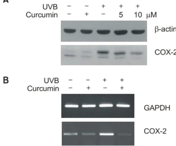
Inhibitory effect of curcum in on UVB-induced expression of CO X-2
To investigate whether curcumin inhibits the UVB-in- duced COX-2 expression, HaCaT cells were exposed to UVB (200 mJ/cm
2) with or without curcumin. As shown in Figure 2A, up-regulation of COX-2 by UVB was markedly decreased in a concentration-depen- dent manner. To investigate whether curcumin at- tenuates COX-2 expression by UVB at transcriptional level, we performed RT-PCR analysis using COX-2 specific primers. As shown in Figure 2B, increased
Figure 1. Up-regulation of COX-2 in UVB-irradiated HaCaT cells. The cells were cultured to 90 % confluence in DMEM supplemented with 10% fetal bovine serum at 37oC and in 5% CO2. The cells were then serum-starved for 24 h. Before UVB irradiation, the cells were washed with PBS and were exposed to 200 mJ/cm2 of UVB. After UVB irradiation cells were continuously cultured in serum-free media. The cells were harvested at indicated times and the cell lysates were prepared for Western blot (A, B) and RT-PCR analysis (C). Similar results were shown in two different experiments.50
C
GAPDH
0 100 200 mJ/cm2
COX-2
A
h 6
0 12
[200 mJ/cm ]2
β-actin COX-2
h 6
0 12
[200 mJ/cm ]2
B
β-actin COX-2
expression of COX-2 mRNA by UVB was clearly inhibited in the presence of curcumin, indicating that COX-2 expression is regulated at transcriptional level by curcumin.
Effect of curcum in on UVB-stim ulated activation of M APKs
Accumulating data suggest that UVB-irradiated HaCaT
cells show an increased expression of COX-2 by MAPKs-dependent pathways, such as p38 MAPK and JNK. However it is largely unknown whether curcumin modulates the expression of COX-2 by inhibition of these MAPKs pathways in UVB-irradiated HaCaT cells. We examined the effect of UVB on activations of p38 MAPK and JNK in HaCaT cells. As shown in Figure 3A, irradiation of UVB (200 mJ/cm
2) resulted in phosphorylation (activation) of both kinases. Acti- vation of both kinases became apparent at 5 min following irradiation of UVB. Stripping and reprobing the same membrane with antibodies against p38 MAPK and JNK revealed no change in total protein levels of each kinase, indicating that UVB induced activation of preexisting p38 MAPK and JNK. To confirm the p38 MAPK-dependent and JNK-depen- dent COX-2 up-regulation, HaCaT cells were pre- treated with SB203580 (20 µM) and SP600125 (20 µM) and then the cells were irradiated by UVB. p38 inhibitor SB203580 and JNK inhibitor SP600125 specifically inhibited the activation of p38 and JNK, respectively (Figure 3B). We next investigated whe- ther curcumin (10 µM) inhibits the activation of p38 MAPK and JNK in UVB-irradiated HaCaT cells. As shown in Figure 3C, phosphorylation of p38 MAPK and JNK by UVB was markedly decreased in curcu- min-treated HaCaT cells. Furthermore UVB-induced COX-2 expressions were dramatically attenuated by pretreatment of SB203580, SP600125, or curcumin (Figure 3D), indicating that curcumin attenuates the UVB-induced COX-2 expression through inhibition of p38 MAPK and JNK activation. However, curcumin
Figure 2. Inhibition of UVB-induced COX-2 expression by curcumin.The cells were irradiated by UVB (200 mJ/cm2) with or without curcumin. The expressions of COX-2 protein and mRNA were examined by Western blot (A) or RT-PCR analysis (B), respectively.
Similar results were shown in two experiments.
A
β-actin COX-2
B
UVB
Curcumin 5 10 µM
CurcuminUVB
GAPDH
COX-2
Figure 3. Inhibition of UVB-induced COX-2 expression by MAPKs inhi- bitors. (A) After UVB irradiation (200 mJ/cm2), the cells were har- vested at indicated times. At each time, whole cell lysates were pre- pared and used for p-p38 MAPK, p-JNK, p38 MAPK, or JNK Wes- tern with respective antibodies. (B, C) The cells were pretreated with 20 µM p38 inhibitor SB203580 (SB) or 20 µM JNK inhibitor SP 600125 (SP), or 10 µM curcumin for 30 min and then treated with UVB (200 mJ/cm2). Whole cell lysates were prepared and used for p-p38 MAPK, p-JNK, p38 MAPK, or JNK Western with re- spective antibodies. (D) These same concentrations of MAPK inhibitors or curcumin were also treated to UVB-irradiated cells and then cell lysates were subjected to COX-2 Western. Similar results were shown in two experiments.
5
D
15 30 min
B
UVB (200 mJ/cm )2
p-JNK
A
p-p38
JNK
SB UVB SP UVB
p38
JNK p-JNK
COX-2 p-p38
SP UVB p-JNK p-p38
JNK p38
C
Curcumin UVBp38
β-actin SB
Curcumin
did not completely inhibit the activation of p38 MAPK or JNK after UVB irradiation, curcumin showed a partial inhibitory effect on the expression of COX-2 compared to SB203580 or SP600125.
Effect of curcum in on UVB-induced activation of AP-1
AP-1, a down-stream molecule of p38 and JNK, plays a key role as a transcription factor involved in UVB-induced COX-2 expression. To investigate whe- ther curcumin decreases the AP-1 binding activity in UVB-irradiated HaCaT cells, we performed an EMSA using 21 bp consensus oligonucleotides. Exposure of HaCaT cells to UVB (200 mJ/cm
2) led to an activation of AP-1 dramatically as compared to control (Figure 4A). However, in the presence of curcumin, UVB- induced AP-1 activation was dramatically inhibited, evidenced by decreased AP-1 binding activity (Figure 4B). These results suggest that UVB-induced p38 and JNK activations are inhibited by curcumin treatment, and subsequently resulting in decreased activation of AP-1 in nucleus, thereby leading the down-regulation of COX-2 expression in HaCaT cells.
D iscussion
COX-2 plays important roles in the development of carcinogenesis as well as inflammation in UVB- irradiated skin (Grandjean-Laquerriere A et al., 2002;
Surh YJ, 2003; Wilgus et al., 2003; Cui et al., 2004;
Nijsten et al., 2004). Curcumin is known to exert its anti-inflammatory action in various cell lines through inhibition of COX-2 expression (Chun et al., 2003;
Kang et al., 2004). Thus, it is important to develop an efficient strategy using curcumin to down-regulate the expression of COX-2 in UVB-irradiated skin (Wolf
et al., 2003).Accumulating evidences suggest that curcumin in- hibits the expression of COX-2 in several experi- mental models (Zhang et al., 1999; Chun et al., 2003;
Surh, 2003; Kang et al., 2004). Consistent with these, through the present study, we have demonstrated that curcumin also effectively inhibits the expressions of COX-2 mRNA and protein induced by UVB irradiation in a human keratinocyte cell line HaCaT, which seems to be, to our knowledge, the first report. Ac- cordingly, the expression of COX-2 has been shown to be affected by various intracellular signaling proteins such as p38 MAPK and JNK (Goel et al., 2001; Surh et al., 2001; Liu et al., 2003; Lin et al., 2004; Kang et al., 2004). Interestingly, it has been recently shown that UVB induces the expression of COX-2 by activation of p38 MAPK in HaCaT cells (Chen et al., 2001) or by activations of p38 MAPK and JNK in artificial epidermis (Mahns et al., 2004).
In this study, we have observed that p38 MAPK and JNK were dramatically activated in UVB-irradiated HaCaT cells. Of importance, we have found in this study that pretreatment with curcumin effectively
suppresses UVB-induced activation of p38 MAPK and JNK as well as expression of COX-2, thus suggesting that curcumin may exert its inhibitory effect on UVB-induced COX-2 expression by inhibiting p38 MAPK and JNK activities. In most cases, p38 MAPK and JNK activations in response to various extra- cellular stimuli are linked to activation of AP-1 (Minden et al., 1994; Davis, 1995; Minden and Karin, 1997). AP-1 is known to regulate the expression of COX-2 in many systems (Dori and Aggarwal, 2004;
Lin et al., 2004; Kang et al., 2004). In this study, we have observed that UVB is able to increase AP-1 DNA binding activity in HaCaT cells that is attenuated by treatment with curcumin. These results may sug- gest that curcumin may suppress AP-1 activity pro-
Figure 4. Suppression of UVB-induced AP-1 activation by curcumin.(A) After UVB irradiation (200 mJ/cm2), nuclear extracts were prepared and AP-1 DNA binding activity was determined by EMSA. (B) The cells were also pretreated with curcumin (10 µM) and were exposed to UVB (200 mJ/cm2). Nuclear extracts were prepared and AP-1 DNA binding activity was determined by EMSA.
A
AP-1
B
0
UVB (200 mJ/cm )2
Arbitrary unit
AP-1
0 1 2 3 4
6 8
4
2
30 60 min
1 2 3 4
UVB Curcumin
AP-1
bably by inhibition of p38 MAPK and JNK activation in HaCaT cells. Taken together, our data suggest that suppressions of p38 MAPK, JNK, and AP-1 activity are an important molecular mechanism underlying curcumin-mediated down-regulation of COX-2 in UVB-irradiated HaCaT cells. It should be noted that curcumin can inhibit phorbol ester-induced expression of COX-2 in mouse skin through suppression of ERK activity (Chun et al., 2003). However, in this study, we have observed that though UVB is able to activate ERK in HaCaT cells, curcumin does not prevent the activation of ERK in response to UVB irradiation in these cells (data not shown). Thus, molecular me- chanisms underlying suppression of COX-2 expres- sion by curcumin may be dependent on the contexts of cells or kinds of stimuli treated to cells.
In conclusion, findings of the present study demon- strate for the first time that curcumin inhibits the expression of COX-2 mRNA and protein in UVB- irradiated HaCaT cells, and indicate that the inhibitory effect of curcumin on UVB-induced COX-2 expression is likely to be, at least in part, associated with suppression of JNK, p38 MAPK, and AP-1, thereby suggesting that curcumin may be used as a promising sunscreen substance.
Acknow ledgem ent
This study was supported by a grant (02-PJ9-PG1- CO04-009) of the Oriental Medicine R&D Project, Ministry of Health and Welfare, Republic of Korea
R eferences
Chen W, Tang Q, Gonzales MS, Bowden GT. Role of p38 MAP kinases and ERK in mediating ultraviolet-B induced cyclooxygenase-2 gene expression in human keratinocytes.
Oncogene 2001;20:3921-6
Cho JW, Jeong YW, Kim KS, Oh JY, Park JC, Lee JC, Baek WK, Suh SI, Suh MH. P21WAF1 is associated with CDK2 and CDK4 protein during HL-60 cell differentiation by TPA treatment. Cell Prolif 2001;34:267-4
Chun KS, Keum YS, Han SS, Song YS, Kim SH, Surh YJ.
Curcumin inhibits phorbol ester-induced expression of cyclooxygenase-2 in mouse skin through suppression of extracellular signal-regulated kinase activity and NF-kappaB activation. Carcinogenesis 2003;24:1515-24
Cui Y, Kim DS, Park SH, Yoon JA, Kim SK, Kwon SB, Park KC. Involvement of ERK AND p38 MAP kinase in AAPH- induced COX-2 expression in HaCaT cells. Chem Phys Lipids 2004;129:43-52
Davis RJ. Transcriptional regulation by MAP kinases. Mol Reprod Dev 1995;42:459-67
De Fabo EC, Noonan FP, Fears T, Merino G. Ultraviolet B but not ultraviolet A radiation initiates melanoma. Cancer Res 2004;64:6372-6
de Gruijl FR. Photocarcinogenesis: UVA vs. UVB radiation.
Skin Pharmacol Appl Skin Physiol 2002;15:316-20
Dorai T, Aggarwal BB. Role of chemopreventive agents in cancer therapy. Cancer Lett 2004;215:129-40
Goel A, Boland CR, Chauhan DP. Specific inhibition of cyclooxygenase-2 (COX-2) expression by dietary curcumin in HT-29 human colon cancer cells. Cancer Lett 2001;172:
111-8
Grandjean-Laquerriere A, Gangloff SC, Le Naour R, Tren- tesaux C, Hornebeck W, Guenounou M. Relative contribution of NF-kappaB and AP-1 in the modulation by curcumin and pyrrolidine dithiocarbamate of the UVB-induced cytokine expression by keratinocytes. Cytokine 2002;18:168-77 Granstein RD, Matsui MS. UV radiation-induced immunosup- pression and skin cancer. Cutis 2004;74(5 Suppl):4-9 Heck DE, Vetrano AM, Mariano TM, Laskin JD. UVB light stimulates production of reactive oxygen species: unexpected role for catalase. J Biol Chem 2003;278:22432-6
Hong J, Bose M, Ju J, Ryu JH, Chen X, Sang S, Lee MJ, Yang CS. Modulation of arachidonic acid metabolism by curcumin and related beta-diketone derivatives: effects on cytosolic phospholipase A(2), cyclooxygenases and 5-lipoxy- genase. Carcinogenesis 2004;25:1671-9
Kang G, Kong PJ, YuhYJ, Lim SY, Yim SV, Chun W, Kim SS. Curcumin suppresses lipopolysaccharide-induced cyclo- oxygenase-2 expression by inhibiting activator protein 1 and nuclear factor kappab bindings in BV2 microglial cells. J Pharmacol Sci 2004;94:325-8
Kulms D, Zeise E, Poppelmann B, Schwarz T. DNA damage, death receptor activation and reactive oxygen species contribute to ultraviolet radiation-induced apoptosisin an essential and independent way. Oncogene 2002;21:5844-51 Lee KS, Lee WS, Suh SI, Kim SP, Lee SR, Ryoo YW, and Kim BC. Melatonin reduces ultraviolet-B induced cell damages and polyamine levels in human skin fibroblasts in culture. Exp Mol Med 2003;35:263-8
Lin SK, Kok SH, Yeh FT, Kuo MY, Lin CC, Wang CC, Gold- ring SR, Hong CY. MEK/ERK and signal transducer and activator of transcription signaling pathways modulate oncostatin M-stimulated CCL2 expression in human oste- oblasts through a common transcription factor. Arthritis Rheum 2004;50:785-93
Liu G, Ma WY, Bode AM, Zhang Y, Dong Z. NS-398 and piroxicam suppress UVB-induced activator protein 1 activity by mechanisms independent of cyclooxygenase-2. J Biol Chem 2003;278:2124-30
Mahns A, Wolber R, Stab F, Klotz LO, Sies H. Contribution of UVB and UVA to UV-dependent stimulation of cyclo- oxygenase-2 expression in artificial epidermis. Photochem Photobiol Sci 2004;3:257-62
Matsumura Y, Ananthaswamy HN. Toxic effects of ultraviolet radiation on the skin. Toxicol Appl Pharmacol 2004;195:298- 308
Minden A, Lin A, Smeal T, Derijard B, Cobb M, Davis R, Karin M. c-Jun N-terminal phosphorylation correlates with activation of the JNK subgroup but not the ERK subgroup of mitogen-activated protein kinases. Mol Cell Biol 1994;14:
6683-8
Minden A, Karin M. Regulation and function of the JNK
subgroup of MAP kinases. Biochim Biophys Acta 1997;1333:
85-104
Nijsten T, Colpaert CG, Vermeulen PB, Harris AL, Van Marck E, Lambert J. Cyclooxygenase-2 expression and an- giogenesis in squamous cell carcinoma of the skin and its precursors: a paired immunohistochemical study of 35 cases.
Br J Dermatol 2004;151:837-45
Ramos J, Villa J, Ruiz A, Armstrong R, Matta J. UV dose determines key characteristics of nonmelanoma skin cancer.
Cancer Epidemiol Biomarkers Prev 2004;13:2006-11 Rhee SJ. Redox signaling: hydrogen peroxide as intracellular messenger. Exp Mol Med 1999;31:53-9
Surh YJ, Chun KS, Cha HH, Han SS, Keum YS, Park KK, Lee SS. Molecular mechanisms underlying chemopreventive activities of anti-inflammatory phytochemicals: down-regula- tion of COX-2 and iNOS through suppression of NF-kappa B activation. Mutat Res 2001;480-481:243-68
Surh YJ. Cancer chemoprevention with dietary phytoche- micals. Nature Rev Cancer 2003;3:768-80
Torres M, Forman HJ. Redox signaling and the MAP kinase pathways. Biofactor 2003;17:287-96
Wilgus TA, Koki AT, Zweifel BS, Kusewitt DF, Rubal PA, Oberyszyn TM. Inhibition of cutaneous ultraviolet light B- mediated inflammation and tumor formation with topical celecoxib treatment. Mol Carcinog 2003;38:49-58
Wolf R, Matz H, Orion E, Lipozencic J. Sunscreens-the ultimate cosmetic. Acta Dermatovenerol Croat 2003;11:158- 62
Zhang F, Altorki NK, Mestre JR, Subbaramaiah K, Dan- nenberg AJ. Curcumin inhibits cyclooxygenase-2 transcription in bile acid- and phorbol ester-treated human gastrointestinal epithelial cells. Carcinogenesis 1999;20:445-51
Zhu YG, Chen XC, Chen ZZ, Zeng YQ, Shi GB, Su YH, Peng X. Curcumin protects mitochondria from oxidative da- mage and attenuates apoptosis in cortical neurons. Acta Pharmacol Sin 2004;25:1606-12