© 2012 The Korean Society of Pathologists/The Korean Society for Cytopathology pISSN 1738-1843
eISSN 2092-8920 75
Benign peripheral nerve sheath tumors (PNST) consist of schwannomas, neurofibromas, and perineuriomas.
1Schwanno- mas and perineuriomas are composed of uniform populations of Schwann cells and perineurial cells, respectively. Unlike these PNSTs, neurofibromas consist of diverse cell types, including Schwann cells, fibroblasts, perineurial cells, and entrapped axo- ns. Perineuriomas are far less common than neurofibromas and schwannomas, accounting for approximately 1% of PNST.
2Peri- neuriomas cannot be diagnosed by light microscopy alone, there- fore, have been recently recognized by the immunoreactivity for epithelial membrane antigen (EMA) of the perineurial cells.
3Hybrid peripheral nerve sheath tumors, which are distinct entities of PNST, have been recently reported. These hybrid PNSTs include hybrid schwannoma-neurofibromas, hybrid schwannoma-perineuriomas and hybrid neurofibroma-perineu- riomas.
4,5These hybrid tumors usually arise in the dermis and subcutis, and they occur over a wide range of age and anatomi- cal distribution.
5The pathogenesis of these hybrid tumors re- mains unknown, but might be associated with a localized mi- croenvironmental change or clonal genetic alteration of the primitive tumor cells.
4In this report, we describe two interesting cases of a pure form
of soft tissue perineurioma, arising in the skin of a 56-year-old female and a hybrid perineurioma and schwannoma of the pos- terior mediastinum of a 53-year-old male.
CASE REPORTS
Case 1
A 56-year-old female presented with a palpable skin nodule on the anterior chest wall. The patient was not associated with neurofibromatosis type 1 or 2. Excision of the skin nodule was performed. The nodule was well-defined, ovoid, pale tan, and rubbery firm, and measured 2.2
×1.5
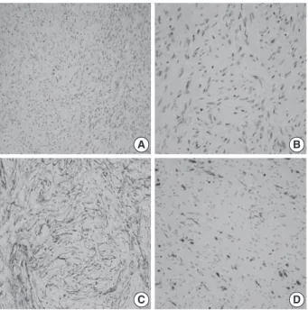
×1.5 cm. On microscopic examination, the nodule was well-circumscribed and composed of slender spindle cells arranged in loose storiform or short fas- cicular patterns. The tumor cells had wavy nuclei and long stre- amer-like cytoplasmic processes (Fig. 1A, B). No mitosis or ne- crosis was found. The tumor cells were immunohistochemically diffusely and strongly positive for EMA and claudin-1. The S- 100 protein was only focally positive (Fig. 1C, D). An electron microscopic study failed to reveal any detailed findings due to inappropriately preserved formalin-fixed paraffin-embedded tis- sue.
A Soft Tissue Perineurioma and a Hybrid Tumor of Perineurioma and Schwannoma
Ji Young Park · Nam Jo Park Sang Pyo Kim · Kun Young Kwon Sang Sook Lee
Department of Pathology, Keimyung University School of Medicine, Daegu, Korea
Perineuriomas are composed of differentiated perineurial cells. Perineuriomas have been recently recognized by the immunoreactivity for epithelial membrane antigen (EMA). Microscopically, peri- neuriomas show proliferation of spindle cells with wavy nuclei and delicate elongated bipolar cy- toplasmic processes. The tumor cells are usually negative for the S-100 protein. Ultrastructurally, perineurial cells reveal slender, nontapered processes containing pinocytic vesicles and discon- tinuous basal lamina. Interestingly, hybrid tumors of benign peripheral nerve sheath tumor (PNST) have been recently reported by using immunohistochemical and ultrastructural investigations. Her- ein, we report a case of soft tissue perineurioma arising in the skin of a 56-year-old female; an- other case of a hybrid tumor of perineurioma and schwannoma in the posterior mediastinum oc- curred in a 53-year-old male, which is the first case of the hybrid PNST tumor reported in Korea.
Key Words: Soft tissue; Perineurioma; Nerve sheath neoplasms; Hybrid tumor Received: January 21, 2011
Revised: April 5, 2011 Accepted: April 7, 2011 Corresponding Author Sang Sook Lee, M.D.
Department of Pathology, Keimyung University School of Medicine, 1095 Dalgubeoldae-ro, Dalseo-gu, Daegu 704-701, Korea Tel: +82-53-580-3811 Fax: +82-53-580-3823 E-mail: [email protected]
The Korean Journal of Pathology 2012; 46: 75-78
http://dx.doi.org/10.4132/KoreanJPathol.2012.46.1.75 ▒ BRIEF CASE REPORT ▒
76 • Park JY, et al.
http://www.koreanjpathol.org http://dx.doi.org/10.4132/KoreanJPathol.2012.46.1.75
the schwannoma-looking area, but did not stain for EMA or claudin-1 (Fig. 2E, F).
Specimens were sampled separately from the two representa- tive areas of the formalin-fixed paraffin-embedded block for ul- trastructural studies. The cells of the perineurioma-like area showed slender and long-tapered processes containing pinocytic vesicles and discontinuous external lamina (Fig. 3A). In con- trast, the schwannoma-looking area exhibited tumor cells with attenuated cell processes invested by continuous basal lamina (Fig. 3B). The pathological diagnosis was a hybrid perineurio- ma and schwannoma.
DISCUSSION
PNSTs include common schwannomas and neurofibromas, and, rare perineuriomas.
Schwannomas are composed of Schwann cells and have char- acteristic cellular features, including Antoni A and B areas, nu- clear palisadings (Verocay bodies), and hyalinized vessels with perivascular hemosiderin depositions. Neurofibromas consist of an admixture of Schwann cells, fibroblasts, perineurial cells, and scattered axons.
1Perineuriomas are exclusively composed of differentiated peri- neurial cells. Perineuriomas can be divided into four categories, rare intraneural perineuriomas, more common soft tissue (extra- neural) perineuriomas, and sclerosing and reticular variants.
2,6Intraneural perineuriomas, formerly known as ‘localized hy- pertrophic neuropathy’ usually occur in the extremities of young individuals.
1They microscopically show concentric prolifera- tion of perineurial cells around axons and Schwann cells, form- ing “onion bulbs” that stain positive for EMA.
1Sclerosing peri- neuriomas occur exclusively in the acral areas of young adults and are characterized by cords of bland epithelioid cells embed- ded in sclerotic collagen.
1Reticular perineuriomas are a recently recognized variant of perineuriomas, which are characterized by a predominantly lace-like or reticular growth pattern composed of fusiform cells with bipolar cytoplasmic processes.
5Soft tissue perineuriomas primarily affect adults and involve the superficial soft tissues of the extremities and trunk; however, approximately 30% can develop in deep soft tissue and, rarely, visceral locations.
2Soft tissue perineuriomas show microscopic proliferation of spindle cells with wavy nuclei and elongated bi- polar cytoplasmic processes arranged in a storiform or short fas- cicular pattern.
1Perineurioimas are usually positive for EMA and can also ex- press claudin-1, GLUT-1, and CD34.
7Electron microscopy
Fig. 1. A soft tissue perineurioma arising in the skin. Micrographs (A, B) show proliferation of spindle cells with wavy nuclei and deli- cate elongated bipolar cytoplasmic processes. Immunohistochem- ical staining reveals proliferating spindle cells, diffusely and strongly positive for epithelial membrane antigen (C) and focally positive for the S-100 protein (D).
A
C
B
D
Case 2
A 53-year-old male visited our out-patient clinic for the eval- uation of the posterior mediastinal mass, incidentally found dur- ing a regular health check-up. No remarkable medical history was noted. Thoracoscopic excision of the mass was performed.
The gross specimen consisted of multiple fragments of pale tan, translucent, and relatively firm soft tissue, measuring 5.0
×5.2
×2.0 cm in aggregates.
The mediastinal mass, microscopically, revealed two distinct spindle cell areas. The majority of the mass was composed of spindle cells arranged in short fascicular or parallel cords in a fi- bromyxoid matrix as seen in soft tissue perineuriomas. The spin- dle cells had wavy nuclei and elongated bipolar cytoplasmic pro- cesses (Fig. 2A). Another spindle cell area was noted, manifest- ing as a typical schwannoma, including Antoni A and B areas, nuclear palisading, and hyalinized blood vessels (Fig. 2D). Nu- clear atypia, necrosis or mitosis was not observed in both areas.
The perineurioma-like areas constituted about 80-85% of the tumor volume.
The perineurioma-like area of the mediastinal tumor was dif-
fusely and strongly positive for EMA and claudin-1, but nega-
tive for S-100 protein (Fig. 2B, C). However, the tumor cells
were evenly and strongly immunoreactive for S-100 protein in
Perineuroma and Hybrid Tumor of PNST • 77
http://www.koreanjpathol.org http://dx.doi.org/10.4132/KoreanJPathol.2012.46.1.75
findings are most useful for diagnosing perineuromas, which are characterized by showing perineurial cells with slender bi- polar cytoplasmic processes containing pinocytic vesicles and discontinuous external lamina.
1They generally behave in a be- nign fashion, and surgical removal is curative.
1Although the tumors are not associated with neurofibroma- tosis type 1 or 2, a case of soft tissue perineurioma in a patient with neurofibromatosis type 2 has been reported.
3Additionally, some studies have described that fluorescence in situ hybridiza- tion and molecular analysis demonstrate deletions or point mu- tations on the chromosome 22q11 in the region of the NF2 gene and chromosome 10. These findings suggested that there might be diverse genetic mechanisms underlying the development of perineuriomas.
6These benign PNSTs are currently distinct entities. However, small numbers of the hybrid form of PNST have been recently
reported. These include hybrid schwannoma-neurofibromas, hybrid schwannoma-perineuriomas, and hybrid neurofibroma- perineuriomas.
4,5,7Feany et al.
4and Hornick et al.
5reported nine cases of schwan- nomas and neurofibromas hybrids and 42 cases of hybrid sch- wannomas and perineuriomas. They reported that hybrid tu- mors have well-documented histological differences with im- munohistochemical and electron microscopic features and sug- gested that the pathogenesis of the diverse differentiation re- mains unknown, but could result from incomplete differentia- tion or genetic alteration of primitive tumor cells, or in respon se to a localized microenvironmental change.
4,5,7Hybrid tumors usually arise in the dermis and subcutis over a wide range of age and anatomical distribution and seem to pursue a benign course.
5Nuclear atypia is relatively common in the hybrid tumors, which seems degenerative in nature, includ-
Fig. 2. A hybrid perineurioma and schwannoma in the posterior mediastinum. Central circle shows two distinct representative areas of the hybrid tumor. The perineurioma-looking area (A) is composed of spindle cells with wavy nuclei and elongated bipolar cytoplasmic processes.
The perineurioma area is also diffusely and strongly positive for epithelial membrane antigen (EMA) (B) but, negative for S-100 protein (C). An- other spindle area manifests as a typical schwannoma (D), including Antoni A and B areas and nuclear palisading. In the schwannoma-look- ing area, the tumor cells are evenly and strongly immunoreactive for S-100 protein (F), but do not stain for EMA (E). H&E, hamatoxylin and eosin.
A
D
B
E
C
F
H&E EMA S-100 protein
78 • Park JY, et al.
http://www.koreanjpathol.org http://dx.doi.org/10.4132/KoreanJPathol.2012.46.1.75
ing smudged chromatin and occasional nuclear pseudoinclu- sions. The differential diagnoses of the hybrid tumors may in- clude other benign nerve sheath tumors and low grade malig- nant peripheral nerve sheath tumors (MPNST). MPNST should be considered, if nuclear atypia is prominent.
5As information and clinical follow-up data on hybrid tumors are limited, additional studies are needed to understand the pa- thogenesis and clinicopathological significance of hybrid tumors.
In conclusion, we described two interesting cases of a pure form of soft tissue perineurioma, which stained positively for EMA and claudin-1, and a hybrid perineurioma and schwanno- ma documented by immunohistochemical and ultrastructural features. To our knowledge, this is the first report of a hybrid peripheral nerve sheath tumor in Korea.
Conflicts of Interest
No potential conflict of interest relevant to this article was reported.
REFERENCES
1. Skovronsky DM, Oberholtzer JC. Pathologic classification of pe- ripheral nerve tumors. Neurosurg Clin N Am 2004; 15: 157-66.
2. Kim JM, Choi JH. Soft tissue perineurioma: a case report. Korean J Pathol 2009; 43: 266-70.
3. Pitchford CW, Schwartz HS, Atkinson JB, Cates JM. Soft tissue peri- neurioma in a patient with neurofibromatosis type 2: a tumor not previously associated with the NF2 syndrome. Am J Surg Pathol 2006; 30: 1624-9.
4. Feany MB, Anthony DC, Fletcher CD. Nerve sheath tumours with hybrid features of neurofibroma and schwannoma: a conceptual challenge. Histopathology 1998; 32: 405-10.
5. Hornick JL, Bundock EA, Fletcher CD. Hybrid schwannoma/peri- neurioma: clinicopathologic analysis of 42 distinctive benign nerve sheath tumors. Am J Surg Pathol 2009; 33: 1554-61.
6. Brock JE, Perez-Atayde AR, Kozakewich HP, Richkind KE, Fletcher JA, Vargas SO. Cytogenetic aberrations in perineurioma: variation with subtype. Am J Surg Pathol 2005; 29: 1164-9.
7. Kazakov DV, Magro G, Yu Orlov A, et al. Benign schwannoma with perineurioma-like areas: a clinicopathologic study of 11 cases. Int J Surg Pathol 2006; 14: 320-5.
Fig. 3. Transmission electron micrographs of the hybrid perineurioma and schwannoma. In the perineurioma area (A), the tumor cells show slender cytoplasmic processes containing pinocytic vesicles (indicated by arrows in the inset) and discontinuous external lamina (arrowheads).
In contrast, the schwannoma area (B) exhibits attenuated cell processes invested by continuous basal lamina (×4,000).
A B